. 2011 Jan 14;286(2):1292-300.
doi: 10.1074/jbc.M110.154849. Epub 2010 Nov 9.

Mannose trimming is required for delivery of a glycoprotein from EDEM1 to XTP3-B and to late endoplasmic reticulum-associated degradation steps

Affiliations

Mannose trimming is required for delivery of a glycoprotein from EDEM1 to XTP3-B and to late endoplasmic reticulum-associated degradation steps

Bella Groisman et al. J Biol Chem. .

Abstract

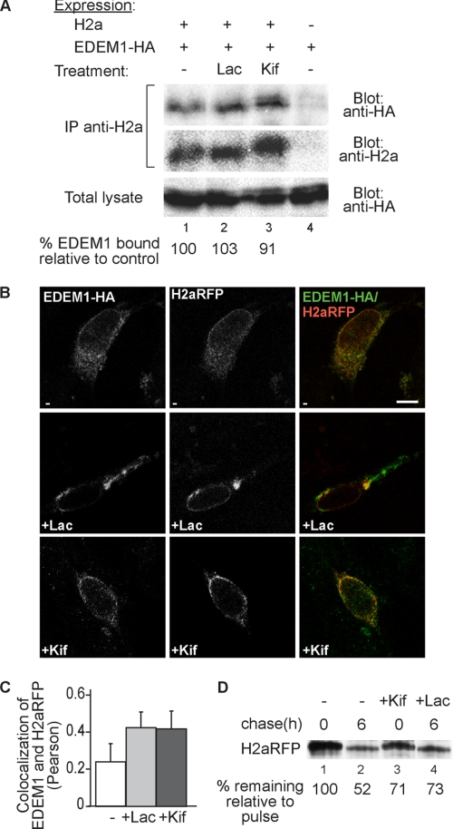
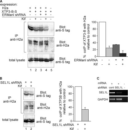
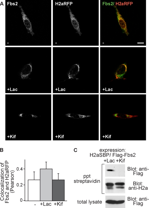
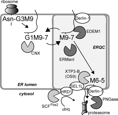
Although the trimming of α1,2-mannose residues from precursor N-linked oligosaccharides is an essential step in the delivery of misfolded glycoproteins to endoplasmic reticulum (ER)-associated degradation (ERAD), the exact role of this trimming is unclear. EDEM1 was initially suggested to bind N-glycans after mannose trimming, a role presently ascribed to the lectins OS9 and XTP3-B, because of their in vitro affinities for trimmed oligosaccharides. We have shown before that ER mannosidase I (ERManI) is required for the trimming and concentrates together with the ERAD substrate and ERAD machinery in the pericentriolar ER-derived quality control compartment (ERQC). Inhibition of mannose trimming prevents substrate accumulation in the ERQC. Here, we show that the mannosidase inhibitor kifunensine or ERManI knockdown do not affect binding of an ERAD substrate glycoprotein to EDEM1. In contrast, substrate association with XTP3-B and with the E3 ubiquitin ligases HRD1 and SCF(Fbs2) was inhibited. Consistently, whereas the ERAD substrate partially colocalized upon proteasomal inhibition with EDEM1, HRD1, and Fbs2 at the ERQC, colocalization was repressed by mannosidase inhibition in the case of the E3 ligases but not for EDEM1. Interestingly, association and colocalization of the substrate with Derlin-1 was independent of mannose trimming. The HRD1 adaptor protein SEL1L had been suggested to play a role in N-glycan-dependent substrate delivery to OS9 and XTP3-B. However, substrate association with XTP3-B was still dependent on mannose trimming upon SEL1L knockdown. Our results suggest that mannose trimming enables delivery of a substrate glycoprotein from EDEM1 to late ERAD steps through association with XTP3-B.


Figures

FIGURE 1.
FIGURE 1.
Mannose trimming and EDEM1 are required for ERAD of H2a. A, HEK 293 cells cotransfected with an H2a cDNA encoding vector were pulse-labeled for 20 min with [35S]cysteine and chased for 0 or 5 h in complete medium in the absence (lanes 1 and 2) or in the presence (lanes 3 and 4) of Kif (100 μm). After the pulse (0 h chase) or chase periods, the cells were lysed, H2a was immunoprecipitated, and the immunoprecipitates were separated in 12% SDS-PAGE followed by phosphorimaging. Bands corresponding to the H2a precursor and the naturally occurring cleaved fragment are indicated on the left. For the pulse samples, two bands can be seen for the H2a precursor and also for the fragment, with the lower ones corresponding to underglycosylated species (one of the three glycosylation sites unoccupied). B and C, similar to the pulse-chase experiment in A but performed with untreated cells transiently cotransfected with an H2a cDNA encoding vector together with either a GFP-containing pSUPER-retro vector (B and C, lanes 1 and 2) or with pSUPER encoding anti-EDEM1 shRNA (B, lanes 3 and 4) or with an EDEM1-HA containing pCMVsport2 plasmid (C, lanes 3 and 4). D, the bar graph shows the average percent H2a remaining after chase relative to the corresponding pulse, calculated from phosphorimaging quantitations of all H2a species (precursor and fragment) from three independent experiments similar to each of the ones shown in A–C. Error bars indicate S.D. between experiments. Student's t test renders p < 0.02 for values in all chase samples compared with control chase. E, in parallel with B, HEK 293 cells were transfected with either pSUPER (control, lane 1) or the same plasmid encoding anti-EDEM1 shRNA (lane 2). RNA was extracted 48 h post-transfection and used for RT-PCR with primers for EDEM1 mRNA (upper panel) compared with GAPDH (lower panel). Lane 3 shows a sample with no RNA template.
FIGURE 2.
FIGURE 2.
Inhibition of mannose trimming does not preclude EDEM1 interaction with H2a nor their subcellular colocalization. A, HEK 293 cells were cotransfected with a vector encoding for HA-tagged EDEM1 (EDEM1-HA) with or without an H2a encoding vector. Two days after transfection, the cells were incubated for 3 h in the absence/presence of 25 μm Lac or 100 μm Kif. The cells were lysed in 1% Nonidet P-40, 50 mm Tris/HCl (pH 8), 150 mm NaCl, and 10% of the lysates were run on 10% SDS-PAGE and immunoblotted with anti-HA antibodies (bottom panel). The rest of the lysates were immunoprecipitated (IP) with anti-H2a and protein A-Sepharose. Eluted samples were subjected to 10% SDS-PAGE and immunobloted with anti-HA (upper panel) or anti-H2a (middle panel). H2a (35-kDa fragment) (see Fig. 1) appeared as a very weak band in immunoblots and is therefore not shown. Quantitations of the relative amounts of EDEM1 associated with H2a are shown at the bottom as percent of EDEM1-HA coprecipitated with H2a relative to the mock transfection. The results are normalized by dividing the intensity of the EDEM1 band in the upper panel by that of its corresponding band in the lower panel and by that of H2a in the middle panel. B, NIH 3T3 cells were transiently cotransfected with H2aRFP together with HA-tagged EDEM1. 24 h after transfection, cells were incubated for 3 h in the absence (upper panels) or presence of 25 μm Lac (middle panels) or 100 μm Kif (lower panels), fixed, permeabilized, and incubated with mouse anti-HA followed by probing with FITC conjugated goat-anti mouse IgG. The samples were analyzed in an LSM confocal microscope. Representative optical slices are shown. Colocalization of FITC with RFP appears yellow. Bar, 10 μm. C, the graph shows colocalization analysis (Pearson) performed using the ImageJ program (average of 30 cells from three independent experiments are presented; error bars are S.D.); p < 0.045 for treated samples compared with untreated. D, similar to the pulse-chase experiment in Fig. 1A but performed with cells transiently transfected with an H2aRFP cDNA-encoding vector in the absence or in the presence of Kif (100 μm) or Lac (25 μm).
FIGURE 3.
FIGURE 3.
ERManI activity is not required for association of H2a with EDEM1. A, HEK 293 cells were cotransfected with a vector encoding for HA-tagged EDEM1 (EDEM1-HA) with or without an H2a encoding vector and with or without a pSUPER plasmid encoding for anti-ERManI shRNA (shERManI). 24 h post-transfection, cells were lysed and processed, and results were quantitated and normalized as in Fig. 2A. B, in parallel with A, HEK 293 cells were transfected with the plasmid encoding anti-ERManI shRNA used in A or with pSUPER encoding anti-LacZ shRNA (control). RNA was extracted 24 h post-transfection and used for RT-PCR with primers for ERManI mRNA (upper panel) compared with GAPDH (lower panel). IP, immunoprecipitation.
FIGURE 4.
FIGURE 4.
Association of the ERAD substrate to XTP3-B requires mannose trimming, even after knockdown of SEL1L. A, plasmids encoding for S-tagged XTP3-B and H2a cDNAs were cotransfected into HEK-293 cells with or without ERManI shRNA in a pSUPER plasmid. 24 h after transfection, cells were incubated for 5 h in the absence/presence of 100 μm Kif. Cells were lysed in 1% Nonidet P-40, 50 mm Tris/HCl (pH 8), 150 mm NaCl and 10% of the lysates were run on 10% SDS-PAGE and immunoblotted with anti-S tag antibodies (bottom panel). The rest of the lysates were immunoprecipitated (IP) with anti-H2a and protein A-Sepharose. Eluted samples were subjected to 10% SDS-PAGE and immunoblotted with anti-S tag (upper panel) or anti-H2a (middle panel). Results were quantitated and normalized as in Fig. 2A; values are averages of three independent experiments, error bars are S.D.; p < 0.04 for all coimmunoprecipitations compared with the control. B, experiment similar to that in A, except for SEL1L shRNA that was used instead of ERManI shRNA (p = 0.03). C, in parallel with B, HEK-293 cells were transfected with the plasmid encoding anti-SEL1L shRNA used in B or with pSUPER encoding anti-LacZ shRNA (control, cont). RNA was extracted 24 h post-transfection and used for RT-PCR with primers for SEL1L mRNA (upper panel) compared with GAPDH (lower panel).
FIGURE 5.
FIGURE 5.
The E3 ligases HRD1 and SCFFbs2 are involved in ERAD of H2a. HEK 293 cells stably expressing H2aSBP were transiently transfected with plasmids encoding dominant negative mutant Myc-tagged HRD1 (mut HRD1) or FLAG-tagged ΔF box-Fbs2 (Fbs2ΔF). Two days after transfection, cells were labeled with [35S]Cys, and each pool of labeled cells was divided into aliquots that were subjected to 0 or 5 h chase. At the end of the pulse or chase periods, the cells were lysed and immunoprecipitated with anti-H2a. The eluted samples were subjected to SDS-PAGE followed by phosphorimaging detection and quantification. Note that H2aSBP is constructed with an uncleavable H2a variant that does not yield fragment. The plotted data provide quantification analysis of the stability of H2a upon coexpression of Fbs2ΔF or mutant HRD1 (average of three independent experiments); p < 0.03 for expression of both dominant negative proteins compared with control.
FIGURE 6.
FIGURE 6.
Subcellular colocalization of H2a with HRD1 is increased by proteasomal inhibition but not by blocking mannose trimming. The latter treatment reduces their association compared with proteasomal inhibition. A, NIH 3T3 cells were transiently cotransfected with H2aRFP together with Myc-tagged mutant (mut) HRD1. 24 h after transfection, cells were incubated for 3 h in the absence (upper panels) or presence of 25 μm Lac (middle panels) or 100 μm Kif (lower panels), fixed, permeabilized, and incubated with mouse anti-Myc and FITC-conjugated goat anti-mouse IgG. Representative confocal optical slices are shown. Bar, 10 μm. B, colocalization analysis (Pearson) of the fluorescence signals shown in A, performed using the ImageJ program (average of 30 cells from three independent experiments are presented); p = 0.004 for Lac-treated compared with untreated and p = 0.0002 for Lac-treated compared with Kif-treated samples. C, Myc-tagged mutant HRD1 was transfected into the H2aSBP expressing the HEK 293 cell line. Two days after transfection, the cells were incubated for 3 h with 25 μm Lac or 100 μm Kif and then lysed in PBS containing 1% Triton X-100 and 0.5% sodium deoxycholate. 10% of the lysates were run on 10% SDS-PAGE and immunoblotted with anti-Myc (bottom panel). The rest of the lysates were precipitated (ppt) with streptavidin beads. Eluted samples were subjected to 10% SDS-PAGE and immunoblotted with anti-Myc (upper panel) or anti-H2a (middle panel).
FIGURE 7.
FIGURE 7.
Association and subcellular colocalization of H2a with Fbs2 also is much reduced by inhibition of mannose trimming compared with the effect of proteasomal inhibition. A and B, experiment similar to that in Fig. 6 (A and B) but with H2aRFP and FLAG-tagged Fbs2ΔF. Mouse anti-FLAG antibodies and FITC-conjugated goat anti-mouse IgG were used to visualize Fbs2. Bar, 10 μm. p = 0.045 for Lac-treated compared with untreated and p = 0.07 for Lac-treated compared with Kif-treated samples. C, FLAG-tagged Fbs2ΔF was transfected into an H2aSBP-expressing HEK 293 cell line. Two days after transfection, the cells were incubated for 3 h with 25 μm Lac or 100 μm Kif. The cells were lysed in 1% Nonidet P-40, 50 mm Tris/HCl (pH 8), 150 mm NaCl, and the same procedure as in Fig. 6C was performed, except that anti-FLAG antibody was used instead of anti-Myc. ppt, precipitation.
FIGURE 8.
FIGURE 8.
Association of H2a with Derlin-1 is not affected by inhibition of mannose trimming. A, experiment similar to that in Fig. 6A, except that endogenous Derlin-1 was visualized using rabbit polyclonal anti-Derlin-1 and Cy2-conjugated goat anti-rabbit IgG. B, experiment similar to that in Fig. 7C, except that Derlin-1 was detected on the blot using rabbit polyclonal anti-Derlin-1. ppt, precipitation.
FIGURE 9.
FIGURE 9.
Model for mannose trimming-mediated delivery of the ERAD substrate from EDEM1 to XTP3-B and to a retrotranslocation complex. Cotranslational glycosylation is followed by trimming of two glucose residues and association with the chaperones/lectins calnexin (CNX) or calreticulin. The glycoprotein is transported to the ERQC (18, 21) and can recycle back to the peripheral ER, undergoing during this process cycles of deglucosylation, reglucosylation by UDP-Glc:glycoprotein glucosyltransferase (4) and trimming of up to 2 α1,2-mannose residues by ERManI (17). Association to Derlin-1 and EDEM1 is followed by trimming of one or two more α1,2-mannose residues, which removes the glycoprotein from the calnexin folding cycle, determining its targeting to ERAD by association with XTP3-B or OS9 and delivery to the ubiquitination and retrotranslocation machinery. All proteins with a lectin activity are in light gray, including the cytosolic SCF E3 ligase component Fbs2, and glycosidases are in dark gray, including EDEM1, for which mannosidase activity has still not been proven in vitro, and peptide N-glycanase (PNGase), which cleaves the sugar chain before proteasomal degradation of the protein.

Similar articles

Cited by

References

    1. Aebi M., Bernasconi R., Clerc S., Molinari M. (2010) Trends Biochem. Sci. 35, 74–82 - PubMed
    1. Hebert D. N., Bernasconi R., Molinari M. (2010) Semin. Cell Dev. Biol. 21, 526–532 - PubMed
    1. Lederkremer G. Z. (2009) Curr. Opin. Struct. Biol. 19, 515–523 - PubMed
    1. D'Alessio C., Caramelo J. J., Parodi A. J. (2010) Semin. Cell Dev. Biol. 21, 491–499 - PMC - PubMed
    1. Määttänen P., Gehring K., Bergeron J. J., Thomas D. Y. (2010) Semin. Cell Dev. Biol. 21, 500–511 - PubMed

Publication types