. 2010 Dec 1;123(Pt 23):4117-27.
doi: 10.1242/jcs.073502. Epub 2010 Nov 9.

Ethanol sensitizes mitochondria to the permeability transition by inhibiting deacetylation of cyclophilin-D mediated by sirtuin-3

Affiliations

Ethanol sensitizes mitochondria to the permeability transition by inhibiting deacetylation of cyclophilin-D mediated by sirtuin-3

Nataly Shulga et al. J Cell Sci. .

Retraction in

Expression of concern in

Abstract

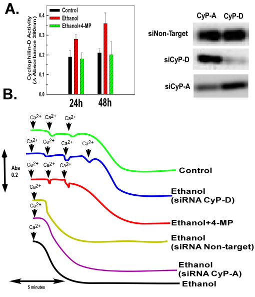
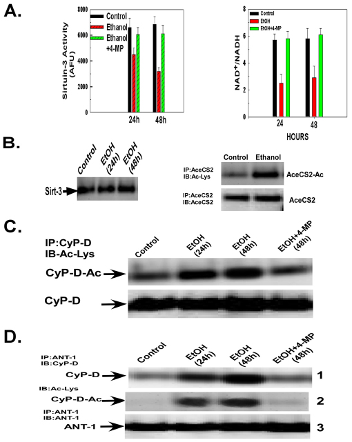
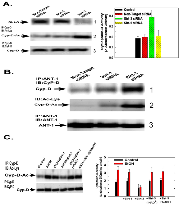
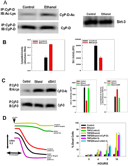
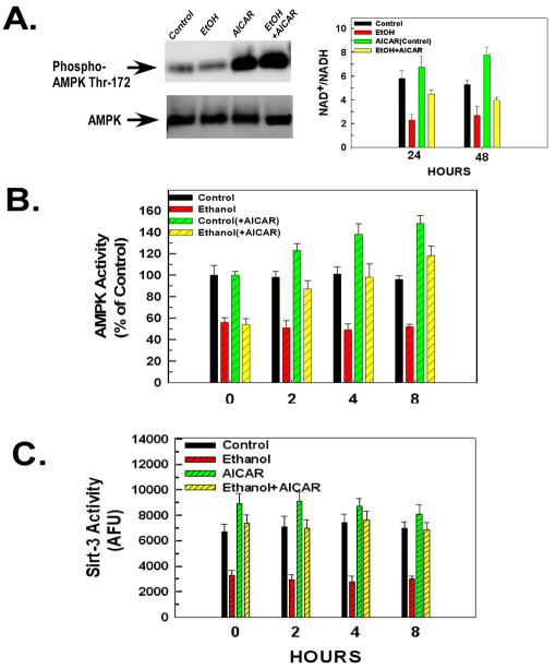
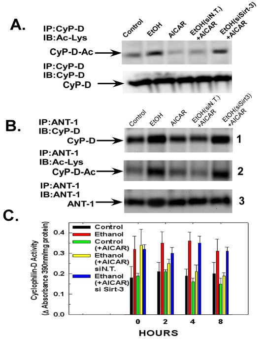
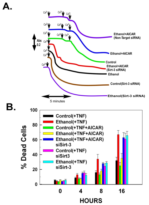
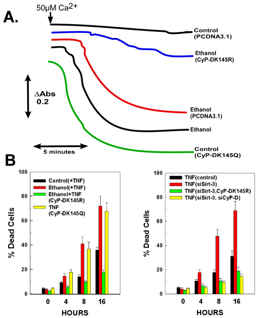
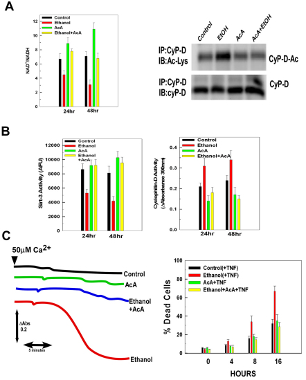
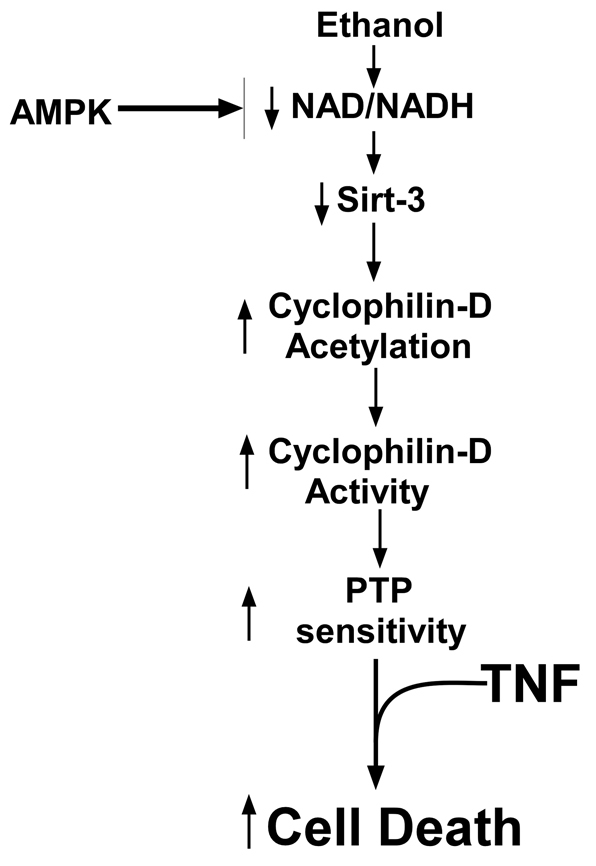
Ethanol increases the vulnerability of mitochondria to induction of the mitochondrial permeability transition (MPT). Cyclophilin-D activity enhances the potential for the permeability transition pore (PTP) to open. In the present study, we demonstrate that ethanol and its metabolism sensitize the PTP to opening, in part by increasing the acetylation and activity of cyclophilin-D. This effect of ethanol is mediated by inhibiting the activity of sirtuin-3, an NAD(+) dependent deacetylase that is localized to the mitochondrial matrix. The ethanol-enhanced acetylation of cyclophilin-D also increases the interaction of cyclophilin-D with the adenine nucleotide translocator-1 (ANT-1) and is dependent on ethanol metabolism. Moreover, activation of AMPK, a known positive modulator of sirtuin activity, prevented the ethanol-induced suppression of sirtuin-3 activity and the attendant increase of cyclophilin-D acetylation, activity and association with ANT-1. Additionally, AMPK reactivation of sirtuin-3 prevented the sensitization to the MPT and the enhancement of cell killing by TNF in cells exposed to ethanol.


Figures

Fig. 1.
Fig. 1.
Ethanol exposure stimulates the peptidyl-prolyl cis-trans isomerase activity of cyclophilin-D and sensitizes the mitochondria to the MPT. (A) H4IIEC3 cells were either left untreated or exposed to 25 mM of ethanol in the absence or presence of 5 mM 4-MP. Following 24 or 48 hours of incubation, the cells were harvested and mitochondria isolated. Alternatively, cells were transfected with siRNA targeting CyP-A or CyP-D. The western blot on the left shows mitochondrial extracts that were assessed for cyclophilin-D activity and cyclophilin-D or A expression. The graph on the right shows the quantification of these experiments; values are the means from triplicate samples, and the error bars indicate standard deviations. P<0.05 for control versus ethanol and ethanol versus ethanol+4-MP by one way ANOVA and Scheffe's post-hoc test. (B) H4IIEC3 cells were either left untreated or transfected with 50 nM of a non-target siRNA or an siRNA targeting sirtuin-3. After 24 hours, cells were either left untreated or exposed to 25 mM ethanol in the absence or presence of 4-MP. After 48 hours, the cells were harvested and the mitochondria isolated. Mitochondrial respiration was initiated by the addition of 1 mM malate and 1 mM glutamate. To trigger mitochondrial swelling, 50 μM Ca2+ was added at time points indicated. The change in absorbance was measured spectrophotometrically.
Fig. 2.
Fig. 2.
Ethanol exposure inhibits sirtuin-3 activity and promotes cyclophilin-D acetylation and binding to the adenine nucleotide translocator-1. (A) H4IIEC3 cells were left untreated or exposed to 25 mM of ethanol for 24 or 48 hours in the absence or presence of 4-MP. The cells were harvested and the NAD+:NADH and sirtuin-3 activity was determined in whole-cell and mitochondrial extracts, respectively. The values are the means from triplicate samples, and the error bars indicate standard deviations. P<0.05 for control versus ethanol and ethanol versus ethanol+4-MP by one-way ANOVA and Scheffe's post-hoc test. (B) H4IIEC3 cells were left untreated or exposed to 25 mM of ethanol for 24 or 48 hours in the absence or presence of 4-MP. The cells were harvested and the mitochondria isolated. Sirtuin-3 expression was determined by western blotting. Acetyl-CoA sythetase 2 (AceCS2) was immunoprecipitated from mitochondrial extracts. The western blots of the immunoprecipitates were probed with antibody against acetylated lysine, stripped and then re-probed with antibody against AceCS2. (C) H4IIEC3 cells were left untreated or exposed to 25 mM of ethanol for 24 or 48 hours in the absence or presence of 4-MP. The cells were harvested and the mitochondria isolated. Cyclophilin-D was immunoprecipitated from mitochondrial extracts. The western blots of the immunoprecipitates were probed with antibody against acetylated lysine, stripped and then re-probed with antibody against cyclophilin-D. (D) H4IIEC3 cells were either left untreated or exposed to 25 mM of ethanol for 24 or 48 hours. The cells were harvested and mitochondria isolated. ANT-1 was immunoprecipitated from mitochondrial extracts. The western blots of the immunoprecipitates were probed with antibodies against cyclophilin-D or ANT-1. To access cyclophilin-D acetylation, the blots were stripped and then re-probed with antibody against acetylated lysine.
Fig. 3.
Fig. 3.
Suppression of sirtuin-3 expression recapitulates the effects of ethanol exposure on cyclophilin-D acetylation, activity and binding to the ANT-1. (A) H4IIEC3 cells were transfected with 50 nM siRNA targeting sirtuin-1, 3 or a non-targeting control. Following 48 hours of incubation, the cells were harvested and mitochondria isolated. Mitochondrial extracts were separated by SDS-PAGE and then electroblotted onto PVDF membranes. Western blots were probed with antibody against sirtuin-3 (panel 1). Alternatively, cyclophilin-D was immunoprecipitated from mitochondrial extracts. The immunoprecipitates were separated by SDS-PAGE and electroblotted onto PVDF membranes. The western blots were probed with antibody against acetylated lysine, then stripped and re-probed with an antibody against cyclophilin-D (panels 2 and 3). Cyclophilin-D activity was determined fluorescently in mitochondrial extracts. The values are the means of three samples, and error bars indicate standard deviations. P<0.05 for non-target siRNA versus sirtuin-3 siRNA. (B) H4IIEC3 cells were transfected with 50 nM of siRNA targeting sirtuin-1, 3 or a non-targeting control. Following 48 hours of incubation, cells were harvested and mitochondria isolated. ANT-1 was immunoprecipitated from mitochondrial extracts. The immunoprecipitates were separated by SDS-PAGE and electroblotted onto PVDF membranes. Western blots were then probed with antibodies against cyclophilin-D or ANT-1. To access cyclophilin-D acetylation, the cyclophilin-D blots were stripped and then re-probed with antibody against acetylated lysine. (C) (Left) Western blot of H4IIEC3 cells that were either left untreated or exposed to 25 mM of ethanol for 48 hours. Cyclophilin-D was immunoprecipitated from mitochondrial extracts and incubated with recombinant sirtuins. The immunoprecipitates were then run out on SDS-PAGE gels and electroblotted onto PVDF membranes. Blots were then probed with antibody against acetylated lysine, stripped and re-probed with antibody against cyclophilin-D. (Right) Quantification of cyclophilin-D activity. Cyclophilin-D immunoprecipitates that had been incubated with recombinant sirtuins. Cyclophilin-D activity was determined fluorescently as described in Materials and Methods. Values are the means from triplicate samples, and error bars indicate standard deviations. P<0.05 for control versus ethanol and ethanol versus ethanol+sirtuin-3 by one-way ANOVA and Scheffe's post-hoc test.
Fig. 4.
Fig. 4.
Increased cyclophilin-D acetylation and decreased sirtuin-3 activity in mitochondria isolated from ethanol-fed rats and mouse hepatocytes exposed to ethanol. (A) Western blots of mitochondria that had been isolated from the liver of control or ethanol-fed rats. Cyclophilin-D was immunoprecipitated from mitochondrial extracts. The immunoprecipitates were separated by SDS-PAGE and electroblotted onto PVDF membranes. Blots were probed with antibody against acetylated lysine, then stripped and re-probed with an antibody against cyclophilin-D. Alternatively, mitochondrial extracts were used to determine the expression of sirtuin-3 by using anti-sirtuin-3 antibody. (B) Quantification of cyclophilin-D and sirtuin-3 activity. Mitochondria that had been isolated from the livers of control or ethanol-fed rats. Cyclophilin-D or sirtuin-3 activity was determined fluorescently in mitochondrial extracts. The values are the means from triplicate samples, and the error bars indicate standard deviations. P<0.05 for control versus ethanol. (C) (Left) Western blots (left) of mouse hepatocytes that had been left untreated or were transfected with siRNA targeting sirtuin-3 or exposed to ethanol. Following 48 hours, the hepatocytes were harvested and mitochondria isolated. Cyclophilin-D was immunoprecipitated from mitochondrial extracts. The immunoprecipitates were then separated by SDS-PAGE and electroblotted. Blots were then probed with antibody against acetylated lysine, stripped and re-probed with antibody against cyclophilin-D. (Right) Quantification of sirtuin-3 and cyclophilin-D activity. Mouse hepatocytes were untreated or exposed to ethanol for 48 hours in the absence or presence of 4-MP. Sirtuin-3 or cyclophilin-D activity was determined fluorescently in mitochondrial extracts as described in Materials and Methods. The values are the means from triplicate samples, and the error bars indicate standard deviations. P<0.05 for control versus ethanol and ethanol versus ethanol+4-MP by one-way ANOVA and Scheffe's post-hoc test. (D) (Left) Mouse hepatocytes were transfected with 50 nM of siRNA targeting sirtuin-3, cyclophilin-D or a non-targeting control; 24 hours after transfection, cells were left untreated or exposed to 25 mM of ethanol for 48 hours. The cells were then harvested and mitochondria isolated. Where shown (arrows), 50 μM Ca2+ was added. The change in absorbance was measured spectrophotometrically at 540 nm. (Right) At 24 hours after transfection, mouse hepatocytes were left untreated or exposed to 25 mM of ethanol for 48 hours. Cells were then treated with 10 ng/ml of TNF. At the times indicated, the viability of the cells was assessed. Values are the means of three samples, and the error bars indicate standard deviations. P<0.05 for TNF(control) versus TNF(ethanol), TNF(ethanol) versus TNF(ethanol-siCyP-D) and TNF(control) versus TNF(siSirt-3) by one-way ANOVA and Scheffe's post-hoc test.
Fig. 5.
Fig. 5.
Activation of AMPK by AICAR reverses the inhibitory effect of ethanol exposure on sirtuin-3 activity. (A) (Left) Western blots of H4IIEC3 cells that had been either left untreated or exposed to 25 mM of ethanol for 48 hours. Where indicated, cells had been treated with 0.5 mM of AICAR for another 8 hours. The cells were harvested and cell extracts were separated by SDS-PAGE and electroblotted onto PVDF membranes. Blots were probed with antibodies against AMPK or specific for AMPK phosphorylated on Thr172. (Right) Quantification of the NAD+:NADH ratio. Cell extracts were used to determine the NAD+:NADH ratio fluorescently as described in Materials and Methods. Values are the means of three samples, error bars indicate standard deviations. P<0.05 for control versus ethanol and ethanol versus ethanol+AICAR by one-way ANOVA and Scheffe's post-hoc test. (B) Quantification of AMPK activity. H4IIEC3 cells were either left untreated or exposed to 25 mM of ethanol for 48 hours. Where indicated cells were subsequently treated with 0.5 mM of AICAR. At the time points indicated the cells were harvested and AMPK activity was determined. The values are the means of three samples, error bars indicate standard deviations. P<0.05 for control versus ethanol, control versus control+AICAR and ethanol versus ethanol+AICAR by one-way ANOVA and Scheffe's post-hoc test. (C) Quantification of sirtuin-3 activity. H4IIEC3 cells were either left untreated or exposed to 25 mM of ethanol for 48 hours. Where indicated, cells were treated with 0.5 mM of AICAR. At the time points indicated, the cells were harvested and mitochondria isolated. Sirtuin-3 activity was measured in mitochondrial extracts. Values are the means of three samples, error bars indicate standard deviations. P<0.05 for control versus ethanol and ethanol versus ethanol+AICAR by one-way ANOVA and Scheffe's post-hoc test.
Fig. 6.
Fig. 6.
Sirtuin-3 is necessary for AICAR to reverse the ethanol-induced activation, acetylation and binding of cyclophilin-D to ANT-1. (A) Western blots of H4IIEC3 cells that had been transfected with non-targeting siRNA or siRNA against sirtuin-3. After 24 hours, the cells were either left untreated or exposed to 25 mM of ethanol for 48 hours. Following ethanol exposure, were indicated, the cells were treated with 0.5 mM of AICAR for 8 hours. The mitochondria were then isolated and mitochondrial extracts were immunoprecipitated with cyclophilin-D antibody. The immunoprecipitates were separated by SDS-PAGE and electroblotted onto PVDF membranes. Blots were probed with antibody against acetylated lysine, then stripped and re-probed with an antibody against cyclophilin-D. (B) Western blots of H4IIEC3 cells that had been transfected with non-targeting siRNA or siRNA against sirtuin-3. After 24 hours, the cells were either left untreated or exposed to 25 mM of ethanol for 48 hours. Where indicated, cells were subsequently treated with 0.5 mM of AICAR for 8 hours. Mitochondrial extracts were immunoprecipitated with antibody against ANT-1. The immunoprecipitates were separated by SDS-PAGE and electroblotted onto PVDF membranes. Blots were then probed with antibodies against cyclophilin-D or ANT-1. To access cyclophilin-D acetylation, the cyclophilin-D blots were stripped and then re-probed with antibody against acetylated lysine. (C) Quantification of cyclophilin-D activity. H4IIEC3 cells were transfected with non-target control siRNA or siRNA against sirtuin-3. After 24 hours, the cells were either left untreated or exposed to 25 mM of ethanol for 48 hours. Subsequently, where indicated, the cells were treated with 0.5 mM of AICAR. At the times indicated, cells were harvested and mitochondria isolated. Cyclophilin-D activity was measured in mitochondrial extracts as described in Materials and Methods. The values are the means of three samples, error bars indicate standard deviations. P<0.05 for control versus ethanol, ethanol versus ethanol+AICAR siN.T. and ethanol+AICAR siN.T. versus ethanol+AICAR siSirt-3 by one-way ANOVA and Scheffe's post-hoc test.
Fig. 7.
Fig. 7.
Sirtuin-3 is required for AMPK activation to reverse the ethanol-induced sensitization to onset of the MPT and TNF-induced cytotoxicity. (A) H4IIEC3 cells were transfected with non-target control siRNA or siRNA against sirtuin-3. After 24 hours, the cells were left untreated or exposed to 25 mM of ethanol for 48 hours. Where indicated, the cells were subsequently treated with 0.5 mM of AICAR for 8 hours. Mitochondria were isolated. Where shown, 50 μM of Ca2+ was added. The change in absorbance was measured at 540 nm. (B) H4IIEC3 cells were transfected with non-target control siRNA or siRNA against sirtuin-3. After 24 hours, the cells were left untreated or exposed to 25 mM of ethanol for 48 hours. Where indicated, the cells were subsequently treated with 0.5 mM of AICAR for 8 hours. Cells were then treated with 10 ng/ml of TNF. At the times indicated, the viability of the cells was assessed. The values are the means of three samples, and error bars indicate standard deviations. P<0.05 for control(+TNF) versus ethanol(+TNF), ethanol(+TNF) versus ethanol(+TNF+AICAR), ethanol(+TNF+AICAR) versus ethanol(+TNF+AICAR)siSirt-3 and control(+TNF) versus control(+TNF)siSirt-3 by one-way ANOVA and Scheffe's post-hoc test.
Fig. 8.
Fig. 8.
Acetylation of the Lys145 residue in cyclophilin-D controls sensitivity to PTP induction and TNF cytotoxicity in ethanol-exposed cells. (A) H4IIEC3 cells expressing CyP-D(K145R) or CyP-D(K145Q) were generated. Cells were then either left untreated or exposed to 25 mM of ethanol for 48 hours. Mitochondria were then isolated. Mitochondrial respiration was initiated by the addition of 1 mM malate and 1 mM glutamate. To trigger mitochondrial swelling, 50 μM Ca2+ was added as indicated. The change in absorbance was measured at 540 nm. (B) (Left) H4IIEC3 cells stably expressing CyP-D(K145R) or CyP-D(K145Q) were exposed to 25 mM of ethanol for 48 hours. Cells were then treated with 10 ng/ml TNF. Alternatively, cells expressing CyP-D(K145R) were transfected with siRNA against sirtuin-3. Following 48 hours, the cells were treated with TNF. (Right) Cells were transfected with siRNA targeting sirt-3 and Cyp-D. Following 48 hours, cells were treated with 10 ng/ml TNF. At the times indicated, the viability of the cells was assessed. The values are the means of three samples, and error bars indicate standard deviations. P<0.05 for control+TNF versus ethanol+TNF, ethanol+TNF versus ethanol+TNF(CyP-DK145R), control+TNF versus TNF(CyP-DK145Q) and TNF(siSirt-3) versus TNF(siSirt-3,CyP-DK145R), TNF(siSirt-3) versus TNF(siSirt-3, siCyP-D) by one-way ANOVA and Scheffe's post-hoc test.
Fig. 9.
Fig. 9.
Maintenance of the NAD+:NADH ratio prevents the ethanol-induced decline of sirtuin-3 activity. (A) (Left) H4IIEC3 cells were untreated or exposed to 25 mM of ethanol in the absence or presence of 10 mM of acetoacetate (AcA) for 48 hours. Cell extracts were prepared to determine the NAD+:NADH ratio. (Right) Western blots of H4IIEC3 cells treated as above. Cyclophilin-D was immunoprecipitated from mitochondrial extracts. The immunoprecipitates were then separated by SDS-PAGE and electroblotted onto PVDF membranes. Blots were then probed with antibody against cyclophilin-D, stripped and reprobed with antibody against acetylated lysine. The values are the means of three samples, and the error bars indicate standard deviations. P<0.05 for control versus ethanol, ethanol versus ACA and ethanol versus ethanol+AcA by one-way ANOVA and Scheffe's post-hoc test. (B) H4IIEC3 cells were untreated or exposed to 25 mM of ethanol in the absence or presence of 10 mM of AcA. After 24 or 48 hours incubation, cyclophilin-D or sirtuin-3 activity was determined in mitochondrial extracts as described in Materials and Methods. The values are the means of three samples, error bars indicate standard deviations. P<0.05 for control versus ethanol, ethanol versus AcA and ethanol versus ethanol+AcA by one-way ANOVA and Scheffe's post-hoc test. (C) (Left) H4IIEC3 cells were untreated or exposed to 25 mM of ethanol in the absence or presence of 10 mM AcA. Following a 48-hour incubation, mitochondria were isolated. Where shown (arrows), a 50 μM Ca2+ was added. (Right) H4IIEC3 cells were treated with 10 ng/ml TNF. At the times indicated, the viability of the cells was assessed. The values are the means of three samples and the error bars indicate standard deviations. P<0.05 for control(+TNF) versus ethanol(+TNF) and ethanol(+TNF) versus ethanol+TNF+AcA by one-way ANOVA and Scheffe's post-hoc test.
Fig. 10.
Fig. 10.
Ethanol-induced decline of sirtuin-3 activity sensitizes mitochondria to MPT induction by TNF. Ethanol metabolism induces a decline of sirtuin-3 activity, causing an increase in cyclophilin-D acetylation and activity, which results in a sensitization to induction of the permeability transition by TNF.

Comment in

  • Findings of research misconduct.
    [No authors listed] [No authors listed] NIH Guide Grants Contracts (Bethesda). 2016 Jun 10:NOT-OD-16-104. NIH Guide Grants Contracts (Bethesda). 2016. PMID: 27311138 Free PMC article. No abstract available.
  • Findings of Research Misconduct.
    [No authors listed] [No authors listed] Fed Regist. 2016 May 13;81(93):29871. Fed Regist. 2016. PMID: 27737277 Free PMC article. No abstract available.

References

    1. Ahn B. H., Kim H. S., Song S., Lee I. H., Liu J., Vassilopoulos A., Deng C. X., Finkel T. (2008). A role for the mitochondrial deacetylase Sirt3 in regulating energy homeostasis. Proc. Natl. Acad. Sci. USA 105, 14447-14452 - PMC - PubMed
    1. Ajmo J. M., Liang X., Rogers C. Q., Pennock B., You M. (2008). Resveratrol alleviates alcoholic fatty liver in mice. Am. J. Physiol. Gastrointest. Liver Physiol. 295, G833-G842 - PMC - PubMed
    1. Baines C. P., Kaiser R. A., Purcell N. H., Blair N. S., Osinska H., Hambleton M. A., Brunskill E. W., Sayen M. R., Gottlieb R. A., Dorn G. W., et al. (2005). Loss of cyclophilin D reveals a critical role for mitochondrial permeability transition in cell death. Nature 434, 658-662 - PubMed
    1. Basso E., Fante L., Fowlkes J., Petronilli V., Forte M. A., Bernardi P. (2005). Properties of the permeability transition pore in mitochondria devoid of Cyclophilin D. J. Biol. Chem. 280, 18558-18561 - PubMed
    1. Bauer M. K., Schubert A., Rocks O., Grimm S. (1999). Adenine nucleotide translocase-1, a component of the permeability transition pore, can dominantly induce apoptosis. J. Cell Biol. 147, 1493-1502 - PMC - PubMed

Publication types

LinkOut - more resources