. 2010 Dec 1;123(Pt 23):4128-44.
doi: 10.1242/jcs.072041. Epub 2010 Nov 9.

Xenopus Kazrin interacts with ARVCF-catenin, spectrin and p190B RhoGAP, and modulates RhoA activity and epithelial integrity

Affiliations

Xenopus Kazrin interacts with ARVCF-catenin, spectrin and p190B RhoGAP, and modulates RhoA activity and epithelial integrity

Kyucheol Cho et al. J Cell Sci. .

Abstract

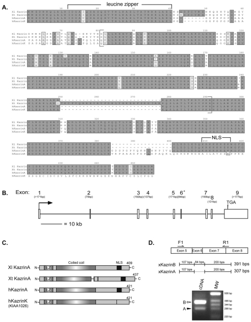
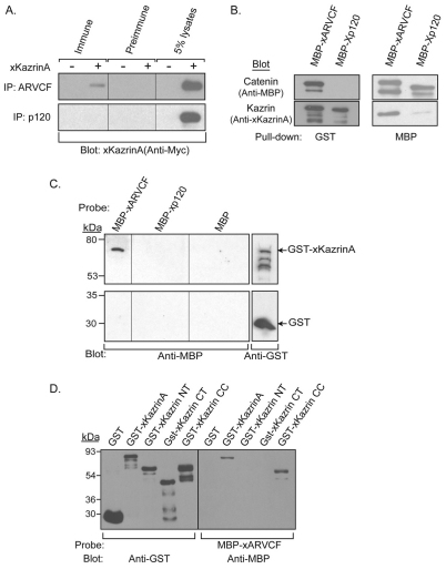
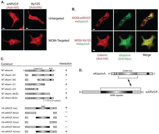
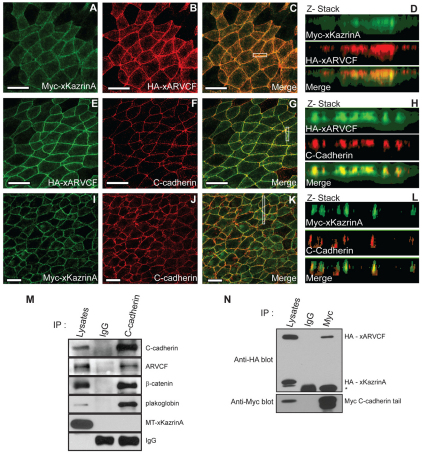
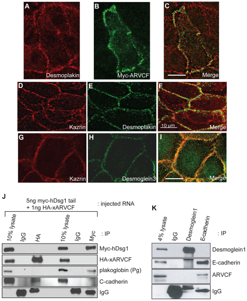
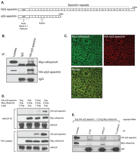
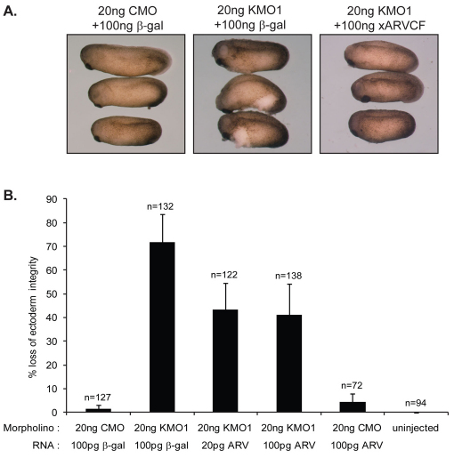
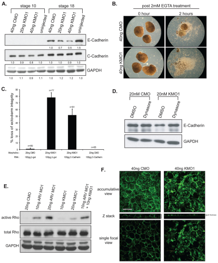
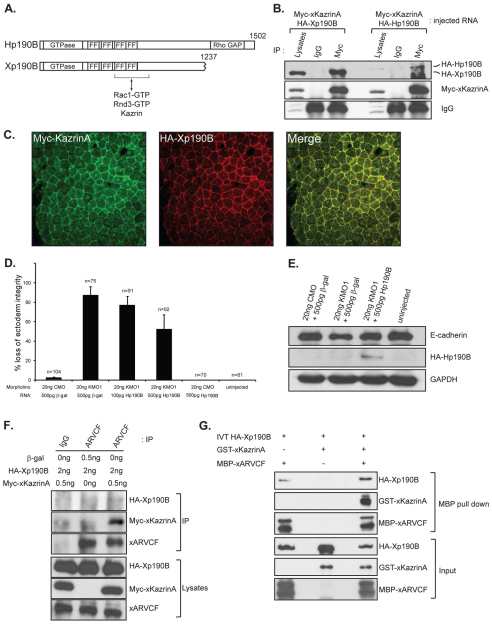
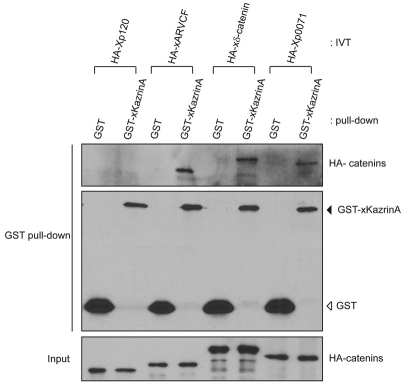
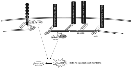
In common with other p120-catenin subfamily members, Xenopus ARVCF (xARVCF) binds cadherin cytoplasmic domains to enhance cadherin metabolic stability or, when dissociated, modulates Rho-family GTPases. We report here that xARVCF binds and is stabilized by Xenopus KazrinA (xKazrinA), a widely expressed conserved protein that bears little homology to established protein families, and which is known to influence keratinocyte proliferation and differentiation and cytoskeletal activity. Although we found that xKazrinA binds directly to xARVCF, we did not resolve xKazrinA within a larger ternary complex with cadherin, nor did it co-precipitate with core desmosomal components. Instead, screening revealed that xKazrinA binds spectrin, suggesting a potential means by which xKazrinA localizes to cell-cell borders. This was supported by the resolution of a ternary biochemical complex of xARVCF-xKazrinA-xβ2-spectrin and, in vivo, by the finding that ectodermal shedding followed depletion of xKazrin in Xenopus embryos, a phenotype partially rescued with exogenous xARVCF. Cell shedding appeared to be the consequence of RhoA activation, and thereby altered actin organization and cadherin function. Indeed, we also revealed that xKazrinA binds p190B RhoGAP, which was likewise capable of rescuing Kazrin depletion. Finally, xKazrinA was found to associate with δ-catenins and p0071-catenins but not with p120-catenin, suggesting that Kazrin interacts selectively with additional members of the p120-catenin subfamily. Taken together, our study supports the essential role of Kazrin in development, and reveals the biochemical and functional association of KazrinA with ARVCF-catenin, spectrin and p190B RhoGAP.


Figures

Fig. 1.
Fig. 1.
Comparison of Xenopus and human Kazrin protein sequences. (A) Sequence alignment of X. laevis Kazrin (Xl Kazrin), X. tropicalis Kazrin (Xt Kazrin) and two isoforms of human Kazrin (hKazrinA and hKazrinK). Identical and similar residues are highlighted in black or grey, respectively. The central coiled-coil region (brackets), conserved potential leucine zipper and the predicted nuclear localization sequence (NLS) are indicated. (B) Genomic structure of X. tropicalis Kazrin. Genomic information was obtained from Ensembl database (Ensembl ID #ENSXETG00000013469 and scaffold 257). Asterisk indicates an alternatively spliced exon in X. laevis. (C) Comparison of Xenopus and human Kazrin isoforms. The coiled coil and polylysine tract (NLS) are respectively depicted as grey or black shaded areas. LZ indicates the putative leucine zipper region. Alternative splicing at the C-terminus of the human KazrinA isoform results in additional isoform human KazrinK without the polylysine tract. The total number of amino acids of each protein is indicated. (D) Identification of xKazrin isoforms using primers (F1 and R1) binding to the 5′- and 3′-flanking regions of exon 6. cDNA was synthesized from embryo stage 18 genomic RNA. Open arrowhead indicates PCR product of xKazrinB cDNA and closed arrowhead indicates that of xKazrinA. MW signifies molecular weight markers.
Fig. 2.
Fig. 2.
Association of full-length xKazrinA with xARVCF. (A) Xenopus embryos expressing HA–xARVCF or HA–Xp120 with (+) or without (−) coexpressed Myc–xKazrinA were harvested at gastrula stage (stage 10.5). Lysates were immunoprecipitated using pre-immune, xARVCF or Xp120 polyclonal antisera, and interactions with xKazrinA detected by immunoblotting using anti-Myc antibody. Total lysates (5%) were also blotted. (B) In vitro co-precipitation of GST–xKazrinA with MBP–xARVCF. GST–xKazrinA was incubated with MBP–xARVCF or MBP–Xp120, precipitated using glutathione–agarose (left panels) or amylose–agarose (right panels) beads and the samples subjected to SDS-PAGE and immunoblotting using anti-MBP (top panels) or anti-Kazrin (bottom panels) antibodies. (C) Direct interaction of purified xARVCF with xKazrinA using blot overlay. Top panel: Duplicate lanes of purified GST–xKazrinA were electrophoresed and immobilized on nitrocellulose then probed using purified MBP–xARVCF, MBP–Xp120 or MBP. Protein complexes were then identified using anti-MBP antibodies (top-left panel). Presence of GST–xKazrinA was detected using anti-GST antibodies (top-right panel). As a negative control, purified GST was immobilized on nitrocellulose and processed in an identical manner (bottom panels). (D) Blot overlay of GST–xKazrinA deletion constructs. Purified GST–xKazrinA, GST–xKazrinA N-terminal (NT), GST–xKazrinA C-terminal (CT), GST–xKazrinA coiled-coil (CC), and GST proteins were electrophoresed and immobilized on nitrocellulose. Total loading was detected using anti-GST antibodies (left panel). The right panel was probed with MBP–xARVCF and the resulting protein complexes detected using anti-MBP antibodies.
Fig. 3.
Fig. 3.
Mitochondrial colocalization of xKazrinA with xARVCF in Xenopus A6 cells and binding domain mapping. (A) HA-tagged xARVCF or Xp120 were transfected into Xenopus kidney epithelial A6 cells, where they each displayed predominant cytoplasmic localization (top panels). Once fused to a peptide sequence tag from human Bcl-Xl directing their ectopic localization to the MOM, xARVCF and Xp120 displayed a punctate pattern characteristic of mitochondrial localization (bottom panels). (B) Coexpression of Myc–xKazrinA with MOM-targeted HA–xARVCF or HA–Xp120 (top and bottom panels, respectively). Cells were coimmunostained for localization of the catenin (HA-epitope, left panels) and for xKazrinA (Myc-epitope, center panels). Co-localization of xKazrinA was observed with xARVCF but not with Xp120, as shown in the merged images (right panels). (C) List of Myc-tagged (MT) xKazrinA and HA-tagged xARVCF constructs (see text for details) and their interaction status. Asterisk indicates mutation of lysine to glutamine; LZ, leucine zipper; mom, mitochondrial outer membrane; ARM, Armadillo domain. The co-relocalization of xKazrinA deletion mutants was tested in the presence of full-length MOM-targeted xARVCF (upper set). Conversely, full-length xKazrinA was tested for co-relocalization with deletion constructs of MOM-targeted xARVCF (lower set). A positive score (+) indicates that numerous cells coexpressing the indicated xKazrinA or xARVCF deletion constructs showed colocalization of xKazrinA with the MOM-targeted xARVCF construct. (D) Representation of the xARVCF–xKazrinA interaction, indicating the putative regions of association. Scale bars: 20 μm.
Fig. 4.
Fig. 4.
Cellular localization of xKazrinA in ectodermal explants of Xenopus blastula (stage 10) embryos. (A–D) Localization and colocalization of coexpressed Myc–xKazrinA and HA–xARVCF in the outer ectodermal layer, detected using anti-Myc and anti-HA antibodies, respectively. The area outlined by a white rectangle in C, was viewed using Z-stack section images (shown in D). (E–H) Localization and colocalization of HA–xARVCF and C-cadherin, detected using anti-Myc and anti-C-cadherin antibodies, respectively. (I–L) Localization and colocalization of Myc–xKazrinA and xC-cadherin. (M) Co-immunoprecipitation of Myc–xKazrinA (MT-xKazrinA) with adherens junction components. Myc–xKazrinA was expressed in early embryos lysed at stage 9–10 and endogenous xC-cadherin was immunoprecipitated. Co-precipitated proteins were detected by immunoblotting using the indicated antibodies. (N) Co-immunoprecipitation tests following the coexpression of Myc–xC-cadherin tail, HA–xARVCF and HA–xKazrinA. Myc–xC-cadherin tail was precipitated using anti-Myc antibodies. Co-associated HA–xARVCF and HA–xKazrinA were detected (versus not) using anti-HA antibody. Asterisk indicates migration of IgG heavy chains (50 kDa). Scale bars: 50 μm.
Fig. 5.
Fig. 5.
Kazrin and ARVCF localization in desmosome. (AI) Human keratinocytes were immunolabeled for the endogenous desmosomal marker desmoplakin (A,E), Myc-tagged xARVCF (B), or endogenous Kazrin (D,G). In addition, Kazrin localization was compared to that of endogenous desmoglein3 (H), a desmosomal cadherin. Merged images (F,I) show partial colocalization of Kazrin with desmoplakin and extensive colocalization with desmoglein3. Colocalization between desmoplakin and ARVCF is shown in C. Photos represent projection images of deconvolved Z-stacks. Scale bars: 10 μm. (J) Interaction between desmosomal cadherin desmoglein1 (Dsg1) and xARVCF in Xenopus embryo lysates. Indicated amounts of in vitro transcribed RNAs were microinjected into one-cell stage embryos. Then, early gastrula embryos were lysed and human desmoglein1 tail or xARVCF were immunoprecipitated, followed by SDS-PAGE and western blotting. (K) Endogenous desmoglein1 or E-cadherin were immunoprecipitated from confluent A431 human epithelial carcinoma cells. Bound endogenous ARVCF was detected with human ARVCF-specific antibody.
Fig. 6.
Fig. 6.
Association of xKazrinA and xβ2-spectrin, and ternary complex formation with xARVCF. (A) Comparison of human β2-spectrin (gene bank ID # NM_003128) and Xenopus partial β2-spectrin (BC046267). Actin-binding domain (ABD) and putative Kazrin-binding region are depicted. (B) In vivo binding of xKazrinA and xβ2-spectin. Enzymatically synthesized mRNAs encoding Myc–xKazrinA and HA–xβ2-spectrin were co-injected into one-cell stage embryos, and the blastula embryo lysates were HA-immunoprecipitated followed by Myc immunoblotting. (C) Colocalization of xKazrinA and xβ2-spectrin in blastula ectoderm. Animal caps were isolated from blastula embryos (stage 9–10) expressing Myc–xKazrinA and HA–xβ2-spectrin, followed by their respective (Myc and HA) immunofluorescent detection. (D) Formation of xARVCF, xβ2-spectrin and xKazrinA ternary complex. Blastula lysates from embryos injected with indicated amounts of each mRNA were immunoprecipitated with xARVCF antibody, followed by immunoblotting for Myc–xKazrinA and HA–xβ2-spectrin. (E) ARVCF-specific ternary complex. Lysates from embryos expressing xβ2-spectrin and xKazrinA were immunoprecipitated using the indicated antibodies. Co-precipitating xβ2-spectrin or xKazrinA were detected by immunoblotting.
Fig. 7.
Fig. 7.
Rescue of loss of ectoderm integrity, induced upon xKazrin depletion, through ectopic expression of xARVCF. (A) The indicated morpholinos and mRNAs were co-injected into one-cell stage embryos. After vitelline membrane removal, representative tailbud stage embryos were imaged (stage 20–22). (B) xARVCF rescue of xKazrin depletion effects upon ectoderm integrity. Two concentrations of xARVCF-encoding mRNA (ARV) were used in the rescues, with similar results being observed from four independent experiments. Error bars represent standard error of the mean (s.e.m.). Numerical results are shown in Table 3.
Fig. 8.
Fig. 8.
Effects of xKazrin depletion on cadherin expression, cell–cell adhesion, Rho activity and actin organization. (A) Decrease of cadherin protein levels after xKazrin depletion. Embryos injected at the one-cell stage with different amounts of the indicated morpholinos were lysed at early gastrula (stage 10) and neurula (stage 18) stages. xE-cadherin and xC-cadherin levels were visualized by immunoblotting. Band intensity was measured with AlphaEaseFC software (Alpha Innotech Corporation). (B) Cell dissociation assay using animal cap explants. Blastula ectoderm (animal caps; stage 9–10) from embryos injected with CMO or KMO1 were dissociated in the presence of 2 mM EGTA. Photos were taken at 0 hour and 2 hours following EGTA addition. (C) Rescue of ectodermal integrity, reduced upon xKazrin depletion, through expression of xC-cadherin. Embryos injected with the indicated morpholino and mRNA were assessed for ectoderm shedding at tailbud stages (stage 20–22). Error bars represent standard error of the mean (s.e.m.). Numerical results are shown in supplementary material Table S2. (D) Effect of inhibiting clathrin-mediated endocytosis on the reduced xE-cadherin levels observed following xKazrin depletion. One-cell stage embryos were injected with the indicated morpholinos. Embryos were subsequently (32–64 cell stages) incubated with 50 mM Dynasore (versus DMSO solvent). Lysates from neurula (stage 18) embryos were immunoblotted for xE-cadherin and GAPDH. (E) Rho activity in response to xARVCF and/or xKazrin depletion. The indicated morpholinos were injected into one-cell stage embryos, and lysates from late blastula (stage 9–10) embryos were tested for Rho activity in vitro. (F) Actin staining of blastula ectoderm after Kazrin depletion. Animal cap were isolated as mentioned in B and filamentous actin in deep (inner) cells was visualized using phalloidin-Alexa 488 fluorescence. Scale bar: 50 μm.
Fig. 9.
Fig. 9.
Interaction of xKazrinA with Xp190B, and the role of xKazrinA in xARVCF–Xp190B RhoGAP association. (A) Comparison of human p190B (GeneBank ID # NM_001030055) and Xenopus p190B (BC084299.1). Protein domains were predicted using the SMART website (http://smart.emblheidelberg.de/). The predicted binding region of Rnd proteins and Kazrin is shown. (B) In vivo binding of xKazrinA to human and Xenopus p190B. The indicated mRNAs were injected into one-cell embryos. Immunoprecipitates of Myc–xKazrinA were obtained from early gastrula (stage 10–11) extracts, followed by immunoblotting for p190Bs (HA antibody). (C) Colocalization of xKazrinA and Xp190B in blastula ectoderm. Blastula ectoderm (stage 9–10) coexpressing Myc–xKazrinA and HA–Xp190B was isolated, fixed and visualized by confocal fluorescent imaging for Myc and HA. (D) Partial rescue of ectoderm integrity, reduced following xKazrin depletion, upon expression of human p190B. Embryos were injected with the indicated amounts of morpholino and mRNA at the one-cell stage. Ectoderm integrity was evaluated at tailbud stage (stage 20–22) in three independent experiments. Error bars indicate standard error of the mean (s.e.m.). Numerical results are shown in supplementary material Table S3. (E) Ectopic p190B rescues xE-cadherin levels, reduced following xKazrin depletion. The indicated morpholinos and mRNAs were injected into one-cell embryos. Neurulation (stage 18) extracts were immunoblotted as indicated. (F) In vivo ternary complex formation of xARVCF–xKazrinA–Xp190B. Extracts from gastrula-stage embryos expressing the indicated proteins and endogenous xARVCF were precipitated for xARVCF. Co-precipitated xKazrinA and Xp190B were detected using antibodies directed against Myc and HA, respectively. (G) In vitro ternary protein complex of xARVCF–xKazrinA–Xp190B. In vitro transcribed and translated (IVT) Xp190B was incubated with bacterially purified GST–xKazrinA and MBP–xARVCF. Pull-down of xARVCF occurred using amylose–agarose beads, followed by immunoblotting as indicated.
Fig. 10.
Fig. 10.
In vitro binding of Xenopus p120 subfamily catenins to xKazrinA. (A) In vitro transcribed and translated (IVT) Xenopus p120 subfamily catenins (HA-tagged) were tested for association with GST–xKazrinA following GST pull-down and immunoblotting as indicated.
Fig. 11.
Fig. 11.
Working model of xARVCF–xKazrinA complex localization and function at the plasma membrane. Kazrin (Kaz) facilitates formation of the trimeric ARVCF–p190B RhoGAP complex, which localizes to the plasma membrane via association of Kazrin with spectrin. This novel RhoGAP complex locally reduces RhoA activity at the plasma membrane and stabilizes the actin cytostructure, supporting cell–cell contacts. An interaction of p120-catenin with p190A RhoGAP at adherens junctions was previously reported (Wildenberg et al., 2006).

Similar articles

Cited by

References

    1. Aho S., McLean W. H., Li K., Uitto J. (1998). cDNA cloning, mRNA expression, and chromosomal mapping of human and mouse periplakin genes. Genomics 48, 242-247 - PubMed
    1. Akhtar N., Hotchin N. A. (2001). RAC1 regulates adherens junctions through endocytosis of E-cadherin. Mol. Biol. Cell 12, 847-862 - PMC - PubMed
    1. Anastasiadis P. Z. (2007). p120-ctn: A nexus for contextual signaling via Rho GTPases. Biochim. Biophys. Acta 1773, 34-46 - PubMed
    1. Anastasiadis P. Z., Moon S. Y., Thoreson M. A., Mariner D. J., Crawford H. C., Zheng Y., Reynolds A. B. (2000). Inhibition of RhoA by p120 catenin. Nat. Cell Biol. 2, 637-644 - PubMed
    1. Aono S., Nakagawa S., Reynolds A. B., Takeichi M. (1999). p120(ctn) acts as an inhibitory regulator of cadherin function in colon carcinoma cells. J. Cell Biol. 145, 551-562 - PMC - PubMed

Publication types

MeSH terms