. 2010 Nov;9(11):2970-81.
doi: 10.1158/1535-7163.MCT-10-0780. Epub 2010 Nov 9.

Expression and silencing of the microtubule-associated protein Tau in breast cancer cells

Affiliations

Expression and silencing of the microtubule-associated protein Tau in breast cancer cells

Tatiana Spicakova et al. Mol Cancer Ther. 2010 Nov.

Abstract

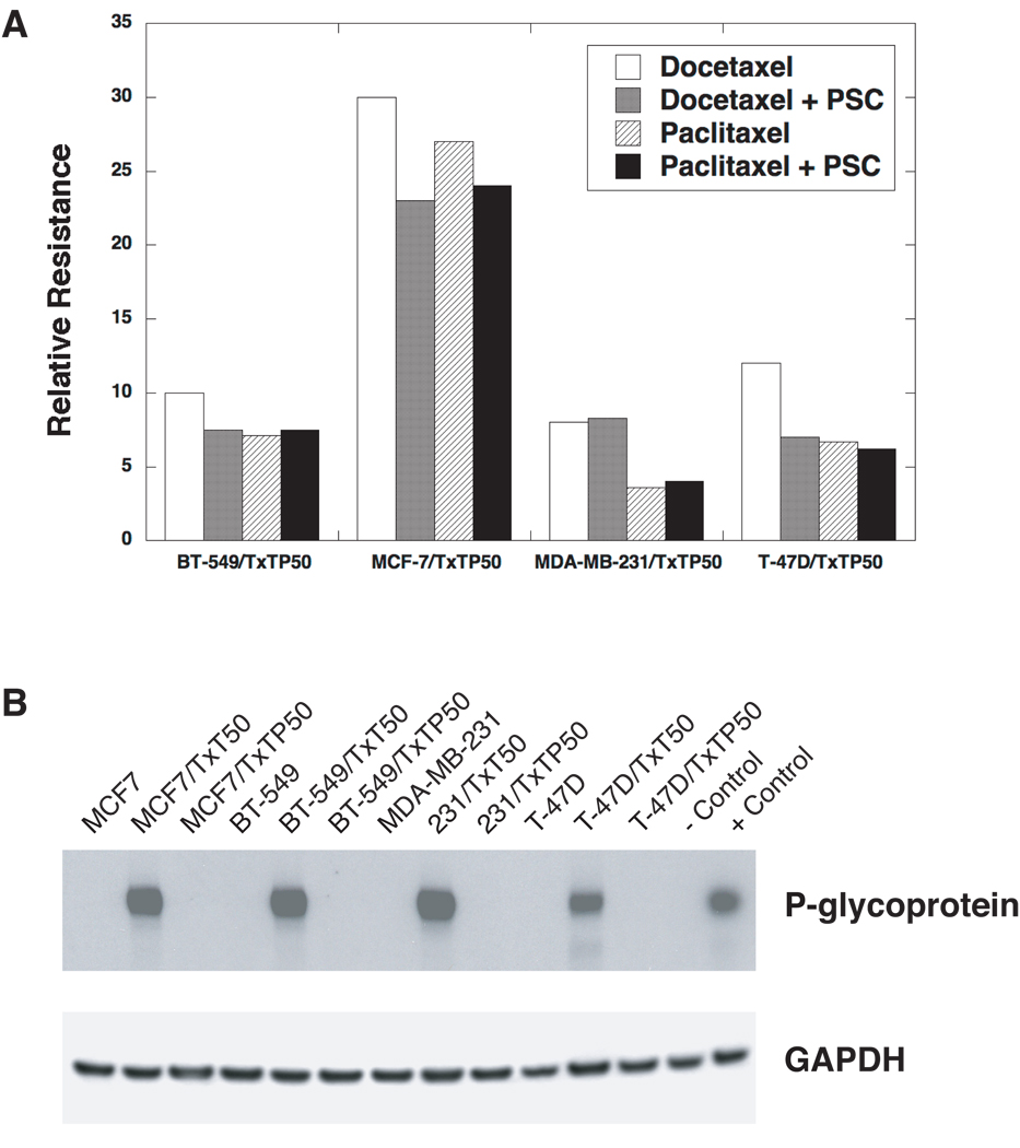
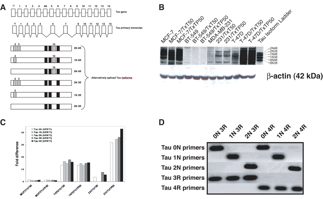
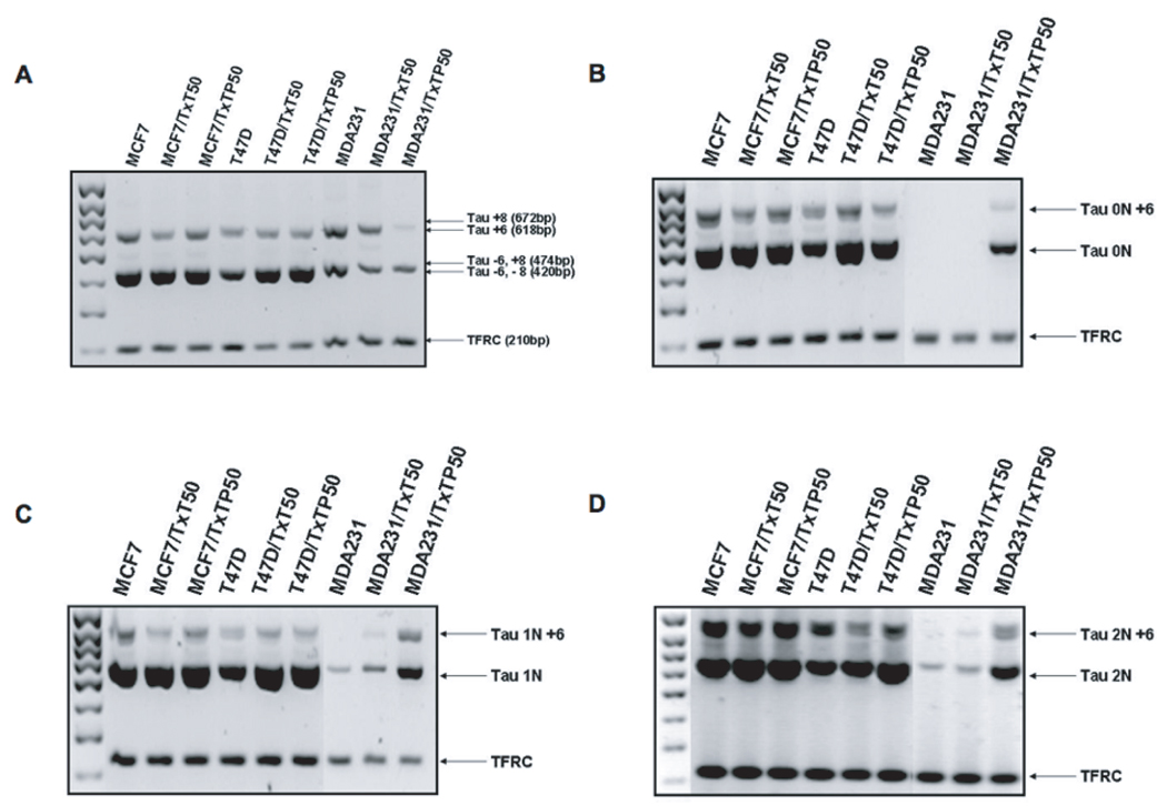
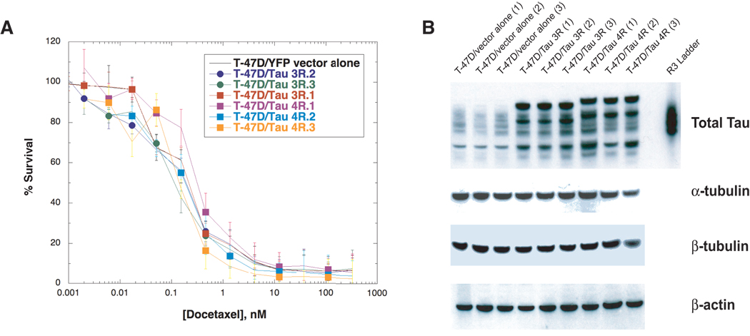
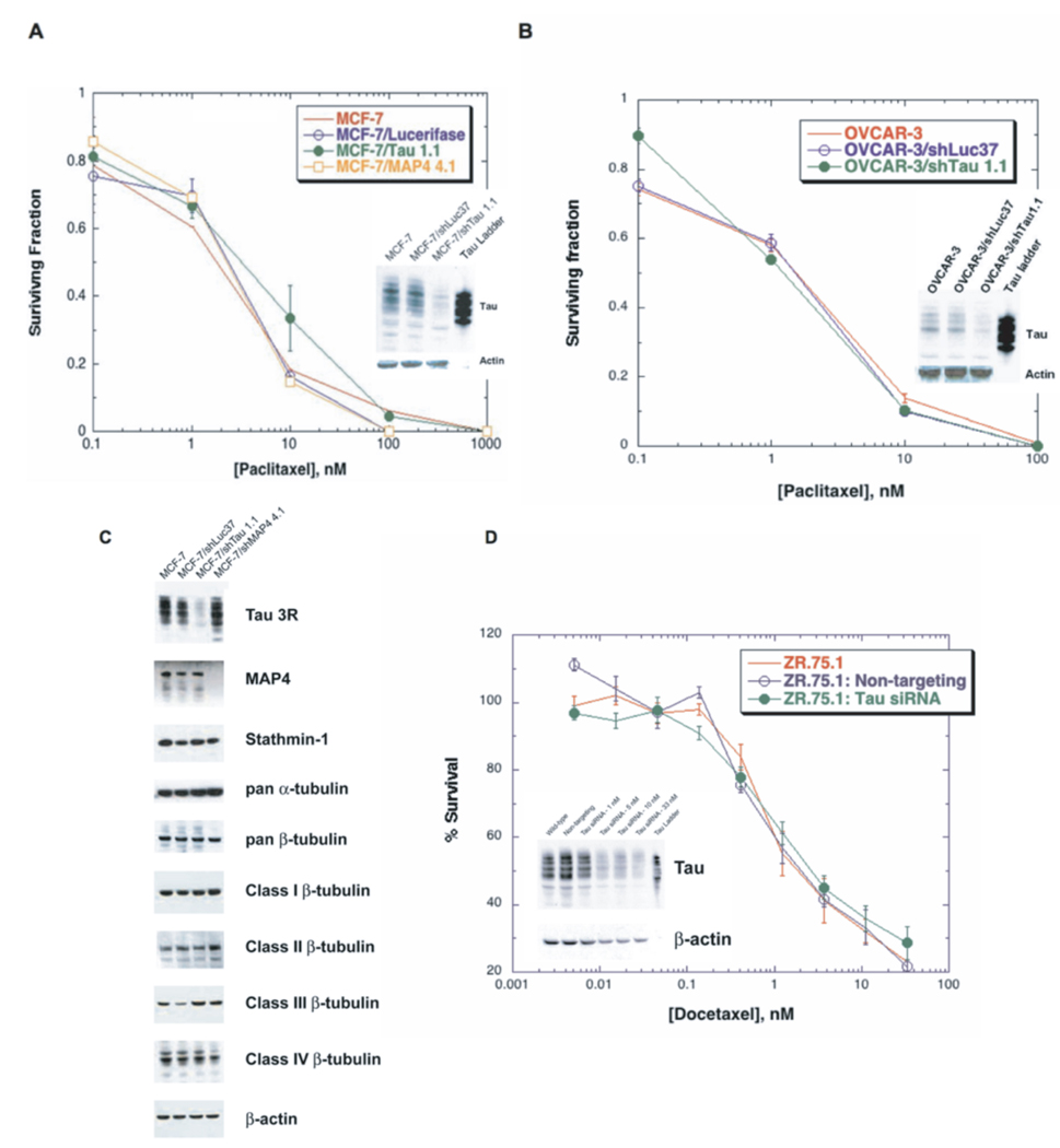
The microtubule-associated protein Tau has been reported to be a predictive factor for clinical response to taxanes in metastatic breast cancer. We generated a panel of eight taxane-resistant variants from four human breast cancer cell lines (MCF-7, T-47D, MDA-MB-231, and BT-549). Four variants had higher levels of Tau compared with their T-47D and MDA-MB-231 parental cells. Using isoform-specific primers, we found that Tau 0N, 1N, 2N, 3R, and 4R isoforms are overexpressed in the resistant variants, as is Tau exon 6 but not exons 4A or 8. To determine whether Tau overexpression produces resistance to taxanes, we derived three independent T-47D clones stably overexpressing Tau 3R and 4R isoforms. Tau overexpression did not result in taxane resistance compared with parental cells transfected with vector alone. We then knocked down Tau expression in three cell lines that expressed Tau constitutively (MCF-7 and ZR-75-1 breast cancer cells, and OVCAR-3 ovarian cancer cells). Lentivirus-mediated silencing of Tau expression in MCF-7 and OVCAR-3 cells did not result in increased taxane sensitivity compared with luciferase short hairpin RNA-infected cells and uninfected parental cells. Transient silencing using Tau-specific small interfering RNAs also did not alter taxane sensitivity relative to nontargeting controls in both MCF-7 and ZR-75-1 cells. These results show that neither overexpression nor depletion of Tau modulates cellular sensitivity to taxanes. Although Tau overexpression has been reported to be a predictive marker of taxane resistance, it is not likely to be a direct mechanism of taxane resistance in breast cancer.


Figures

Figure 1
Figure 1. Chemical structures of (A) docetaxel and paclitaxel, and (B) PSC-833
Figure 2
Figure 2. Development of taxane resistant breast cancer variants
(A) The relative resistance of four breast cancer variants co-selected by stepwise exposure to docetaxel (T×T), with and without the P-gp inhibitor PSC-833 (PSC). The highest T×T concentration used in the step-wise selections was 50 nM. Hence, the BT-549 variant selected with T×T at 50 nM drug concentration and PSC-833 at 2 µM is designated “BT-549/T×TP50”. The resistance levels are expressed as the ratios of the IC50’s of the variants to the parental cell lines respectively. These IC50 values were determined by a 72 hour SRB assay. (B) P-glycoprotein expression in parental and taxane resistant variants. The cells were selected with T×T, with and without PSC (designated as “P”). Protein extracts were prepared from the parental and taxane resistant cell lines, resolved by PAGE, and transferred to nitrocellulose membranes. The membranes were probed with an antibody against P-gp (C219) and an anti-GAPDH antibody was used as a loading control. All of the docetaxel alone selections resulted in the activation of the MDR1 gene and these variants were positive for P-gp expression. All of the variants co-selected with PSC were P-gp negative and drug accumulation assays determined that the taxane resistance observed was non-transporter mediated (data not shown). The doxorubicin-selected human uterine sarcoma MES-SA/D×5 variant was used as a positive control for P-gp expression.
Figure 3
Figure 3. Schematic diagram of the Tau gene, primary transcript and alternatively spliced isoforms, and expression of Tau isoforms in taxane resistant breast cancer variants
(A) The human Tau gene encodes 16 exons but the human brain primary transcript lacks exons 4A, 6 and 8. Exons 2, 3 and 10 are alternatively spliced, giving rise to six major isoforms. The inclusion or exclusion of exon 10 determines whether the isoform will be a 4R-Tau (with exon 10) or a 3R-Tau (without exon 10). The inclusion or exclusion of exons 2 and 3 determines whether the isoform will be 0N-Tau (without exons 2 and 3), 1N-Tau (with exon 2 but without exon 3), or 2N-Tau (with both exons 2 and 3). Expression of Tau was assessed in the panel of breast cancer variants (B) at the protein level using Western blot analysis with an antibody recognizing all Tau isoforms; and (C) at the mRNA level using quantitative real time PCR analysis with Tau isoform-specific primers. The ratio of Tau isoforms was calculated between taxane resistant variants and parental cells after normalization to HPRT1 internal control. The presented data is representative of at least three independent experiments. (D) Specificity of primer oligonucleotides was confirmed in a PCR reaction using six plasmids, each encoding a specific Tau isoform.
Figure 4
Figure 4. Expression of Tau exons 4A, 6 and 8
Total RNA was extracted from each cell line, reverse transcribed to cDNA and amplified by polymerase chain reaction to test for the presence of (A) exon 6 and 8 using primers spanning exons 5 and 9; (B) exon 6 in Tau 0N isoforms; (C) exon 6 in Tau 1N isoforms; (D) exon 6 in Tau 2N isoforms. Each PCR reaction co-amplified housekeeping gene TFRC as an internal and loading control. The arrows point to all possible products given the particular primer pair.
Figure 5
Figure 5. Survival assay of T-47D cells overexpressing Tau after docetaxel exposure
(A) SRB assays were used to determine cell survival of T-47D cells stably transfected with Tau-3R (three clones), Tau-4R (three clones) and vector alone (three clones) after drug exposure as described in Materials and Methods. Briefly, 8,000 cells were seeded in 96-well tissue culture plates, allowed to attach overnight followed by addition of drug at increasing concentrations for 72 hr. Total proteins were fixed and stained with SRB, plates were washed thoroughly and read in a multi-well spectrophotometer at 570 nm. (B) Each clone was assessed for protein expression of Tau, β-tubulin and α-tubulin by Western blot analysis.
Figure 6
Figure 6. Sensitivity assays in Tau-silenced cells following taxane exposure
Clonogenic survival assays after exposure to paclitaxel were done in (A) MCF-7 cells infected with Tau, luciferase or MAP4 shRNA, and (B) OVCAR-3 cells infected with Tau or luciferase shRNA. Cells were seeded in 6-well tissue culture plates, allowed to attach overnight, exposed to various concentrations of paclitaxel for 24 hr in the presence of 2 µM PSC, and incubated in drug free medium for 14 days. The surviving cells were stained with sulforhodamine B (SRB) and colonies greater than 50 cells were counted and expressed as a percentage of an untreated control. Tau knockdown was confirmed by Western blot analysis (inset in panels A and B). Results were confirmed using an SRB colorimetric assay following a 72 hr taxane incubation. (C) Protein extracts from cells knocked down in Tau and MAP4 were probed for microtubule associated proteins and tubulin isoforms. Actin was used as a loading control. (D) Treatment with a Tau-specific siRNA from Dharmacon resulted in >90% silencing in ZR-75-1 breast cancer cells. Docetaxel sensitivity was not altered in response in Tau knockdown (33 nM siRNA) relative to wild-type and non-targeting controls as determined by an SRB assay. Similar results were obtained in the MCF-7 cell line under identical conditions (data not shown).

Similar articles

Cited by

References

    1. Ring AE, Ellis PA. Taxanes in the treatment of early breast cancer. Cancer Treat Rev. 2005;31:618–627. - PubMed
    1. Bria E, Nistico C, Cuppone F, et al. Benefit of taxanes as adjuvant chemotherapy for early breast cancer: pooled analysis of 15,500 patients. Cancer. 2006;106:2337–2344. - PubMed
    1. Henderson IC, Berry DA, Demetri GD, et al. Improved outcomes from adding sequential Paclitaxel but not from escalating Doxorubicin dose in an adjuvant chemotherapy regimen for patients with node-positive primary breast cancer. J Clin Oncol. 2003;21:976–983. - PubMed
    1. Ghersi D, Wilcken N, Simes RJ. A systematic review of taxane-containing regimens for metastatic breast cancer. Br J Cancer. 2005;93:293–301. - PMC - PubMed
    1. Orr GA, Verdier-Pinard P, McDaid H, Horwitz SB. Mechanisms of Taxol resistance related to microtubules. Oncogene. 2003;22:7280–7295. - PMC - PubMed

Publication types

MeSH terms