

Adamantyl-substituted retinoid-related molecules induce apoptosis in human acute myelogenous leukemia cells
- PMID: 21062916
- PMCID: PMC3058343
- DOI: 10.1158/1535-7163.MCT-10-0546
Adamantyl-substituted retinoid-related molecules induce apoptosis in human acute myelogenous leukemia cells
Abstract
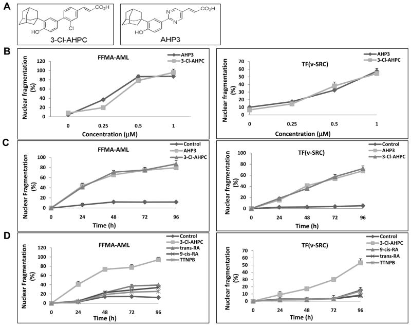
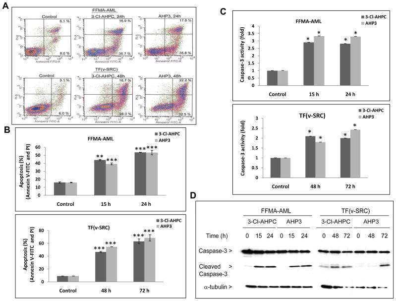
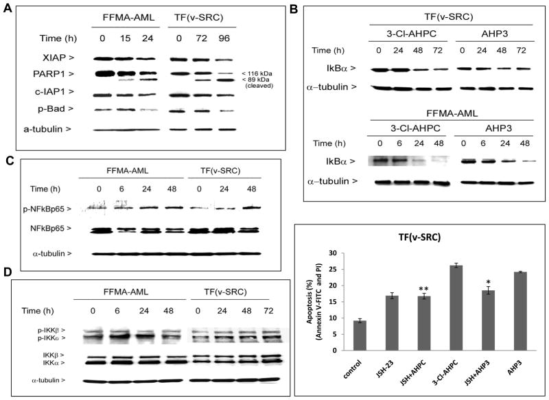
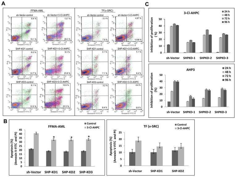
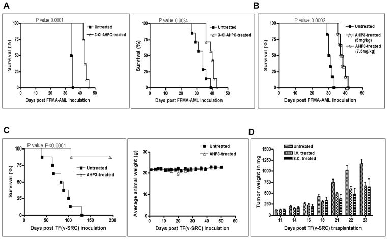
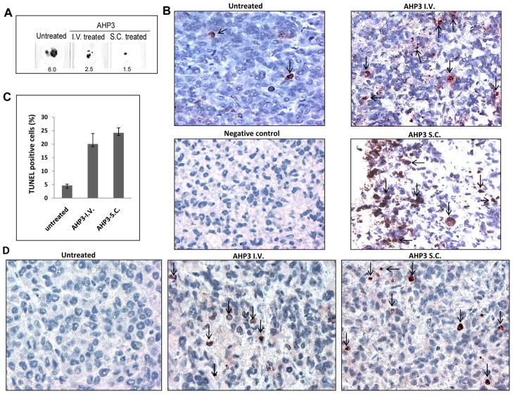
The adamantyl-substituted retinoid-related (ARR) compounds 3-Cl-AHPC and AHP3 induce apoptosis in vitro and in vivo in a newly established human acute myelogenous leukemia (AML) cell line, FFMA-AML, and in the established TF(v-SRC) AML cell line. FFMA-AML and TF(v-SRC) cells displayed resistance to apoptosis mediated by the standard retinoids (including trans-retinoic acid, 9-cis-retinoic acid, and the synthetic retinoid TTNPB) but showed sensitivity to apoptosis mediated by 3-Cl-AHPC- and AHP3 in vitro and in vivo as documented by poly(ADP-ribose) polymerase (PARP) cleavage and apoptosis terminal deoxyribonucleotidyl transferase-mediated dUTP nick end labeling assay. 3-Cl-AHPC or AHP3 exposure in vitro resulted in decreased expression of the antiapoptotic proteins (cellular inhibitor of apoptosis 1, X-linked inhibitor of apoptosis protein) and phospho-Bad and activated the NF-κB canonical pathway. A significant prolongation of survival was observed both in nonobese diabetic severe combined immunodeficient mice carrying FFMA-AML cells and treated with either 3-Cl-AHPC or AHP3 and in severe combined immunodeficient mice carrying TF(v-SRC) cells and treated with AHP3. We have previously shown that ARRs bind to the orphan nuclear receptor small heterodimer partner (SHP) and that the expression of SHP is required for ARR-mediated apoptosis. Induced loss of SHP in these AML cells blocked 3-Cl-AHPC- and AHP3-mediated induction of apoptosis. These results support the further development of 3-Cl-AHPC and AHP3 as potential therapeutic agents in the treatment of AML patients.
©2010 AACR.
Conflict of interest statement
No potential conflicts of interest were disclosed.
Figures
Similar articles
-
Induction of apoptosis in retinoid-refractory acute myelogenous leukemia by a novel AHPN analog.Blood. 2003 Nov 15;102(10):3743-52. doi: 10.1182/blood-2003-01-0108. Epub 2003 Jul 31. Blood. 2003. PMID: 12893763
-
SHP and Sin3A expression are essential for adamantyl-substituted retinoid-related molecule-mediated nuclear factor-kappaB activation, c-Fos/c-Jun expression, and cellular apoptosis.Mol Cancer Ther. 2009 Jun;8(6):1625-35. doi: 10.1158/1535-7163.MCT-08-0964. Epub 2009 Jun 9. Mol Cancer Ther. 2009. PMID: 19509248
-
Adamantyl-substituted retinoid-derived molecules that interact with the orphan nuclear receptor small heterodimer partner: effects of replacing the 1-adamantyl or hydroxyl group on inhibition of cancer cell growth, induction of cancer cell apoptosis, and inhibition of SRC homology 2 domain-containing protein tyrosine phosphatase-2 activity.J Med Chem. 2008 Sep 25;51(18):5650-62. doi: 10.1021/jm800456k. Epub 2008 Aug 30. J Med Chem. 2008. PMID: 18759424 Free PMC article.
-
The peptidomimetic, 1-adamantyl-substituted, and flex-het classes of retinoid-derived molecules: structure-activity relationships and retinoid receptor-independent anticancer activities.Mini Rev Med Chem. 2010 Jun;10(6):455-91. doi: 10.2174/138955710791384045. Mini Rev Med Chem. 2010. PMID: 20370709 Review.
-
Cellular and pharmacological bases of the antitumor activity of a novel adamantyl retinoid, ST1926.J Chemother. 2004 Nov;16 Suppl 4:74-6. doi: 10.1179/joc.2004.16.Supplement-1.74. J Chemother. 2004. PMID: 15688616 Review.
Cited by
-
Adamantyl arotinoids that inhibit IκB kinase α and IκB kinase β.ChemMedChem. 2013 Jul;8(7):1184-98. doi: 10.1002/cmdc.201300100. Epub 2013 May 7. ChemMedChem. 2013. PMID: 23653373 Free PMC article.
-
The lipophilic bullet hits the targets: medicinal chemistry of adamantane derivatives.Chem Rev. 2013 May 8;113(5):3516-604. doi: 10.1021/cr100264t. Epub 2013 Feb 25. Chem Rev. 2013. PMID: 23432396 Free PMC article. Review. No abstract available.
-
Adamantyl Retinoid-Related Molecules Induce Apoptosis in Pancreatic Cancer Cells by Inhibiting IGF-1R and Wnt/β-Catenin Pathways.J Oncol. 2012;2012:796729. doi: 10.1155/2012/796729. Epub 2012 Apr 5. J Oncol. 2012. PMID: 22570653 Free PMC article.
-
Nuclear receptor small heterodimer partner in apoptosis signaling and liver cancer.Cancers (Basel). 2011 Jan 5;3(1):198-212. doi: 10.3390/cancers3010198. Cancers (Basel). 2011. PMID: 24212613 Free PMC article.
-
Inhibition of IκB kinase-β and IκB kinase-α by heterocyclic adamantyl arotinoids.Bioorg Med Chem. 2014 Feb 15;22(4):1285-302. doi: 10.1016/j.bmc.2014.01.006. Epub 2014 Jan 10. Bioorg Med Chem. 2014. PMID: 24457093 Free PMC article.
References
-
- Byrd JC, Mrozek K, Dodge RK, et al. Pretreatment cytogenetic abnormalities are predictive of induction success, cumulative incidence of relapse, and overall survival in adult patients with de novo acute myeloid leukemia; results from Cancer and Leukemia Group B (CALGB 8461) Blood. 2002;100:4325–4336. - PubMed
-
- Grimwade D, Walker H, Harrison G, et al. The predictive value of hieratical cytogenetic classification in older adults with acute myeloid leukemia (AML) analysis of 1065 patients entered into United Kingdom Medical Research Council AML 11 trial. Blood. 2001;98:1312–1320. - PubMed
-
- Mrozek K, Heerema NA, Bloomfield CD. Cytogenetics in acute leukemia. Blood Rev. 2004;18:115–136. - PubMed
-
- Warrell RP, Frankel SR, Miller WH, et al. Differentiation therapy of acute promyelocytic leukemia with tretinoin (all-trans-retinoic acid) N Eng J Med. 1991;324:1385–1393. - PubMed
-
- Melnick A, Licht JD. Deconstructing a disease: RARα, its fusion partners and their roles in the pathogenesis of acute promyelocytic leukemia. Blood. 1999;93:3167–3215. - PubMed
Publication types
MeSH terms
Substances
Grants and funding
LinkOut - more resources
Full Text Sources
Other Literature Sources
Medical
Research Materials
Miscellaneous
