Temporomandibular joint and 3.0 T pseudodynamic magnetic resonance imaging. Part 2: evaluation of articular disc obscurity
- PMID: 21062942
- PMCID: PMC3520210
- DOI: 10.1259/dmfr/92017549
Temporomandibular joint and 3.0 T pseudodynamic magnetic resonance imaging. Part 2: evaluation of articular disc obscurity
Abstract
Objectives: This study examined the relationship between temporomandibular joint (TMJ) dysfunctions and obscurity grades of interpreted anterior and posterior borders of the articular disc (Da and Dp, respectively) by 3.0 T pseudodynamic MRI.
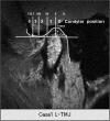
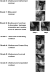
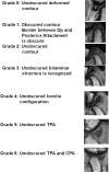
Methods: Da and Dp were classified into seven obscurity grades, and the Dp contour was classified into three types. The grades, types and TMJ function were compared by 3.0 T pseudodynamic MRI.
Results: Unobscured Da images at condylar positions posterior to the articular eminence were associated with normal TMJ function (P = 0.046 < 0.05). Unobscured Dp images at condylar positions anterior to the articular eminence were associated with normal TMJ function (P = 0.033 < 0.05). In addition, unobscured Dp images following flap insertion were associated with normal TMJ function (P = 0.043 < 0.05). There was no statistical relationship between Dp contour types and TMJ movement, but any change observed in the Dp contour during mouth opening was associated with abnormal TMJ function (P = 0.040 < 0.05).
Conclusions: Grading of Da and Dp obscurity based on how well the areas were defined in the images, identifying the condylar positions in relation to the glenoid fossa and articular eminences, and observing the changes in Dp contour types were useful for diagnosing TMJ abnormalities.
Figures


Similar articles
-
Temporomandibular joint and 3.0 T pseudodynamic magnetic resonance imaging. Part 1: evaluation of condylar and disc dysfunction.Dentomaxillofac Radiol. 2010 Dec;39(8):475-85. doi: 10.1259/dmfr/29741224. Dentomaxillofac Radiol. 2010. PMID: 21062941 Free PMC article.
-
Morphological and positional assessments of TMJ components and lateral pterygoid muscle in relation to symptoms and occlusion of patients with temporomandibular disorders.J Oral Rehabil. 2000 Oct;27(10):860-74. doi: 10.1046/j.1365-2842.2000.00622.x. J Oral Rehabil. 2000. PMID: 11065021
-
Inclination of the condylar long axis is not related to temporomandibular disc displacement.J Investig Clin Dent. 2019 Feb;10(1):e12375. doi: 10.1111/jicd.12375. Epub 2018 Nov 26. J Investig Clin Dent. 2019. PMID: 30474234
-
Part II: Temporomandibular Joint (TMJ)-Regeneration, Degeneration, and Adaptation.Curr Osteoporos Rep. 2018 Aug;16(4):369-379. doi: 10.1007/s11914-018-0462-8. Curr Osteoporos Rep. 2018. PMID: 29943316 Review.
-
The Roles of Indian Hedgehog Signaling in TMJ Formation.Int J Mol Sci. 2019 Dec 13;20(24):6300. doi: 10.3390/ijms20246300. Int J Mol Sci. 2019. PMID: 31847127 Free PMC article. Review.
Cited by
-
Comparison of a 32-channel head coil and a 2-channel surface coil for MR imaging of the temporomandibular joint at 3.0 T.Dentomaxillofac Radiol. 2016;45(4):20150420. doi: 10.1259/dmfr.20150420. Epub 2016 Feb 3. Dentomaxillofac Radiol. 2016. PMID: 26837671 Free PMC article.
-
In vivo measurement of the 3D kinematics of the temporomandibular joint using miniaturized electromagnetic trackers: technical report.Med Biol Eng Comput. 2013 Apr;51(4):479-84. doi: 10.1007/s11517-012-1015-4. Epub 2012 Dec 14. Med Biol Eng Comput. 2013. PMID: 23242785
References
-
- Rees LA. The structure and function of the mandibular joint. Br Dent J 1954;96:125–133
-
- Scapino RP. Histopathology of the disc and posterior attachment in disc displacement internal derangement of the TMJ, In:Magnetic resonance of the temporomandibular joint New York:Thieme,1990:63–73
-
- Scapino RP. The posterior attachment: its structure, function, and appearance in TMJ imaging studies. Part 1. Craniomandib Disord Fac Oral Pain 1991;5:83–95 - PubMed
-
- Drace JE, Enzmann DR. Defining the normal temporomandibular joint: closed-, partially open-, and open-mouth MR imaging of asymptomatic subjects. Radiology 1990;177:67–71 - PubMed
Publication types
MeSH terms
LinkOut - more resources
Full Text Sources
Medical

