

Temporomandibular joint and 3.0 T pseudodynamic magnetic resonance imaging. Part 2: evaluation of articular disc obscurity
- PMID: 21062942
- PMCID: PMC3520210
- DOI: 10.1259/dmfr/92017549
Temporomandibular joint and 3.0 T pseudodynamic magnetic resonance imaging. Part 2: evaluation of articular disc obscurity
Abstract
Objectives: This study examined the relationship between temporomandibular joint (TMJ) dysfunctions and obscurity grades of interpreted anterior and posterior borders of the articular disc (Da and Dp, respectively) by 3.0 T pseudodynamic MRI.
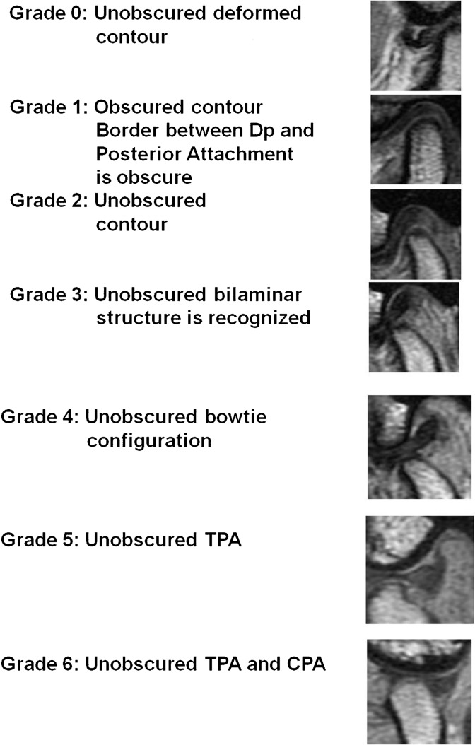
Methods: Da and Dp were classified into seven obscurity grades, and the Dp contour was classified into three types. The grades, types and TMJ function were compared by 3.0 T pseudodynamic MRI.
Results: Unobscured Da images at condylar positions posterior to the articular eminence were associated with normal TMJ function (P = 0.046 < 0.05). Unobscured Dp images at condylar positions anterior to the articular eminence were associated with normal TMJ function (P = 0.033 < 0.05). In addition, unobscured Dp images following flap insertion were associated with normal TMJ function (P = 0.043 < 0.05). There was no statistical relationship between Dp contour types and TMJ movement, but any change observed in the Dp contour during mouth opening was associated with abnormal TMJ function (P = 0.040 < 0.05).
Conclusions: Grading of Da and Dp obscurity based on how well the areas were defined in the images, identifying the condylar positions in relation to the glenoid fossa and articular eminences, and observing the changes in Dp contour types were useful for diagnosing TMJ abnormalities.
Figures


References
-
- Rees LA. The structure and function of the mandibular joint. Br Dent J 1954;96:125–133
-
- Scapino RP. Histopathology of the disc and posterior attachment in disc displacement internal derangement of the TMJ, In:Magnetic resonance of the temporomandibular joint New York:Thieme,1990:63–73
-
- Scapino RP. The posterior attachment: its structure, function, and appearance in TMJ imaging studies. Part 1. Craniomandib Disord Fac Oral Pain 1991;5:83–95 - PubMed
-
- Drace JE, Enzmann DR. Defining the normal temporomandibular joint: closed-, partially open-, and open-mouth MR imaging of asymptomatic subjects. Radiology 1990;177:67–71 - PubMed
Publication types
MeSH terms
LinkOut - more resources
Full Text Sources
Medical
