Review
. 2011 Mar;52(3):419-34.
doi: 10.1194/jlr.R009761. Epub 2010 Nov 9.

Lipid metabolism in myelinating glial cells: lessons from human inherited disorders and mouse models

Affiliations
Review

Lipid metabolism in myelinating glial cells: lessons from human inherited disorders and mouse models

Roman Chrast et al. J Lipid Res. 2011 Mar.

Abstract

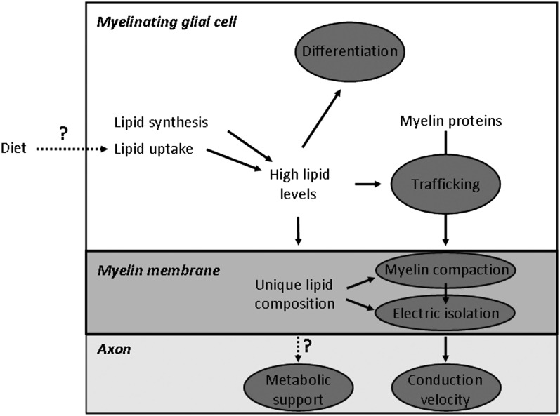
The integrity of central and peripheral nervous system myelin is affected in numerous lipid metabolism disorders. This vulnerability was so far mostly attributed to the extraordinarily high level of lipid synthesis that is required for the formation of myelin, and to the relative autonomy in lipid synthesis of myelinating glial cells because of blood barriers shielding the nervous system from circulating lipids. Recent insights from analysis of inherited lipid disorders, especially those with prevailing lipid depletion and from mouse models with glia-specific disruption of lipid metabolism, shed new light on this issue. The particular lipid composition of myelin, the transport of lipid-associated myelin proteins, and the necessity for timely assembly of the myelin sheath all contribute to the observed vulnerability of myelin to perturbed lipid metabolism. Furthermore, the uptake of external lipids may also play a role in the formation of myelin membranes. In addition to an improved understanding of basic myelin biology, these data provide a foundation for future therapeutic interventions aiming at preserving glial cell integrity in metabolic disorders.


Figures

Fig. 1.
Fig. 1.
Ultrastructural comparison of the membranes of a promyelinating versus a mature myelinating glial cell. Electron micrographs of normal mouse sciatic nerves showing (A) promyelin figure at postnatal day 0 (P0) and (B) adult myelinated fiber (P100). The inner lip (also called inner mesaxon) of the myelinating glial cell, which turns around the axon to form the membranous spiral of myelin, is depicted with an asterisk (*). Scale bar, 0.5 μm. Sciatic nerve isolation and electron microscopy were done as described in (110).
Fig. 2.
Fig. 2.
Fatty acid composition of myelin compared with other membranes. Depicted is the amount of fatty acid in mol percentage of total amount of fatty acids. Bold numbered and gray numbered lipids depict, respectively, strongly higher or lower levels of these fatty acids in myelin compared with hepatocytes or erythrocyte membranes. Myelin membrane of mouse sciatic nerve were isolated as described in (110, 163). Mouse erythrocyte membranes and hepatocyte phospholipids were isolated and analyzed according to (110, 163). It should be noted that under the isolation conditions used, amide bonds in sphingolipids are relatively stable. Hence, the very long-chain fatty acids found in myelin are not reflecting the high level of galactosphingolipids in myelin, but are more likely to result from a particular fatty acyl composition of the glycerophospholipids (phosphatidylcholine, phosphatidylethanolamine, phosphatidylinositol, and phosphatidylserine). In addition, with the detection method used, the bar representing 18:1 fatty acids does not include 18:1 alcohol of plasmalogens, which nevertheless was previously detected to be very low in PNS myelin in mouse (110).
Fig. 3.
Fig. 3.
Human inherited and mouse experimental defects in the cholesterol biosynthesis pathway that cause myelin disorders. Solid line arrows depict a direct link between two steps, dashed line arrows imply intermediate steps that are not shown. Dark gray ovals show the positions of human disease genes; light gray oval shows the position of mutated genes in experimental mouse models. Sqs, squalene synthase; DHCR7, sterol delta-7-reductase; SLOS, Smith-Lemli-Opitz syndrome; CYP27A1, sterol 27-hydroxylase; CTX, Cerebrotendinous xanthomatosis; ABC1, ATP-binding cassette transporter A1; TD, Tangier disease.
Fig. 4.
Fig. 4.
Human inherited and mouse experimental defects in the glycosphingolipids synthesis pathway that cause myelin disorders. Solid line arrows depict direct link between two steps, dashed line arrow implies intermediate steps that are not shown. Dark gray ovals show positions of human diseases; light gray ovals show the position of mutated genes in experimental mouse models. ARSA, arylsulfatase A; ML, Metachromatic leukodystrophy; Cst, cerebroside sulfotransferase; GALC, galactosylceramidase; KD, Krabbe disease; Cgt, UDP-galactose:ceramide galactosyltransferase; Ugcg, UDP-glucose ceramide glucosyltransferase; SMPD1, sphingomyelin phosphodiesterase-1; NPD, Niemann-Pick disease type A; FA2H, fatty acid 2-hydroxylase; DLSP, Dysmyelinating leukodystrophy and spastic paraparesis with or without dystonia; Gal-Cer, galactocerebroside; Glc-Cer: glucocerebroside; 2-OH FA, 2-hydroxy fatty acids; FA, fatty acids.
Fig. 5.
Fig. 5.
Human inherited and mouse experimental defects in fatty acids and plasmalogen metabolism that cause myelin disorders. Solid line arrows depict direct link between two steps, dashed line arrow implies intermediate steps that are not shown. Dashed line rectangles represent various lipid metabolic pathways in the peroxisome. Dark gray ovals show positions of human disease genes; light gray ovals show the position of mutated genes in experimental mouse models. Dhapat, dihydroxyacetone phosphate acyltransferase; PHYH, phytanoyl-CoA hydroxylase; RD, Refsum disease; PEX1-12, peroxins; PBD, peroxisome biogenesis disorders; ALDH3A2, fatty aldehyde dehydrogenase; SLS, Sjogren-Larsson syndrome; ABCD1, ATP-binding cassette transporter D1; ALD, adrenoleukodystrophy; VLCFA, very long-chain fatty acids.
Fig. 6.
Fig. 6.
Human inherited and mouse experimental defects in general lipid metabolism that cause myelin disorders. Solid line arrows depict direct link between two steps, dashed line arrows implies intermediate steps that are not shown. Dashed line circle represents the citric acid cycle in mitochondria. Dark gray oval shows positions of human disease genes; light gray oval shows the position of mutated genes in an experimental mouse model. PC, pyruvate carboxylase; PCD, pyruvate carboxylase deficiency; ASPA, aspartoacetylase; CD, Canavan disease; NAA, N-acetylaspartic acid; FA, fatty acids.
Fig. 7.
Fig. 7.
Schematic diagram of the various roles of lipids in myelinating glial cells. Gray ovals show the processes that are affected by lipid metabolism and involved in myelinated fiber function. High lipid levels, which are required for synthesis of a full myelin membrane, are ensured by both the endogenous synthesis of lipids as by the uptake of lipids from the extracellular environment. Lipids influence myelinating glial cell differentiation and trafficking of myelin proteins to the myelin membrane. The unique lipid composition of the myelin membrane is required for proper myelin membrane wrapping (compaction) and electrostatic isolation of the axon from extracellular environment, thereby promoting saltatory conduction and conduction velocity. Question marks show processes with a potential effect on myelin fiber function: the influence of lipid-specific diets on myelin lipid dependent processes, and the possible role of myelin lipids in metabolic support of the axon. See text for further explanation.

Similar articles

Cited by

References

    1. Barres B. A. 2008. The mystery and magic of glia: a perspective on their roles in health and disease. Neuron. 60: 430–440. - PubMed
    1. Franz D. N., Iggo A. 1968. Conduction failure in myelinated and non-myelinated axons at low temperatures. J. Physiol. 199: 319–345. - PMC - PubMed
    1. Waxman S. G. 1980. Determinants of conduction velocity in myelinated nerve fibers. Muscle Nerve. 3: 141–150. - PubMed
    1. Hartline D. K., Colman D. R. 2007. Rapid conduction and the evolution of giant axons and myelinated fibers. Curr. Biol. 17: R29–R35. - PubMed
    1. Norton W. T., Poduslo S. E. 1973. Myelination in rat brain: method of myelin isolation. J. Neurochem. 21: 749–757. - PubMed

Publication types