. 2010 Nov 15;70(22):9319-28.
doi: 10.1158/0008-5472.CAN-10-1783. Epub 2010 Nov 9.

Synergistic enhancement of carboplatin efficacy with photodynamic therapy in a three-dimensional model for micrometastatic ovarian cancer

Affiliations

Synergistic enhancement of carboplatin efficacy with photodynamic therapy in a three-dimensional model for micrometastatic ovarian cancer

Imran Rizvi et al. Cancer Res. .

Abstract

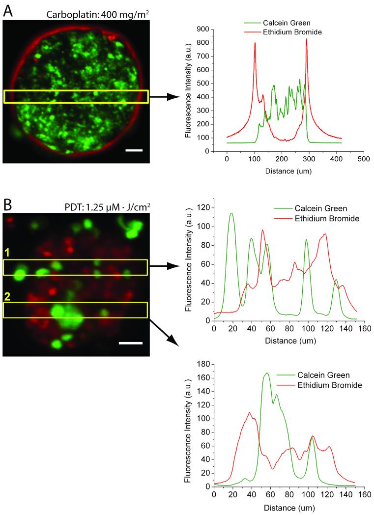
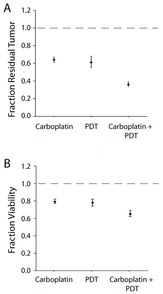
Metastatic ovarian cancer (OvCa) frequently recurs due to chemoresistance, highlighting the need for nonoverlapping combination therapies that mechanistically synergize to eradicate residual disease. Photodynamic therapy (PDT), a photochemistry-based cytotoxic modality, sensitizes ovarian tumors to platinum agents and biologics and has shown clinical promise against ovarian carcinomatosis. We introduce a three-dimensional (3D) model representing adherent ovarian micrometastases and high-throughput quantitative imaging methods to rapidly screen the order-dependent effects of combining benzoporphyrin-derivative (BPD) monoacid A-based PDT with low-dose carboplatin. 3D ovarian micronodules grown on Matrigel were subjected to BPD-PDT either before or after carboplatin treatment. We developed custom fluorescence image analysis routines to quantify residual tumor volume and viability. Carboplatin alone did not eradicate ovarian micrometastases at a dose of 400 mg/m2, leaving surviving cores that were nonsensitive or impermeable to chemotherapy. BPD-PDT (1.25 μmol/L·J/cm2) created punctate cytotoxic regions within tumors and disrupted micronodular structure. Treatment with BPD-PDT prior to low-dose carboplatin (40 mg/m2) produced a significant synergistic reduction [P<0.0001, analysis of covariance (ANCOVA)] in residual tumor volume [0.26; 95% confidence interval (95% CI), 0.19-0.36] compared with PDT alone (0.76; 95% CI, 0.63-0.92) or carboplatin alone (0.95; 95% CI, 0.83-1.09), relative to controls. This synergism was not observed with the reverse treatment order. Here, we demonstrate for the first time the use of a 3D model for micrometastatic OvCa as a rapid and quantitative reporter to optimize sequence and dosing regimens of clinically relevant combination strategies. This approach combining biological modeling with high-content imaging provides a platform to rapidly screen therapeutic strategies for a broad array of metastatic tumors.


Figures

Figure 1
Figure 1
Biological characterization of a 3D model for micrometastatic OvCa. A, OVCAR5 cells overlaid on GFR-Matrigel™ formed 3D ovarian micronodules representing adherent micrometastatic disease with B, punctate E-cadherin expression, and C, fibronectin (red) expression (blue: DAPI), markers for poor prognosis. D, mean diameter increased from 34.3μm (±2.6μm) 3 days post-plating (n = 9) to 108.9μm (± 13.0μm) at day 10 (n = 9) and 131.9μm (±25.3μm) at day 17 (n = 3) (E). Scale bars: (B) and (C), 20μm; (D) 150μm.
Figure 2
Figure 2
Monolayer cultures significantly overestimate carboplatin and BPD-PDT efficacy compared to cells in 3D. A, fractional viability of OVCAR5 cells in monolayer (striped) following carboplatin treatment (40 mg/m2) was 0.30 (± 0.082) (n = 10), as compared to 0.90 (± 0.057) in 3D (solid), (n = 6) (p < 0.001, two-tailed t-test). B, left, monolayer cultures treated with 1.25μM·J/cm2 BPD-PDT, had a fractional viability of 0.35 (± 0.04) (n = 12), as compared to 0.75 (±0.04) in 3D (n = 13) (p = 0.003, two-tailed t-test). N-values vary from 6 to 16 for individual treatment groups within each culture condition. B, right, confocal fluorescence images show BPD-MA distribution and autofluorescence from a representative nodule. Scale bars = 25μm.
Figure 3
Figure 3
Differential patterns of cytotoxic response in carboplatin and BPD-PDT treated 3D micronodules. A, left, a representative micronodule treated with 400 mg/m2 carboplatin showing cell death (ethidium bromide) on the periphery of a viable (calcein) tumor core, with A, right, distinct and minimally overlapping fluorescence intensity profiles. B, left, BPD-PDT 1.25μM·J/cm2 treatment leads to nodular disruption and punctate cytoxicity. B, right, intensity profile scans through two regions of interest (yellow boxes) show highly overlapping fluorescence patterns. Scale bars = 25μm.
Figure 4
Figure 4
BPD-PDT synergizes with low-dose carboplatin to reduce residual tumor volume. A, images of residual disease (calcein). B, fraction residual tumor following carboplatin alone or BPD-PDT alone was 0.95 (95% CI=0.83-1.09, n = 11) and 0.76 (95% CI=0.63-0.92 n = 15), respectively, relative to no treatment (grey dashed line). The combination treatment, BPD-PDT followed by carboplatin, produced a synergistic reduction in residual tumor to 0.26 (95% CI=0.19-0.36, n = 11) (p<0.0001, interaction term from ANCOVA). Scale bars = 250μm.
Figure 5
Figure 5
BPD-PDT followed by carboplatin synergistically reduces tumor viability. A, images of tumor viability (calcein and ethidium bromide fluorescence) following treatment. B, mean fraction viability in micronodules treated with either carboplatin alone or BPD-PDT alone was 0.92 (95% CI=0.88-0.97, n = 11) and 0.80 (95%CI=0.74-0.86, n = 15), respectively, relative to no treatment (grey dashed line). The combination treatment, BPD-PDT followed by carboplatin, reduced viability to 0.45 (95% CI=0.38-0.53, n = 11), indicating a significant synergism (p<0.0001, interaction term from ANCOVA). Scale bars = 250μm.
Figure 6
Figure 6
Carboplatin treatment prior to BPD-PDT does not produce a synergistic interaction. No synergism was observed with the reverse treatment order, as evaluated by fraction of residual tumor (A) (p=0.3326), and tumor viability (B) (p=0.1368) (interaction term from ANCOVA for both treatment response metrics), relative to no treatment (grey dashed lines).

References

    1. BAST RC, JR., HENNESSY B, MILLS GB. THE BIOLOGY OF OVARIAN CANCER: NEW OPPORTUNITIES FOR TRANSLATION. NAT REV CANCER. 2009;9:415–28. - PMC - PubMed
    1. CHO KR, SHIH IEM. OVARIAN CANCER. ANNUAL REVIEW OF PATHOLOGY. 2009;4:287–313. - PMC - PubMed
    1. NAORA H, MONTELL DJ. OVARIAN CANCER METASTASIS: INTEGRATING INSIGHTS FROM DISPARATE MODEL ORGANISMS. NAT REV CANCER. 2005;5:355–66. - PubMed
    1. DEL CARMEN MG, RIZVI I, CHANG Y, et al. SYNERGISM OF EPIDERMAL GROWTH FACTOR RECEPTOR-TARGETED IMMUNOTHERAPY WITH PHOTODYNAMIC TREATMENT OF OVARIAN CANCER IN VIVO. JNCI. 2005;97:1516–24. - PubMed
    1. CHI DS, EISENHAUER EL, ZIVANOVIC O, et al. IMPROVED PROGRESSION-FREE AND OVERALL SURVIVAL IN ADVANCED OVARIAN CANCER AS A RESULT OF A CHANGE IN SURGICAL PARADIGM. GYNECOL ONCOL. 2009;114:26–31. - PubMed

Publication types

MeSH terms