. 2010;26(4-5):749-60.
doi: 10.1159/000322342. Epub 2010 Oct 29.

Acid sphingomyelinase deficiency attenuates bleomycin-induced lung inflammation and fibrosis in mice

Affiliations

Acid sphingomyelinase deficiency attenuates bleomycin-induced lung inflammation and fibrosis in mice

Rajwinder Dhami et al. Cell Physiol Biochem. 2010.

Abstract

Background/aims: The sphingomyelin/ceramide signaling pathway is an important component of many cellular processes implicated in the pathogenesis of lung disease. Acid sphingomyelinase (ASM) is a key mediator of this pathway, but its specific role in pulmonary fibrosis has not been previously investigated. Here we used the bleomycin model of pulmonary fibrosis to investigate fibrotic responses in normal and ASM knockout (ASM(-/-)) mice, and in NIH3T3 fibroblasts with and without ASM siRNA treatment.

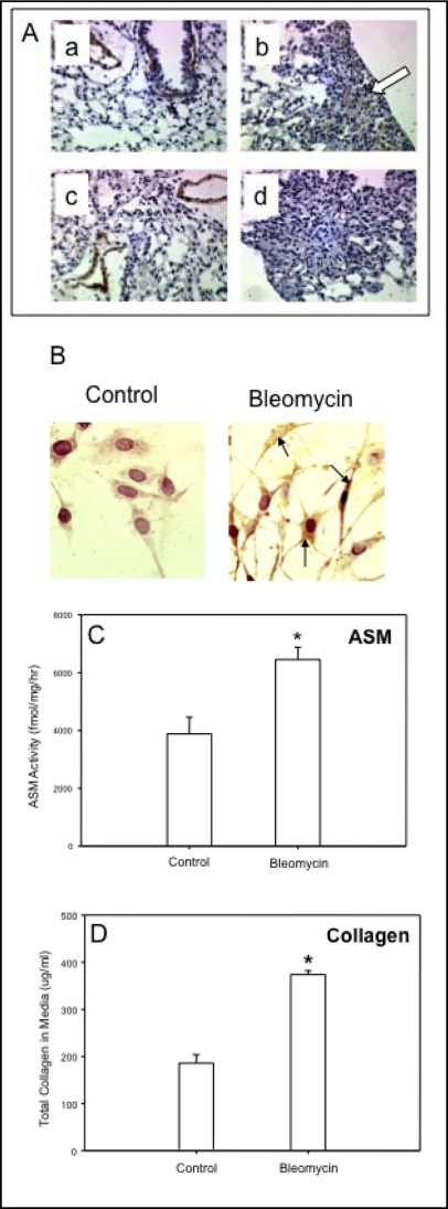
Methods: Mice and cells with and without ASM activity were treated with bleomycin, and the effects on lung inflammation, formation of collagen producing myofibroblasts, and apoptosis were assessed.

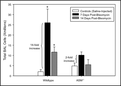
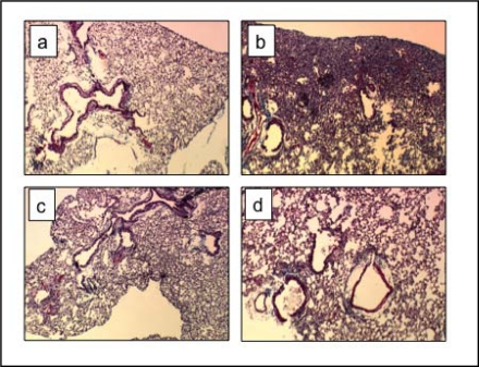
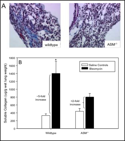
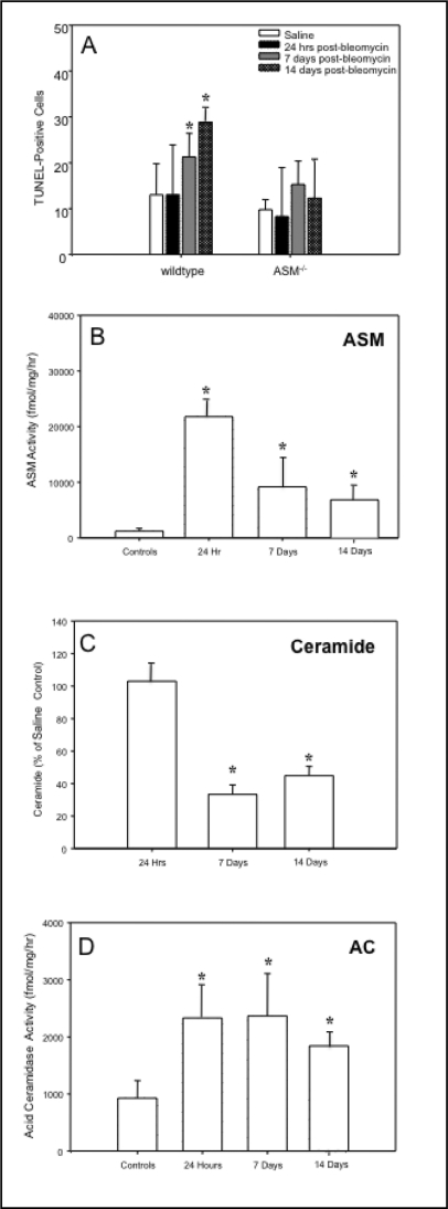
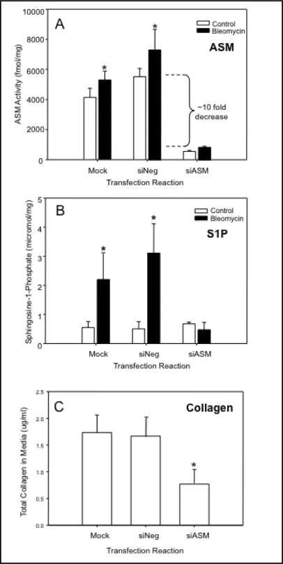
Results: The development of bleomycin-induced inflammation and fibrosis in wildtype mice correlated with the rapid activation of ASM, and was markedly attenuated in the absence of ASM activity. Along with the elevated ASM activity, there also was an elevation of acid ceramidase (AC) activity, which was sustained for up to 14 days post-bleomycin treatment. Studies in NIH3T3 fibroblasts confirmed these findings, and revealed a direct effect of ASM/AC activation on the formation of myofibroblasts. Cell studies also showed that a downstream effect of bleomycin treatment was the production of sphingosine-1-phosphate.

Conclusions: These data demonstrate that the sphingomyelin/ceramide signaling pathway is involved in the pathogenesis of bleomycin-induced pulmonary fibrosis, and suggest that inhibition of ASM may potentially slow the fibrotic process in the lung.


Figures

Fig. 1
Fig. 1
The total number of inflammatory cells recovered from the pulmonary airspaces of 8–10 week old wildtype and ASM' mice by bronchoalveolar lavage (n=10 per group). By 7 days after bleomycin instillation, the numbers of cells in the lungs of wildtype mice increased 14-fold and remain significantly elevated at 14 days. In ASM' mice, the total cells in the airspaces were only elevated ∼2-fold and returned to pre- bleomycin levels by 14 days. * indicates the statistically significant increase (p-value < 0.05) in bleomycin-treated wildetype animals as compared with controls (saline injected).
Fig. 2
Fig. 2
Histological assessment of lungs 14 days after bleomycin exposure. Significant fibrosis is observed in lung parenchyma of bleomycin-exposed wildtype mice (b) compared with saline-treated wildtype mice (a). The lungs of ASM−/− mice after bleomycin exposure (d) were similar to the lungs of saline-treated counterparts (c). Lungs were stained with Trichrome. Images are shown at 40X magnification. Sections from 6 animals/group were analyzed. Representative images are shown.
Fig. 3
Fig. 3
Extracellular matrix deposition in bleomycin-instilled lungs. Trichrome staining (blue) highlights areas of excessive collagen deposition at fibrotic lesions seen in wildtype mice after bleomycin treatment that were not evident in ASM−/− mice (A). Total soluble collagen measured in whole lungs was increased ∼5-fold in wildtype mice 14 days after bleomycin instillation and less than 2-fold in ASM−/− mice compared with their saline-treated counterparts (B). * indicates the statistically significant increase in wildtype animals compared with saline controls with a p-value < 0.05. n=10 per group.
Fig. 4
Fig. 4
The sphingomyelin/ceramide apoptosis pathway in bleomycin-instilled lungs. The numbers of TUNEL positive lung cells were significantly increased in wildtype mice starting at 7 days post-bleomycin treatment, whereas there was no significant change in the numbers of apoptotic cells in ASM−/− mouse lungs (A). Sections from 6 animals/group were analyzed. Representative images are shown. ASM activity also was significantly elevated in wildtype mice 24 hrs after bleomycin instillation and while reduced by 7 and 14 days, remained significantly higher than control (saline injected) mice (B). The ceramide levels in the lungs of bleomycin-treated wildtype mice were equivalent to the saline-injected controls at 24 hr, and at 7 and 14 days were significantly reduced compared with saline- treated controls (C). The acid ceramidase (AC) activity in wildtype lungs was increased at 24 hr and remained significantly elevated for up to 14 days (D). * indicates significant differences from control (saline-injected) and bleomycin-treated animals (p value <0.05).
Fig. 5
Fig. 5
(A) Alpha-smooth muscle actin (SMA)-expressing cells were increased in the parenchyma of lungs of wildtype mice 14 days after bleomycin exposure (e.g., b, arrow showing brown staining), but not in ASM−/− mice (d). As expected, both wildtype (a) and ASM-deficient (c) saline-treated control mice showed alpha-SMA expression only in smooth muscle cells around blood vessels, and not in the parenchyma. Sections from 6 animals/group were analyzed. Representative images are shown. (B) NIH3T3 fibroblasts exposed to bleomycin for 24 hours showed a marked increase in alpha-SMA expression as assessed by immunohistochemistry (black arrows point to intensely-staining cells within the cell body). There was a concomitant increase in ASM activity in NIH3T3 fibroblasts after exposure to bleomycin for 24 hr (C) and, at the same time a significant increase in collagen secretion (D). * indicates significant differences between control (saline injected) and bleomycin treated mice with a p-value <0.05.
Fig. 6
Fig. 6
ASM siRNA transfection significantly reduced endogenous ASM activity in NIH3T3 fibroblasts (A, white boxes) compared to mock and Negative (siNeg) transfected cells, and prevented the bleomycin-induced increase in this activity (A, black boxes). ASM siRNA transfection also significantly prevented the bleomycin-induced increases in sphingosine-1-phosphate (S1P) levels in these cells (B). The release of collagen also was reduced in cells transfected with ASM siRNA, in comparison with mock and siNeg-transfected cells (C). Note that due to the siRNA transfection assay, the number of cells used to detect collagen secretion in these experiments was significantly smaller than that used in Figure 5D, explaining the differences in the total collagen determined. * indicates significant differences (p-value of <0.05). Each experiment was replicated at least three times.

Similar articles

Cited by

References

    1. Maher TM, Wells AU, Laurent GJ. Idiopathic pulmonary fibrosis: multiple causes and multiple mechanisms? Eur Respir J. 2007;30:835–839. - PubMed
    1. Hauber HP, Blaukovitsch M. Current and future treatment options in idiopathic pulmonary fibrosis. Inflamm Allergy Drug Targets. 2010 in press. - PubMed
    1. Chua F, Gauldie J, Laurent GL. Pulmonary fibrosis: searching for model answers. Am J Respir Cell Mol Biol. 2005;33:9–13. - PubMed
    1. Tager AM, LaCamera P, Shea BS, Campanella GS, Selman M, Zhao A, Polosukhin V, Wain J, Karimi-Shah B, Kim ND, Hart WK, Pardo A, Blackwell TS, Xu Y, Chun J, Luster AD. The lysophosphatidic acid receptor LPA1 links pulmonary fibrosis to lung injury by mediating fibroblast recruitment and vascular leak. Nat Med. 2008;14:45–54. - PubMed
    1. Scotton CJ, Chambers RC. Molecular targets in pulmonary fibrosis: the myofibroblast in focus. Chest. 2007;132:1311–1321. - PubMed

Publication types

MeSH terms