

Confocal endomicroscopic examination of malignant biliary strictures and histologic correlation with lymphatics
- PMID: 21063210
- PMCID: PMC3572850
- DOI: 10.1097/MCG.0b013e3181fbdc38
Confocal endomicroscopic examination of malignant biliary strictures and histologic correlation with lymphatics
Abstract
Background and aims: Current methods to diagnose malignant biliary strictures are of low sensitivity. Confocal endomicroscopy is a new approach that may improve the diagnosis of indeterminate biliary strictures. The purpose of this study was to evaluate indeterminate biliary strictures using probe-based confocal laser endomicroscopy and to understand the histologic basis for the confocal images.
Methods: Fourteen patients with indeterminate biliary strictures underwent endoscopic retrograde cholangiopancreatography with examination of their common bile duct with fluorescein-aided probe-based confocal laser endomicroscopy. Standard brushings and biopsies were performed. In parallel, rat bile ducts were examined either with conventional staining and light microscopy or with multiphoton microscopy.
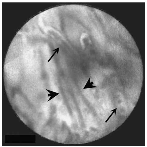
Results: Earlier published criteria were used to evaluate possible malignancy in the confocal images obtained in the 14 patients. None of the individual criteria were found to be specific enough for malignancy, but a normal-appearing reticular pattern without other putative markers of malignancy was observed in all normal patients. Multiphoton reconstructions of intact rat bile ducts revealed that the reticular pattern seen in normal tissue was in the same focal plane but was smaller than blood vessels. Special stains identified the smaller structures in this network as lymphatics.
Conclusions: Our limited series suggests that a negative confocal imaging study of the biliary tree can be used to rule out carcinoma, but there are frequent false positives using individual earlier published criteria. An abnormal reticular network, which may reflect changes in lymphatics, was never seen in benign strictures. Better correlation with known histologic structures may lead to improved accuracy of diagnoses.
Figures


References
-
- Lazaridis KN, Gores GJ. Cholangiocarcinoma. Gastroenterology. 2005;128:1655–1667. - PubMed
-
- Khan SA, Thomas HC, Davidson BR, et al. Cholangiocarcinoma. Lancet. 2005;366:1303–1314. - PubMed
-
- Ahrendt SA, Nakeeb A, Pitt HA. Cholangiocarcinoma. Clin Liver Dis. 2001;5:191–218. - PubMed
-
- Bennett JJ, Green RH. Malignant masquerade: dilemmas in diagnosing biliary obstruction. Surg Oncol Clin N Am. 2009;18:207–214. vii. - PubMed
Publication types
MeSH terms
Grants and funding
LinkOut - more resources
Full Text Sources
Medical
