. 2011 Apr;35(4):411-21.
doi: 10.1097/SHK.0b013e31820239ee.

Heparin use in a rat hemorrhagic shock model induces biologic activity in mesenteric lymph separate from shock

Affiliations

Heparin use in a rat hemorrhagic shock model induces biologic activity in mesenteric lymph separate from shock

Yong Qin et al. Shock. 2011 Apr.

Abstract

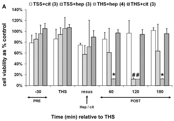
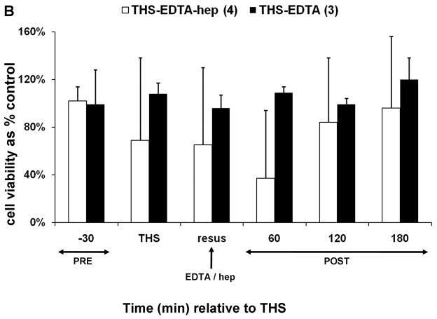
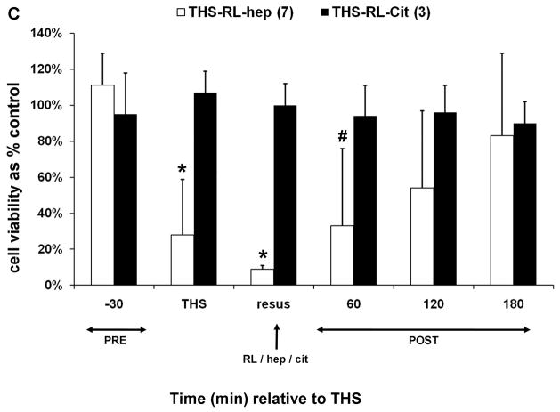
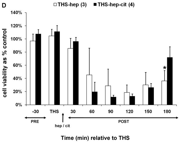
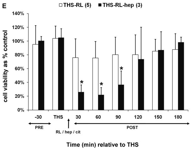
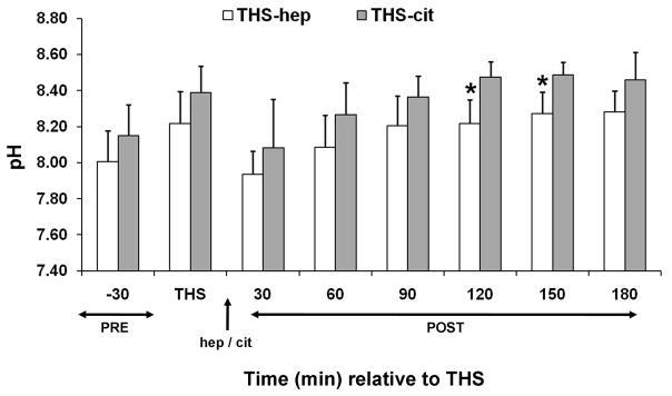
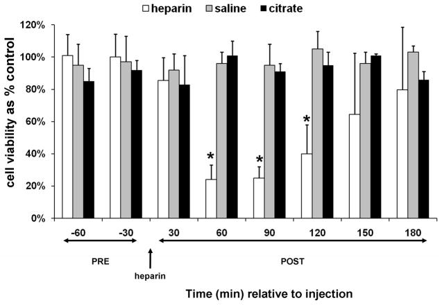
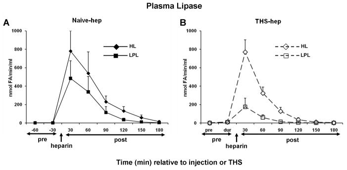
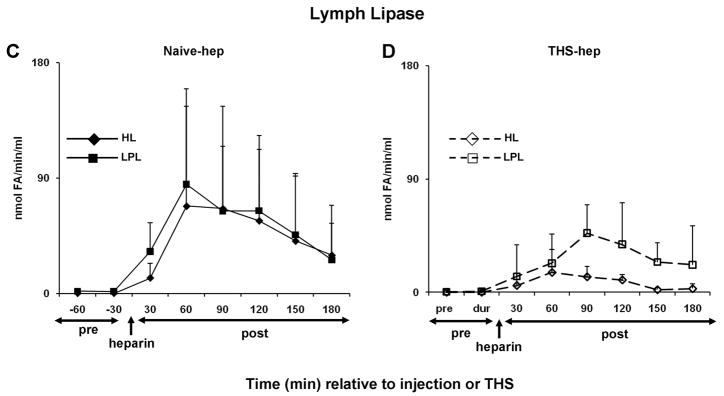
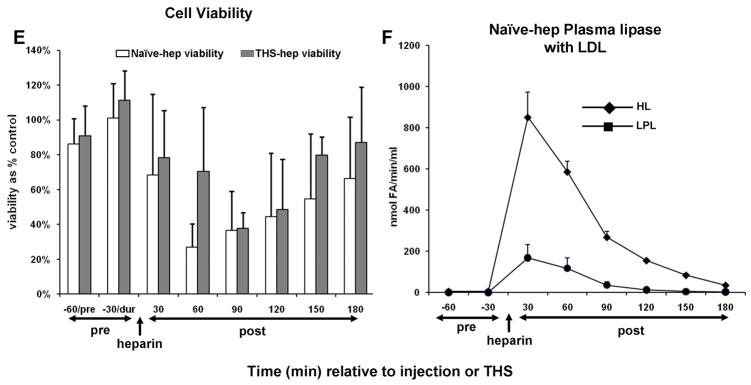
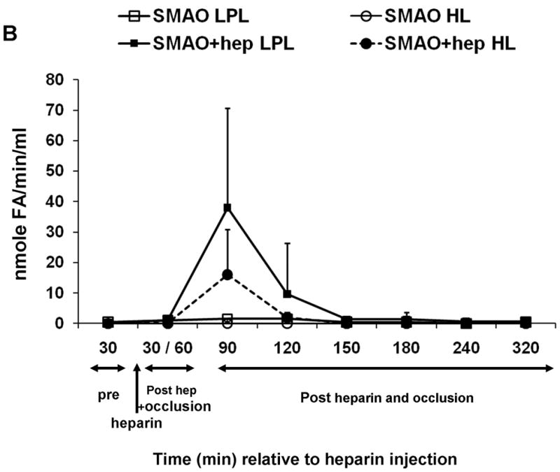
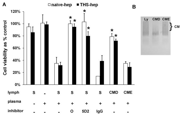
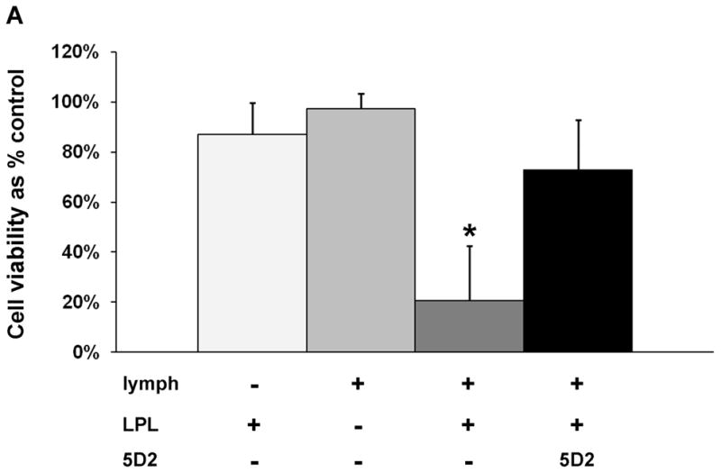
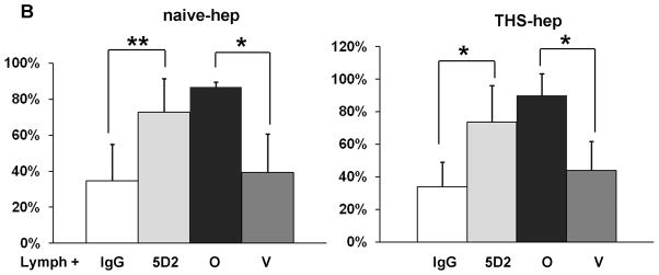
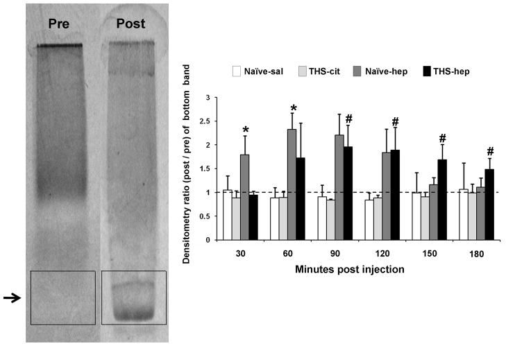
Experimental data have shown that mesenteric lymph from rats subjected to trauma-hemorrhagic shock (THS) but not trauma-sham shock induces neutrophil activation, cytotoxicity, decreased red blood cell (RBC) deformability, and bone marrow colony growth suppression. These data have led to the hypothesis that gut factors produced from THS enter the systemic circulation via the mesenteric lymphatics and contribute to the progression of multiple organ failure after THS. Ongoing studies designed to identify bioactive lymph agents implicated factors associated with the heparin use in the THS procedure. We investigated if heparin itself was responsible for reported toxicity to human umbilical vein endothelial cells (HUVECs). Human umbilical vein endothelial cell toxicity was not induced by lymph when alternate anticoagulants (citrate and EDTA) were used in THS. Human umbilical vein endothelial cell toxicity was induced by lymph after heparin but not saline or citrate injection into trauma-sham shock and naive animals and was dose dependent. Activities of both heparin-releasable lipases (lipoprotein and hepatic) were detected in the plasma and lymph from THS and naive animals receiving heparin but not citrate or saline. Lymph-induced HUVEC toxicity correlated with lymph lipase activities. Finally, incubation of HUVECs with purified lipoprotein lipase added to naive lymph-induced toxicity in vitro. These data show that heparin, not THS, is responsible for the reported lymph-mediated HUVEC toxicity through its release of lipases into the lymph. These findings can provide alternative explanations for several of the THS effects reported in the literature using heparin models, thus necessitating a review of previous work in this field.


Conflict of interest statement

Conflict-of-Interest: None

Figures

Fig. 1
Fig. 1
Trauma hemorrhagic-shock (THS) mesenteric lymph does not induce endothelial cell toxicity in the absence of heparin. Animals were subjected to THS as described in Materials and Methods using either 100U of heparin (THS-hep, in 100ul, N=4), 1.5ml of 0.105M sodium citrate (THS-cit, from three BD Vacutainer tubes, N=3) or sodium EDTA (21.6mg in 200ul of saline from two BD Vacutainer tubes) in the syringe used for blood withdrawal. Animals were resuscitated (resus) with shed blood (A,B,D) or 3volumes of Lactated Ringer’s solution (RL, C,D). Trauma sham-shocked (TSS) animals received the same amount of heparin (TSS-hep) or citrate (TSS-cit) at the same time and rate of blood reperfusion in the shocked animals. Cannulas in the surgical groups were primed with citrated-saline (10.5mM, final concentration). Lymph was collected at designated intervals: 30min prior to THS; THS: 90min during THS period when blood is withdrawn; resus: 15min period when shed blood is returned; post-THS: 30 or 60min intervals following THS. Note: 1h post-THS is 45min post-resuscitation. The numbers of animal in each group are shown in parenthesis in the legend. Lymph was tested using the MTT cell viability assay as originally described (21). Data represent cell viability expressed as mean % of control ± SD. A. Lymph was tested at 5% v/v final concentration for 18h. *THS-hep vs TSS-Cit and THS-Cit; # TSS-hep and THS-hep vs TSS-Cit and THS-Cit (One-way ANOVA with Tukey post hoc test at overall significance at P=0.05). B and C. Lymph was incubated with cells at 5% v/v for 18h. *P ≤ 0.001 and #P < 0.05, Student’s t test compared with Lactated Ringer’s with heparin. D and E. Animals were subjected to THS using a fixed volume method. Heparin, citrate and RL were in quantities described above and were administered in combination either through shed blood (D. THS-hep, THS-hep-cit) or by RL (E. THS-RL, THS-RL-hep). Lymph was incubated with cells at 3% v/v for 3h (THS-hep, THS-hep-cit) and 10% v/v for 18h (THS-RL) or 3h (THS-RL-hep). The values for THS-RL are an average of three independent measures. *P < 0.05, Student’s t test.
Fig. 1
Fig. 1
Trauma hemorrhagic-shock (THS) mesenteric lymph does not induce endothelial cell toxicity in the absence of heparin. Animals were subjected to THS as described in Materials and Methods using either 100U of heparin (THS-hep, in 100ul, N=4), 1.5ml of 0.105M sodium citrate (THS-cit, from three BD Vacutainer tubes, N=3) or sodium EDTA (21.6mg in 200ul of saline from two BD Vacutainer tubes) in the syringe used for blood withdrawal. Animals were resuscitated (resus) with shed blood (A,B,D) or 3volumes of Lactated Ringer’s solution (RL, C,D). Trauma sham-shocked (TSS) animals received the same amount of heparin (TSS-hep) or citrate (TSS-cit) at the same time and rate of blood reperfusion in the shocked animals. Cannulas in the surgical groups were primed with citrated-saline (10.5mM, final concentration). Lymph was collected at designated intervals: 30min prior to THS; THS: 90min during THS period when blood is withdrawn; resus: 15min period when shed blood is returned; post-THS: 30 or 60min intervals following THS. Note: 1h post-THS is 45min post-resuscitation. The numbers of animal in each group are shown in parenthesis in the legend. Lymph was tested using the MTT cell viability assay as originally described (21). Data represent cell viability expressed as mean % of control ± SD. A. Lymph was tested at 5% v/v final concentration for 18h. *THS-hep vs TSS-Cit and THS-Cit; # TSS-hep and THS-hep vs TSS-Cit and THS-Cit (One-way ANOVA with Tukey post hoc test at overall significance at P=0.05). B and C. Lymph was incubated with cells at 5% v/v for 18h. *P ≤ 0.001 and #P < 0.05, Student’s t test compared with Lactated Ringer’s with heparin. D and E. Animals were subjected to THS using a fixed volume method. Heparin, citrate and RL were in quantities described above and were administered in combination either through shed blood (D. THS-hep, THS-hep-cit) or by RL (E. THS-RL, THS-RL-hep). Lymph was incubated with cells at 3% v/v for 3h (THS-hep, THS-hep-cit) and 10% v/v for 18h (THS-RL) or 3h (THS-RL-hep). The values for THS-RL are an average of three independent measures. *P < 0.05, Student’s t test.
Fig. 2
Fig. 2
The pH of lymph from THS animals receiving heparin (hep) or citrate (cit). The pH was measured in each lymph sample at each time point immediately following cell removal and addition of anti-proteases (N=6 each group). *P = 0.04, Student’s t test.
Fig. 3
Fig. 3
Mesenteric lymph from naïve animals post-heparin injection induces HUVEC toxicity. Cell viability after incubation with the supernatant of lymph collected from animals pre- and post heparin (250U/kg, N=5) or saline (100ul, N=3) or citrate (1.5ml of 0.105M sodium citrate from BD Vacutainer tubes, N=3) injection. The animals were surgically manipulated as in the THS model except cannulation of the femoral artery was omitted. The jugular cannula was primed with citrated-saline (10.5mM, final concentration). Injections were at the same rate as in the THS model. Lymph was collected at seven intervals: two 30min intervals post-trauma/pre-injection (pre) and six 30min intervals following injection of heparin, saline, or citrate (post). All samples were tested at 3% v/v final concentration for 3h. Data are expressed as mean ± SD. *Heparin vs saline and citrate (One-way ANOVA with Tukey post hoc test at overall significance at P=0.05).
Fig. 4
Fig. 4
Hepatic (HL) and lipoprotein (LPL) lipase activities are detected in mesenteric lymph after heparin injection. Animal surgery, heparin injection (250U/kg) and lymph collection were as described in Figure Legend 2. Both HL and LPL were measured within the same plasma (A, N=5, naïve -hep; B, N=3 THS-hep) or lymph (C, N=7, naïve -hep, D, N=5, THS-hep) samples collected pre- and post heparin injection/THS (250U/kg) and are expressed as nmoles of fatty acids released in 1min per 1ml (nmole FA/min/ml). E. Cell viability after incubation with lymph from naïve -hep and THS-hep samples shown in C and D. Lymph was tested at 3% v/v final concentration for 3h. F. Plasma lipase measurements pre-and post heparin injection into an animal in which the lymph duct had been ligated (LDL). Plasma collected from both a heparinized and non-heparinized naïve animal was aliquoted and served as an internal positive and negative control, respectively, for lipase assays and an indicator of daily assay variability. The heparinized control varied by 12% (HL) and 30% (LPL) over all measurements (N=6). No more than 1nmol of fatty acid (FA)/min/ml was ever measured for either lipase in the non-heparinized control. No loss in cell viability was detected after cells were incubated with lymph collected from naïve animals receiving saline (N=3) or THS animals receiving citrate (N=3) injections and no lipase activity was detected at any time point in this lymph (N=2 each group, data not shown). Data are expressed as mean ± SD.
Fig. 4
Fig. 4
Hepatic (HL) and lipoprotein (LPL) lipase activities are detected in mesenteric lymph after heparin injection. Animal surgery, heparin injection (250U/kg) and lymph collection were as described in Figure Legend 2. Both HL and LPL were measured within the same plasma (A, N=5, naïve -hep; B, N=3 THS-hep) or lymph (C, N=7, naïve -hep, D, N=5, THS-hep) samples collected pre- and post heparin injection/THS (250U/kg) and are expressed as nmoles of fatty acids released in 1min per 1ml (nmole FA/min/ml). E. Cell viability after incubation with lymph from naïve -hep and THS-hep samples shown in C and D. Lymph was tested at 3% v/v final concentration for 3h. F. Plasma lipase measurements pre-and post heparin injection into an animal in which the lymph duct had been ligated (LDL). Plasma collected from both a heparinized and non-heparinized naïve animal was aliquoted and served as an internal positive and negative control, respectively, for lipase assays and an indicator of daily assay variability. The heparinized control varied by 12% (HL) and 30% (LPL) over all measurements (N=6). No more than 1nmol of fatty acid (FA)/min/ml was ever measured for either lipase in the non-heparinized control. No loss in cell viability was detected after cells were incubated with lymph collected from naïve animals receiving saline (N=3) or THS animals receiving citrate (N=3) injections and no lipase activity was detected at any time point in this lymph (N=2 each group, data not shown). Data are expressed as mean ± SD.
Fig. 5
Fig. 5
Heparin alone or pre-incubated with naïve lymph does not induce HUVEC toxicity. HUVECs were incubated (3h) with heparin (1, 2, or 4U/ml, final concentration) or with naïve lymph (3% v/v) that had been pre-incubated (pre-inc) with heparin (1, 2, or 4U/ml) for 0, 30, 60, 90, 120 min. The data represent an average of two tests in each group.
Fig. 6
Fig. 6
Superior mesenteric arterial occlusion (SMAO) delays appearance of lymph-induced HUVEC toxicity. (A) Lymph collected from animals pre, during and post occlusion of the superior mesenteric artery either with (N=3) or without (N=3) heparin injection (250U/kg) was incubated with endothelial cells at 3% v/v final concentration for 3h to test for induced HUVEC toxicity. (B) Lipase activities in the same samples were made as described in Fig. 4 legend. Data are expressed as mean ± SD. *P= 0.05, compared with SMAO without heparin (Student’s t test).
Fig. 6
Fig. 6
Superior mesenteric arterial occlusion (SMAO) delays appearance of lymph-induced HUVEC toxicity. (A) Lymph collected from animals pre, during and post occlusion of the superior mesenteric artery either with (N=3) or without (N=3) heparin injection (250U/kg) was incubated with endothelial cells at 3% v/v final concentration for 3h to test for induced HUVEC toxicity. (B) Lipase activities in the same samples were made as described in Fig. 4 legend. Data are expressed as mean ± SD. *P= 0.05, compared with SMAO without heparin (Student’s t test).
Fig. 7
Fig. 7
Toxicity to HUVECs can be recreated in vitro. A. Plasma withdrawn 30min post-heparin injection (naïve -hep) or 1–2h post-THS with heparin use (THS-hep) was incubated with the supernatant from lymph (S) or lymph that had been depleted (CMD) or enriched (CME) with chylomicrons with and without inhibitors (Orlistat (O, 252uM), LPL monoclonal antibody (5D2, 0.08mg/ml), isogenic control (IgG, 0.08mg/ml)). The amount of plasma was normalized to an LPL activity of 115 nmoles of fatty acids released in 1min per 1ml (nmolesFA/min/ml). Data reflect duplicate measures of N=3 naïve -hep plasma and N=2 1h and N=2 2h post-THS-hep plasma. Incubations were for 3hr with 3% lymph final concentration. Viabilities are expressed as mean % of control + SD. *P < 0.05 vs. Lymph + heparinized plasma. B. An agarose lipoprotein gel depicting lymph substrates used in the chylomicron (CM) depletion assays. CMs run as a smear when present in high concentration.
Fig. 8
Fig. 8
Lipoprotein lipase is responsible for lymph-induced HUVEC toxicity. A. Lipoprotein lipase purified from bovine milk was incubated with naïve lymph in the same manner as our experiments with heparinized plasma. The amount of LPL was normalized to an activity of 115 nmolesFA/min/ml. Data are expressed as mean ± SD of three measures. *P < 0.01 compared to all other groups (One-way ANOVA and Tukey post hoc test). B. Eight naïve -hep and nine THS-hep lymph samples (2–3 time point samples from three animals each group) were pre-incubated with the 5D2 monoclonal antibody/isogenic control (0.08mg/ml–0.16mg/ml, final concentration) or Orlistat/vehicle control (252uM, final concentration) for 1h at 4C then incubated with cells for 3–4 h. For proper comparison purposes, femoral artery was cannulated in the naïve -hep animals. **P = 0.002, *P < 0.001, Student’s t test.
Fig. 8
Fig. 8
Lipoprotein lipase is responsible for lymph-induced HUVEC toxicity. A. Lipoprotein lipase purified from bovine milk was incubated with naïve lymph in the same manner as our experiments with heparinized plasma. The amount of LPL was normalized to an activity of 115 nmolesFA/min/ml. Data are expressed as mean ± SD of three measures. *P < 0.01 compared to all other groups (One-way ANOVA and Tukey post hoc test). B. Eight naïve -hep and nine THS-hep lymph samples (2–3 time point samples from three animals each group) were pre-incubated with the 5D2 monoclonal antibody/isogenic control (0.08mg/ml–0.16mg/ml, final concentration) or Orlistat/vehicle control (252uM, final concentration) for 1h at 4C then incubated with cells for 3–4 h. For proper comparison purposes, femoral artery was cannulated in the naïve -hep animals. **P = 0.002, *P < 0.001, Student’s t test.
Fig. 9
Fig. 9
Lymph lipoprotein profiles are altered after heparin injection. The left gel is representative of lymph samples prior to heparin administration and samples post-injection of saline (naïve -sal, N=3) and in post-THS samples in which citrate was used (THS-cit, N=3). The right gel is representative of lymph samples post-heparin (naïve -hep, N=3) and in post-THS samples in which heparin was used (THS-hep, N=4). Data were analyzed by densitometry of boxes drawn around the gel migration area of the fast migrating band appearing in post-heparin samples (shown). Values of each lane were compared to the pre-injection lymph control and these ratios were graphed. The pre-injection control has an inherent value of one and is represented by the dotted line. There was no overall significance in the means in any of the band areas for either the naïve -saline or THS-cit groups. Data are expressed as mean ± SD. *P ≤ 0.05, compared with naïve -hep pre, 120, 150 180min post-heparin; **P ≤ 0.05, ANOVA compared with THS-hep pre, 30min post-THS (One-way ANOVA and Tukey post hoc test).

Similar articles

Cited by

References

    1. Deitch EA. Multiple organ failure: Pathophysiology and potential future therapy. Ann Surg. 1992;216(2):117–134. - PMC - PubMed
    1. Deitch EA, Goodman E. Prevention of Multiple Organ Failure. Surg Clin North Am. 1999;79(6):1471–1488. - PubMed
    1. Glenn TM, Lefer AM. Protective effect of thoracic lymph diversion in hemorrhagic shock. Am J Physiol. 1970;219(5):1305–1310. - PubMed
    1. Papp M, Nemeth E, Feuer I, Fodor I. Effect of an impairment of lymph flow on experimental acute “pancreatitis”. Acta Med Acad Sci Hung. 1957;11(2):203–208. - PubMed
    1. Deitch EA, Xu DZ, Kaiser VL. Role of the gut in development of injury and shock induced SIRS and MODS: the gut-lymph hypothesis, a review. Front Biosci. 2006;11:520–528. - PubMed

Publication types