. 2011 Mar;35(3):229-39.
doi: 10.1097/SHK.0b013e318200032b.

A physiological model for autonomic heart rate regulation in human endotoxemia

Affiliations

A physiological model for autonomic heart rate regulation in human endotoxemia

Panagiota T Foteinou et al. Shock. 2011 Mar.

Abstract

The systemic inflammatory response syndrome often accompanies critical illnesses and can be an important cause of morbidity and mortality. Marked abnormalities in cardiovascular function accompany acute illnesses manifested as sustained tachyarrhythmias, which are but one component of systemic dysregulation. The realization that cardiac pacemaker activity is under control of the autonomic nervous system has promoted the analysis of heart rate (HR) variation for assessing autonomic activities. In acute illnesses, autonomic imbalance manifesting in part as parasympathetic attenuation is associated with increased morbidity in patients who manifest systemic inflammatory response syndrome phenotype. Driven by the premise that biological phenotypes emerge as the outcome of the coordinated action of network elements across the host, a multiscale model of human endotoxemia, as a prototype model of systemic inflammation in humans, is developed that quantifies critical aspects of the complex relationship between inflammation and autonomic HR regulation. In the present study, changes in HR response to acute injury, phenotypically expressed as tachycardia, are simulated as a result of autonomic imbalance that reflects sympathetic activity excess and parasympathetic attenuation. The proposed model assesses both the anti-inflammatory and cardiovascular effects of antecedent stresses upon the systemic inflammatory manifestations of human endotoxemia as well as a series of nonlinear inflammatory relevant scenarios. Such a modeling approach provides a comprehensive conceptual framework linking inflammation and physiological complexity via a multiscale model that may advance the translational potential of systems modeling in clinical research.


Figures

Figure 1
Figure 1. Schematic illustration of the Warner model representing the relationship between stimulation of efferent sympathetic nerve activity to the heart and heart rate (HR)
A1 represents the concentration of sympathetic neurotransmitter (catecholamine) at the nerve ending and f1 represents the frequency stimulates preformed on the nerve; A0 represents the concentration of catecholamine in peripheral tissues (i.e. blood); A2 represents the concentration at the active site on sinoatrial (SA) node which must react with chemical substance B to produce a change in heart rate, adapted from (18).
Figure 2
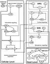
Figure 2. Network topology of the multiscale model of human endotoxemia for the assessment of autonomic heart rate regulation
Elementary pro-inflammatory pathways (i.e. NF-kB signaling module) triggered by the recognition process of endotoxin (LPS) from its signaling receptor (TLR4, R) propagate the effect of LPS signaling on the transcriptional (cellular) response level (P, A, E). Essential modules associated with the release of stress hormones (cortisol (F), epinephrine (EPI)) from neuroendocrine axis (HPA, SNS) coupled with their anti-inflammatory influence on the host are further considered. Finally, at the systemic level, biochemical reactions associated with the release, binding and degradation of cardiac (sympathetic) neurotransmitters (A1, A2) on the SA node are also incorporated. Efferent autonomic outflow is represented by sympathetic (Tsym) and parasympathetic activities (Tpar) that act antagonistically giving rise to changes in heart rate (HR) response assessed by clinical monitoring of vital signs.
Figure 3
Figure 3. Dynamic profiles of the elements that constitute the autonomic heart rate regulation signaling module in human endotoxemia
(A) Simulated concentrations of cardiac (SNS) neurotransmitters at the level of sympathetic nerve ending (A1) and at the active site of sinus node (SA) of the heart (A2); (B) Efferent sympathetic activity during the progression of the acute inflammatory reaction; (C) Simulated efferent parasympathetic (vagal) activity and (D) Heart rate (HR) response to endotoxin induced inflammation. Human experimental data (○ circles) associated with vagal measurements (time domain HRV measure, pNN50) and vital signs (heart rate measurements) are used to calibrate the model. Solid lines (—) represent model predictions under conditions of low-dose endotoxin while ○ circles refer to experimental data expressed as mean ± SEM. The initial condition of the inflammatory stimulus (LPS(t=0hr) = 1) reflects LPS concentration relative to 2-ng/kg body weight.
Figure 4
Figure 4. In silico simulation of the cardiovascular effects of acute epinephrine infusion on the host initiated 3hr prior to endotoxin challenge (t = 0hr) and continued for another 6hr after LPS
Solid lines simulate the host dynamics under conditions of low-dose endotoxin (LPS) while dashed and dotted lines reflect the dynamics of the host pre-exposed to epinephrine infusion at various doses. The acute pre-exposure of the host into epinephrine (wEPI,ex = 1) at increasing values of the parameter Rin,EPI = 6, 12, 24 potentiates (A) circulating levels of epinephrine and (B) the overall efferent sympathetic outflow (Tsym) relative to the responses induced by endotoxin administration while (C) vagal activity is significantly attenuated compared to the effect induced only by LPS and finally (D) heart rate response to endotoxin is further increased due to prior epinephrine infusion.
Figure 5
Figure 5. In silico assessment of the cardiovascular implications associated with acute epinephrine infusion on the host
Human experimental data depicted by □ and ◇ represent mean ± SEM refer to human subjects that received either low dose (2ng/kg BW) LPS or an infusion of epinephrine for 3hr before LPS administration and continued until 6 hours after LPS, respectively. These data are specifically used to validate qualitatively the structure of the proposed human inflammation model employed from (14) and not to train the model. Descriptive statistics in the original experimental study show that there was a significant change in the parasympathetic activity (Tpar) and heart rate response (HR) across the two experimental conditions (□ vs. ◇) from 0hr until 24 hours after LPS exposure. Computationally such situation is captured by the differential predicted responses between dashed and solid lines. We specifically observe that there exists a simulated trajectory of exogenously induced catecholamine excess (represented by dashed line - Rin,EPI = 6) that lies in general agreement with the relevant human experimental data. More details about the statistical assessment of the model in comparison to the data are provided in Appendix (Table 4).
Figure 6
Figure 6. Simulated dose dependent effects of LPS on neuroendocrine immune system interactions
A high concentration of LPS can cause a dysregulation in the host dynamics characterized by abnormal transcriptional and hormonal responses. Temporal responses of critical inflammatory components for various initial conditions of the inflammatory stimulus include: (A) pro-inflammatory response (P); (B) anti-inflammatory response (A) and stress hormones such as (C) cortisol (F) and (D) epinephrine (EPI). Solid lines simulate the progression of a self-limited inflammatory response at increasing LPS doses (LPS(t=0hr) = 0.4 ng/kg, LPS(t=0hr) = 4ng/kg and LPS(t=0hr) = 6ng/kg) that are less than the critical threshold (LPS(t=0hr) = 8ng/kg - four times greater than the nominal value (2ng/kg BW) used to calibrate the model) that gives rise to unresolved inflammatory responses represented by sustained inflammatory markers and hormonal responses (dashed lines).
Figure 7
Figure 7. Simulated dose dependence of LPS effects on systemic (heart) level responses
A high inflammatory challenge disrupts the autonomic control systems which give rise to prolonged elevations in heart rate. (A) Dynamic responses of catecholamines at the sinus node of the heart (A2); (B) efferent sympathetic, (C) parasympathetic activities and (D) heart rate (HR) responses to increasing levels of the inflammatory stimulus (LPS). While — lines represent constrained inflammatory responses, dashed lines refer to a persistent inflammatory response simulated by high LPS concentration given that such situation can be equated to the severely stressed clinical phenotype manifested as sustained elevations in heart rate (cardiovascular instability).
Figure 8
Figure 8. Dynamic inflammatory cellular and physiological responses as a function of time after the administration of high inflammatory challenge (dashed lines) while solid lines represent “virtual” human subjects that receive an infusion of epinephrine
The acute pre-exposure of the host into epinephrine (initiated 3hr before LPS and continued until 6hr after endotoxin, i.e. Rin,EPI = 6) attenuates the pro-inflammatory response (P), relative to the effect mediated by high concentration of LPS, via potentiation of the anti-inflammatory component of the host (A). In addition to the anti-inflammatory role of epinephrine, exogenous up-regulation in circulating levels of epinephrine increase the efferent sympathetic activity (Tsym) which is followed by further reduction in vagal function (Tpar) and these autonomic changes mediate early tachycardia (HR) which is eventually restored within 24 hours.
Figure 9
Figure 9. Modulation of the progression of unresolved inflammatory response due to high LPS concentration under conditions of hydrocortisone infusion
The effect of low-dose steroid administration initiated 6hr before high LPS concentration and continued until 6hr after LPS is simulated in — lines. Such exogenously-induced hypercortisolemia (wFex=1, Rin,F=2.922 – parameters taken from our prior work (13)) potentiates total plasma concentration of cortisol ashown in panel (A) and modulates cytokine and hormonal responses. Circulating levels of epinephrine are attenuated in response to antecedent periods of hypercortisolemia relative to the excessive adrenergic response which is illustrated in panel (B). At the autonomic level such attenuation is expected to reduce efferent sympathetic activity mediating (C) an increase in implied vagal function (Tpar) and finally (D) controlling heart rate as shown by reversibility in the progression of the inflammatory reaction towards homeostasis (baseline) – “recovery phase” (solid lines).

Similar articles

Cited by

References

    1. Cavaillon JM, Annane D. Compartmentalization of the inflammatory response in sepsis and SIRS. J Endotoxin Res. 2006;12:151–70. - PubMed
    1. Bone RC. Immunologic dissonance: a continuing evolution in our understanding of the systemic inflammatory response syndrome (SIRS) and the multiple organ dysfunction syndrome (MODS) Ann Intern Med. 1996;125:680–7. - PubMed
    1. Sharshar T, Hopkinson NS, Orlikowski D, Annane D. Science review: The brain in sepsis--culprit and victim. Crit Care. 2005;9:37–44. - PMC - PubMed
    1. Tracey KJ. The inflammatory reflex. Nature. 2002;420:853–9. - PubMed
    1. Taub DD. Neuroendocrine interactions in the immune system. Cell Immunol. 2008;252:1–6. - PMC - PubMed

Publication types

MeSH terms