. 2011 Jan 5;30(1):130-44.
doi: 10.1038/emboj.2010.276. Epub 2010 Nov 9.

CAMP (C13orf8, ZNF828) is a novel regulator of kinetochore-microtubule attachment

Affiliations

CAMP (C13orf8, ZNF828) is a novel regulator of kinetochore-microtubule attachment

Go Itoh et al. EMBO J. .

Abstract

Proper attachment of microtubules to kinetochores is essential for accurate chromosome segregation. Here, we report a novel protein involved in kinetochore-microtubule attachment, chromosome alignment-maintaining phosphoprotein (CAMP) (C13orf8, ZNF828). CAMP is a zinc-finger protein containing three characteristic repeat motifs termed the WK, SPE, and FPE motifs. CAMP localizes to chromosomes and the spindle including kinetochores, and undergoes CDK1-dependent phosphorylation at multiple sites during mitosis. CAMP-depleted cells showed severe chromosome misalignment, which was associated with the poor resistance of K-fibres to the tension exerted upon establishment of sister kinetochore bi-orientation. We found that the FPE region, which is responsible for spindle and kinetochore localization, is essential for proper chromosome alignment. The C-terminal region containing the zinc-finger domains negatively regulates chromosome alignment, and phosphorylation in the FPE region counteracts this regulation. Kinetochore localization of CENP-E and CENP-F was affected by CAMP depletion, and by expressing CAMP mutants that cannot functionally rescue CAMP depletion, placing CENP-E and CENP-F as downstream effectors of CAMP. These data suggest that CAMP is required for maintaining kinetochore-microtubule attachment during bi-orientation.


Conflict of interest statement

The authors declare that they have no conflict of interest.

Figures
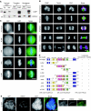
Figure 1
Figure 1
A MAD2L2-interacting protein, CAMP, localizes to chromosomes and the spindle including kinetochores. (A) Coprecipitation of endogenous CAMP with endogenous MAD2L2. Total cell lysates prepared from HeLa cells treated with thymidine or nocodazole were subjected to immunoprecipitation (IP) with anti-CAMP antibody. Lysates and IPs were separated by SDS–PAGE and probed by western blotting with anti-CAMP and anti-MAD2L2 antibodies, respectively. *IgG light chain. (B) Subcellular localization of CAMP during mitosis. HeLa cells were stained with anti-CAMP (red) and anti-tubulin (green) antibodies. DNA was stained with DAPI (blue). Scale bar=10 μm. (C) Subcellular localization of CAMP fragments in metaphase. Localization of GFP-tagged full-length CAMP or each region of CAMP expressed in HeLa cells is shown (green). DNA was stained with DAPI (blue). Scale bar=10 μm. (D) Summary of the subcellular localization of CAMP-deletion mutants. Localization of each mutant on chromosomes or the spindle is schematically shown, as indicated by either ‘+' or ‘−'. Sequences of the SPE, WK, and FPE motifs are shown. (E) Kinetochore localization of CAMP. Localization of GFP-CAMP-ΔC-ZNF expressed in HeLa cells is shown (green). Kinetochores were visualized with CREST serum (red) and DNA was stained with DAPI (blue). Arrowheads indicate spindle poles. Right panels are magnifications of the boxed areas. Scale bar=5 μm.
Figure 2
Figure 2
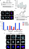
CAMP-depletion causes chromosome misalignment. (A) Depletion of CAMP with siRNAs. Total cell lysates prepared from HeLa cells treated with mock or CAMP siRNA for the indicated periods were separated by SDS–PAGE and probed by western blotting with anti-CAMP or anti-actin antibody. (B) Increased mitotic index in CAMP-depleted cells. The mitotic index for mock-, CAMP siRNA-, or CAMP/MAD2L1 siRNA-treated HeLa cells was measured 48 h after transfection. Error bars represent the s.d. (C) CAMP-depletion induced chromosome misalignment. HeLa cells treated with mock or CAMP siRNA for 48 h were stained with an anti-tubulin antibody (red). DNA was stained with DAPI (blue). Scale bar=10 μm. (D) Expression of RNAi-resistant GFP-CAMP. Lysates were prepared from HeLa cells transfected with mock or CAMP siRNA with or without RNAi-resistant GFP-CAMP. The lysates were separated by SDS–PAGE and probed by western blotting with an anti-CAMP antibody. (E) Quantitative analysis of chromosome misalignment in CAMP-depleted cells. HeLa cells were transfected with RNAi-resistant GFP-CAMP constructs for 24 h, preceded by transfection with CAMP siRNA for 36 h. Cells were treated with MG132 (10 μM) for the final 2 h. The number of misaligned chromosomes per cell was scored. Error bars represent the s.d. (F) MAD2L1 localizes to kinetochores in CAMP-depleted cells. HeLa cells treated with mock or CAMP siRNA for 48 h were stained for MAD2L1 (red) and DNA (blue). Scale bar=10 μm.
Figure 3
Figure 3
CAMP is required for proper kinetochore–microtubule attachment. (A) Live-cell imaging of HeLa cells expressing histone H2B-GFP, 48 h after mock- or CAMP-siRNA transfection. T=0 was defined as the time point at which chromosome condensation became evident (prophase). Scale bar=10 μm. See also Supplementary Movies 1 and 2. (B) Prolonged mitosis in CAMP-depleted cells. HeLa cells were treated as in (A). The time from nuclear envelope breakdown (NEBD) to anaphase onset was measured in live-cell imaging and categorized. The percentages of cells in each category are shown in the graph. (C) Defective kinetochore–microtubule attachment in CAMP-depleted cells. HeLa cells expressing CENP-A-GFP (green) were treated with mock or CAMP siRNA for 48 h, and with MG132 for the final 2 h. Cells were stained for tubulin (red) and DNA (blue). Insets show individual kinetochore–microtubule attachments. Scale bar=5 μm. (D) Quantitative analysis of inter-kinetochore distance in CAMP-depleted cells. HeLa cells expressing GFP-CENP-A were treated as in (C). The distance between CENP-A on sister kinetochores was measured. (E) Instability of kinetochore microtubules in CAMP-depleted cells. HeLa cells were treated as in (C), and then incubated on ice for 10 min before fixation. Cells were stained for kinetochores (CREST, red), tubulin (green) and DNA (blue). Scale bar=5 μm.
Figure 4
Figure 4
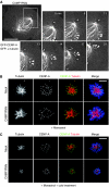
K-fibres cannot resist tension in CAMP-depleted cells. (A) Disruption of kinetochore–microtubule attachment in CAMP-depleted cells. HeLa cells expressing GFP-CENP-A and GFP-α-tubulin were treated with CAMP siRNA for 48 h before live-cell imaging. T=0 was set arbitrarily for the first panel. Arrowheads indicate kinetochores on a sister chromatid pair. Arrows indicate microtubules attached to these kinetochores. Scale bar=10 μm. See also Supplementary Movie 4. (B) K-fibres on a monopolar spindle in CAMP-depleted cells. HeLa cells expressing GFP-CENP-A (green) were treated with mock or CAMP siRNA for 48 h, and with monastrol (100 μM) for the final 2 h before fixation. Cells were stained for tubulin (red) and DNA (blue). Scale bar=5 μm. (C) Instability of K-fibres on monopolar spindles in CAMP-depleted cells. HeLa cells expressing GFP-CENP-A (green) were treated with mock or CAMP siRNA for 48 h and with monastrol (100 μM) for the final 2 h, and then incubated on ice for 10 min before fixation. Cells were stained for tubulin (red) and DNA (blue). Scale bar=5 μm.
Figure 5
Figure 5
The FPE region of CAMP is responsible for chromosome alignment. (A, B) HeLa cells were transfected with RNAi-resistant GFP-CAMP constructs for 24 h, preceded by transfection with CAMP siRNA for 36 h. Cells were treated with MG132 for the final 2 h. The structures of these constructs are the same as shown in Figure 1D. A CAMP-deletion construct lacking each domain was used in (A), while a CAMP fragment containing each domain was used in (B). The percentage of cells with misaligned chromosomes is shown for cells expressing each construct. Experiments were repeated three times and representative data are shown. Oligo 1 was used for RNAi; similar results were obtained for oligo 2 (data not shown).
Figure 6
Figure 6
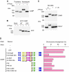
Phosphorylation of CAMP during mitosis is essential for chromosome alignment. (A) CAMP is phosphorylated during mitosis. Total cell lysates prepared from HeLa cells treated with thymidine or nocodazole were incubated in the absence (−) or presence (+) of λ-phosphatase for 30 min at 30°C. Equal amounts of cell lysates were separated by SDS–PAGE and probed by western blotting with an anti-CAMP antibody. (B) CAMP is phosphorylated at serine residues in the SPE/D sequences. Lysates were prepared from nocodazole-treated HeLa cells expressing GFP-tagged CAMP with (GFP-CAMP-22A) or without mutations in the serine residues in the SPE/D sequences. Equal amounts of cell lysates were separated by SDS–PAGE and probed by western blotting with an anti-GFP antibody. (C) CDK1-dependent phosphorylation of CAMP. HeLa cells were treated with nocodazole for 12 h after thymidine treatment, and with RO-3306 (CDK1 inhibitor; 10 μM) for the time indicated. Total cell lysate at T=0 was incubated in the presence (+) or absence (−) of λ-phosphatase for 30 min at 30°C. Equal amounts of cell lysates were separated by SDS–PAGE and probed by western blotting with an anti-CAMP or anti-CDC27 antibody. (D) Mitotic phosphorylation of CAMP is essential for chromosome alignment. HeLa cells were transfected with RNAi-resistant GFP-CAMP constructs for 24 h, preceded by transfection with CAMP siRNA for 36 h. Cells were treated with MG132 for the final 2 h. The structures of these constructs are schematically represented; each serine residue that was replaced with alanine is indicated by ‘A'. The percentage of cells with misaligned chromosomes is shown for cells expressing each construct. Experiments were repeated three times and representative data are presented. Oligo 1 was used for RNAi; similar results were obtained for oligo 2 (data not shown).
Figure 7
Figure 7
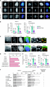
Decreased kinetochore localization of CENP-E and CENP-F correlates with chromosome misalignment by CAMP mutants. (A) Decreased CENP-E and CENP-F on kinetochores in CAMP-depleted cells. HeLa cells were treated with mock or CAMP siRNA for 48 h and with MG132 for the final 2 h. Cells were stained for CENP-E or CENP-F (green), CREST (red), and DNA (blue). Scale bar=10 μm. (B) Quantification of fluorescence intensity (arbitrary unit; AU) of the CENP-E or CENP-F signal at kinetochores on aligned or misaligned chromosomes. Misaligned chromosomes in mock-transfected cells were selected in prometaphase cells. Error bars represent the s.d. (C) Colocalization of CAMP with CENP-E/CENP-F on kinetochores in nocodazole-treated cells. Localization of GFP-CAMP-FPE expressed in HeLa cells treated with nocodazole (2 μM) is shown (green), together with CENP-E/CENP-F (red) and DNA (blue). Right panels are magnifications of the boxed areas. Scale bars=5 μm. (D) Quantitative analysis of chromosome misalignment in cells depleted of CAMP/CENP-E/CENP-F. HeLa cells were transfected with the indicated siRNAs for 48 h, and treated with MG132 (10 μM) for the final 2 h. The percentage of cells with misaligned chromosomes is shown. Error bars represent the s.d. (E) Quantification of fluorescence intensity (arbitrary unit; AU) of the CENP-E or CENP-F signal at kinetochores in cells expressing GFP-CAMP-Full length or GFP-CAMP-Full length-22A. HeLa cells were treated as in Figure 6D. Misaligned chromosomes in GFP-CAMP-Full length-transfected cells were selected in prometaphase cells. Error bars represent the s.d. (F) Recovery of kinetochore levels of CENP-E/CENP-F in cells expressing non-phosphorylatable CAMP mutants. HeLa cells were treated as in Figure 6D. Fluorescence intensity (arbitrary unit; AU) of the CENP-E or CENP-F signal at kinetochores on aligned chromosomes was quantified. Error bars represent the s.d. The results in Figure 6D are schematically shown, as indicated by either ‘+' or ‘−'.
Figure 8
Figure 8
Schematic diagram of CAMP structure with regard to the functions revealed in this study. Each serine residue phosphorylated during mitosis is indicated by ‘P'. See text for details.

Similar articles

Cited by

References

    1. Asbury CL, Gestaut DR, Powers AF, Franck AD, Davis TN (2006) The Dam1 kinetochore complex harnesses microtubule dynamics to produce force and movement. Proc Natl Acad Sci USA 103: 9873–9878 - PMC - PubMed
    1. Ashar HR, James L, Gray K, Carr D, Black S, Armstrong L, Bishop WR, Kirschmeier P (2000) Farnesyl transferase inhibitors block the farnesylation of CENP-E and CENP-F and alter the association of CENP-E with the microtubules. J Biol Chem 275: 30451–30457 - PubMed
    1. Baker DJ, Dawlaty MM, Galardy P, van Deursen JM (2007) Mitotic regulation of the anaphase-promoting complex. Cell Mol Life Sci 64: 589–600 - PMC - PubMed
    1. Bomont P, Maddox P, Shah JV, Desai AB, Cleveland DW (2005) Unstable microtubule capture at kinetochores depleted of the centromere-associated protein CENP-F. EMBO J 24: 3927–3939 - PMC - PubMed
    1. Cahill DP, da Costa LT, Carson-Walter EB, Kinzler KW, Vogelstein B, Lengauer C (1999) Characterization of MAD2B and other mitotic spindle checkpoint genes. Genomics 58: 181–187 - PubMed

Publication types