. 2011 Mar;38(3):552-61.
doi: 10.1007/s00259-010-1637-4. Epub 2010 Nov 10.

Targeted multi-pinhole SPECT

Affiliations

Targeted multi-pinhole SPECT

Woutjan Branderhorst et al. Eur J Nucl Med Mol Imaging. 2011 Mar.

Abstract

Purpose: Small-animal single photon emission computed tomography (SPECT) with focused multi-pinhole collimation geometries allows scanning modes in which large amounts of photons can be collected from specific volumes of interest. Here we present new tools that improve targeted imaging of specific organs and tumours, and validate the effects of improved targeting of the pinhole focus.

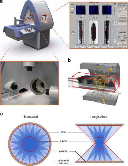
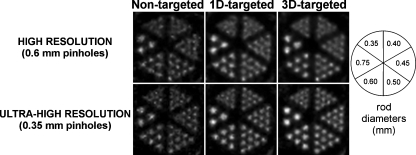
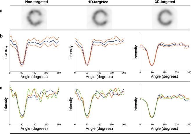
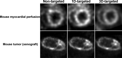
Methods: A SPECT system with 75 pinholes and stationary detectors was used (U-SPECT-II). An XYZ stage automatically translates the animal bed with a specific sequence in order to scan a selected volume of interest. Prior to stepping the animal through the collimator, integrated webcams acquire images of the animal. Using sliders, the user designates the desired volume to be scanned (e.g. a xenograft or specific organ) on these optical images. Optionally projections of an atlas are overlaid semiautomatically to locate specific organs. In order to assess the effects of more targeted imaging, scans of a resolution phantom and a mouse myocardial phantom, as well as in vivo mouse cardiac and tumour scans, were acquired with increased levels of targeting. Differences were evaluated in terms of count yield, hot rod visibility and contrast-to-noise ratio.

Results: By restricting focused SPECT scans to a 1.13-ml resolution phantom, count yield was increased by a factor 3.6, and visibility of small structures was significantly enhanced. At equal noise levels, the small-lesion contrast measured in the myocardial phantom was increased by 42%. Noise in in vivo images of a tumour and the mouse heart was significantly reduced.

Conclusion: Targeted pinhole SPECT improves images and can be used to shorten scan times. Scan planning with optical cameras provides an effective tool to exploit this principle without the necessity for additional X-ray CT imaging.


Figures

Fig. 1
Fig. 1
a U-SPECT-II system with close-ups of a mouse on the animal bed with a transparent heater pad in front of optical cameras and the graphical user interface with optical images and three real-time projection images. b Example of various FOV selection boundaries in three dimensions. c Schematic cross-sections of FOV and CFOV in U-SPECT-II general-purpose collimators
Fig. 2
Fig. 2
Different volume selections applied to obtain a specific SPECT acquisition of (a) a resolution phantom, (b) a mouse myocardial phantom, (c) a mouse tumour, and (d) a mouse heart using an atlas. Red (large) box nontargeted selection, orange (middle) box selection targeted in one dimension only, yellow (small) box selection targeted in three dimensions
Fig. 3
Fig. 3
a Photograph of a mouse myocardial phantom with lesion. b Schematic drawing of a phantom showing the dimensions of the left ventricle and lesion in millimetres. c Reconstructed short-axis slice with profile range used for circumferential profiles. d Different regions used for calculation of noise and contrast (solid areas “non-infarcted” regions, dashed area “infarcted” region)
Fig. 4
Fig. 4
Reconstructed images of micro-hot-rod phantom scans with high-resolution (top) and ultra-high-resolution (bottom) collimators for three different levels of targeting
Fig. 5
Fig. 5
Average defect contrast as a function of average noise in the mouse myocardial phantom. Curves were generated by interpolating values found at different iterations (dash-dots nontargeted, dashes 1D-targeted, solid 3D-targeted). Comparisons at equal contrast and at equal noise, as described in the text, are illustrated using markers connected by dotted lines
Fig. 6
Fig. 6
Images and circumferential profiles of reconstructed short-axis slices of the mouse myocardial phantom for nontargeted acquisition (left), 1D-targeted acquisition (centre) and 3D-targeted acquisition (right) show stronger noise reduction with a higher degree of targeting. a Short-axis slices from reconstruction of one noise realization. b Profiles of mean (solid) and mean ±1SD (dashed), both calculated over ten noise realizations. c Example profiles from reconstructions of three different noise realizations
Fig. 7
Fig. 7
Reconstructed end-diastolic short-axis slices from gated myocardial perfusion scans (top) and transaxial slices from the mouse tumour scans (bottom) for three different levels of targeting

References

    1. Beekman FJ, van der Have F, Vastenhouw B, van der Linden AJA, van Rijk PP, Burbach JPH, Smidt MP. U-SPECT-I: A novel system for submillimeter-resolution tomography with radiolabeled molecules in mice. J Nucl Med. 2005;46(7):1194–1200. - PubMed
    1. Furenlid LR, Wilson DW, Chen YC, Kim H, Pietraski PJ, Crawford MJ, FastSPECT BHH., II A second-generation high-resolution dynamic SPECT imager. IEEE Trans Nucl Sci. 2004;51(3):631–635. doi: 10.1109/TNS.2004.830975. - DOI - PMC - PubMed
    1. Van der Have F, Vastenhouw B, Ramakers RM, Branderhorst W, Krah JO, Ji C, Staelens SG, Beekman FJ. U-SPECT-II: an ultra-high resolution device for molecular small-animal imaging. J Nucl Med. 2009;50(4):599–605. doi: 10.2967/jnumed.108.056606. - DOI - PubMed
    1. Goertzen AL, Jones DW, Seidel J, Li K, Green MV. First results from the high-resolution mouseSPECT annular scintillation camera. IEEE Trans Med Imaging. 2005;24(7):863–867. doi: 10.1109/TMI.2005.843782. - DOI - PubMed
    1. Ishizu K, Mukai T, Yonekura Y, Pagani M, Fujita T, Magata Y, Nishizawa S, Tamaki N, Shibasaki H, Konishi J. Ultra-high-resolution SPECT system using four pinhole collimators for small animal studies. J Nucl Med. 1995;36(12):2282–2287. - PubMed