. 2011 Aug;68(15):2603-13.
doi: 10.1007/s00018-010-0573-6. Epub 2010 Nov 10.

Functional impact of PTP1B-mediated Src regulation on oxidative phosphorylation in rat brain mitochondria

Affiliations

Functional impact of PTP1B-mediated Src regulation on oxidative phosphorylation in rat brain mitochondria

Etienne Hébert Chatelain et al. Cell Mol Life Sci. 2011 Aug.

Abstract

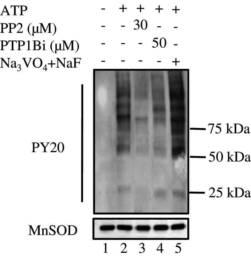
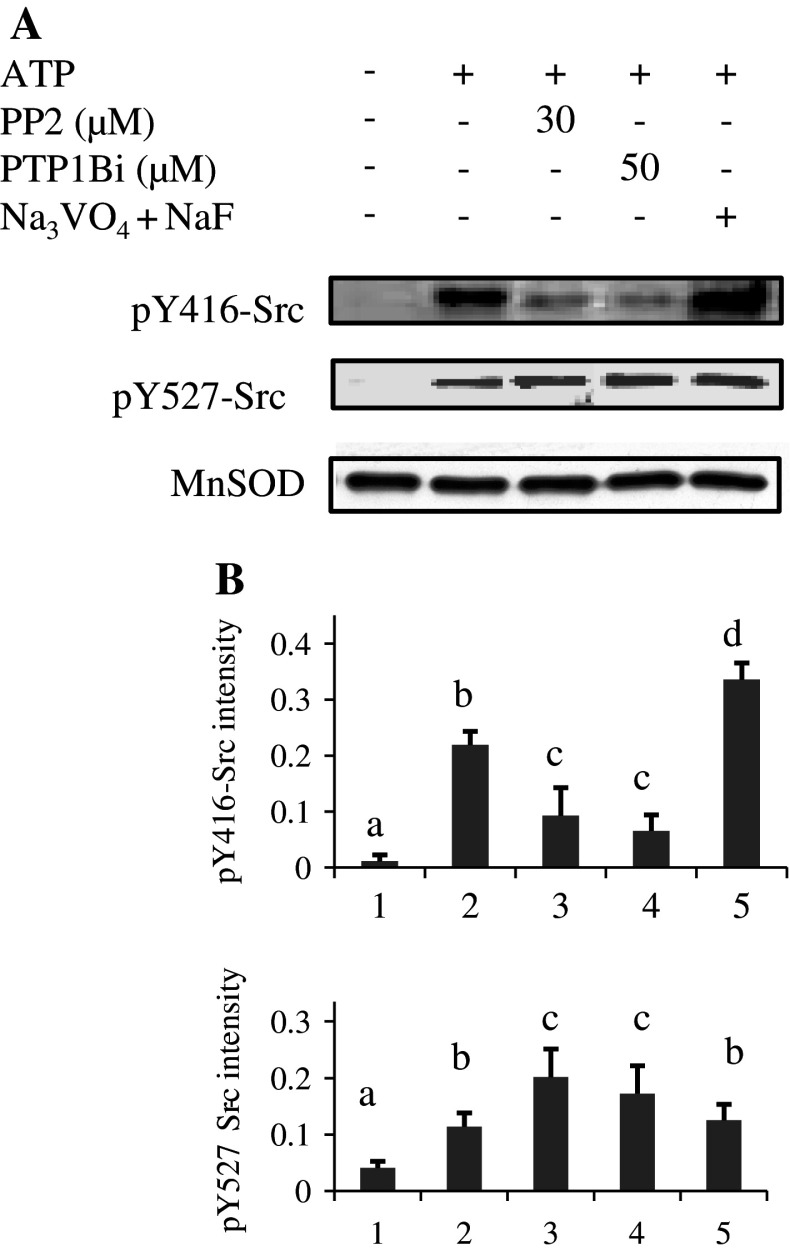
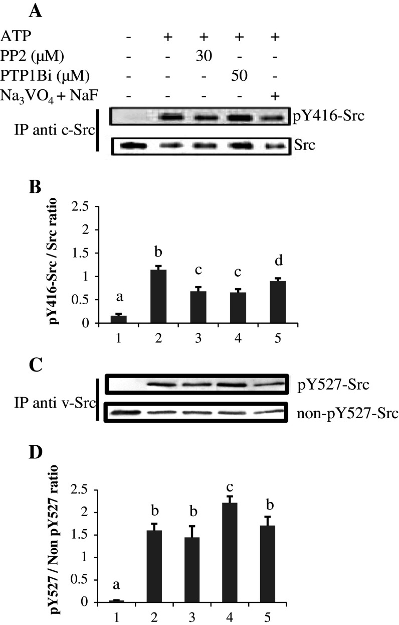
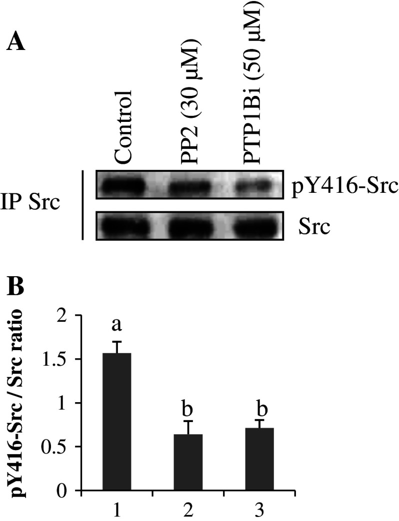
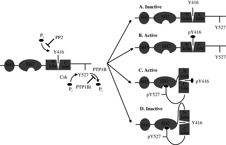
Given the presence of Src and PTP1B within rat brain mitochondria, we have investigated whether PTP1B regulates Src activity in mitochondria as in the cytosol. Results showed that Src was stimulated by in vitro addition of ATP to mitochondria, and this stimulation was reversed by a membrane-permeable allosteric inhibitor of PTP1B and by a potent selective Src inhibitor. They also indicated a direct action of PTP1B on phosphorylated tyrosine 527 residue of Src, thus implicating a role for PTP1B in the modulation of Src activity in mitochondria. Putative Src and PTP1B substrates were identified by liquid chromatography tandem mass spectrometry and two-dimensional blue native/SDS-PAGE. Both inhibitors inhibited ADP-stimulated respirations concurrently with Src activation and complex IV activation by ATP, while having no effect or increasing the activity of the other complexes. Our analysis emphasizes the regulatory function of Src and its modulation by PTP1B on oxidative phosphorylation in mitochondria.


Figures

Fig. 1
Fig. 1
Changes in protein tyrosine phosphorylation of rat brain mitochondria following exposure to ATP, PP2, PTP1Bi, or orthovanadate + NaF. Mitochondria (50 μg) were untreated (lane 1) or treated at 30°C for 10 min with 1 mM ATP alone (lane 2) or after pre-incubation at 30°C for 10 min with 30 μM PP2 (lane 3), 50 μM PTP1Bi (lane 4), or 2 mM orthovanadate plus 10 mM NaF (lane 5). The reaction was stopped by addition of 5× Laemmli buffer. Tyrosine-phosphorylated proteins on immunoblots were detected with an antibody to phosphotyrosine. The membrane was stripped and reprobed with an antibody to MnSOD as a loading control (below). Data are representative of at least three experiments
Fig. 2a, b
Fig. 2a, b
Phosphorylation status of mitochondrial Src in in vitro treatments. a Mitochondria, treated as described in the legend to Fig. 1, were Western blotted and immunodetected first with the anti-pY416-Src antibody, stripped and reprobed with the anti-pY527-Src antibody, stripped again and finally probed with an antibody to MnSOD as a loading control. b The intensity of pY416-Src and pY527-Src labeling was normalized over the MnSOD signal for each treatment. Data are representative of at least three experiments. Means that are not significantly different (P > 0.05) according to the Tukey test share the same letters
Fig. 3a–d
Fig. 3a–d
pY416-Src and pY527-Src labelings in immunoprecipitated Src from mitochondria in in vitro experiments. a Src was immunoprecipitated from mitochondria treated as described in the legend to Fig. 1, and immunoblotted first with the anti-pY416-Src antibody, stripped and reprobed with anti-Src antibody. b Normalizations of pY416-Src labeling versus Src signal. c For analysis of pY527-Src, Src was immunoprecipitated with an antibody to v-Src that recognizes an epitope in the SH3 region, and immunoblotted first with an antibody to pY527-Src, stripped and reprobed with an antibody to non-pY527-Src (see “Materials and methods”). d Normalizations of pY527-Src labeling versus non-pY527-Src signal. Data are representative of three experiments. Means that are not significantly different (P > 0.05) according to the Tukey test share the same letters
Fig. 4a–c
Fig. 4a–c
Identification of Src-dependent phosphorylation sites among mitochondrial proteins. Mitochondria were treated as described in the legend of Fig. 1. Proteins were then solubilized with n-dodecyl-maltoside (2.5 mg/mg protein), and 100 to 400 μg per lane was subjected to BN-PAGE (5–13%). a The BN-PAGE was directly transferred to PVDF membranes, and tyrosine-phosphorylated proteins were detected with an antibody to phosphotyrosine (PY20). Membranes were then stripped and reprobed with antibodies against one subunit of each OxPhos complex. b Individual lanes corresponding to each treatment (CTL, ATP, ATP + PP2, ATP + PTP1Bi) were cut, soaked with 1% sodium dodecyl sulfate (w/v) and 1% (v/v) β-mercaptoethanol, and subjected to a second-dimension SDS-PAGE (12.5%) for immunodetection of tyrosine phosphorylation. For each lane, one second-dimension gel was kept for Coomassie blue staining. c Spots (arrows) detected both in the Coomassie blue-stained gel and in the immunoblot corresponding to the ATP treatment were analyzed by LC-MS/MS. Data are representative of at least three experiments
Fig. 5a, b
Fig. 5a, b
pY416-Src labeling in immunoprecipitated Src from oxygraphic assays. a Mitochondrial proteins were sampled from the oxygraphic chamber and immunoprecipitation of Src was immediately performed, as described in “Materials and methods.” The phosphorylation status of Src was detected with the anti-pY416-Src antibody. The membrane was stripped and reprobed with anti-Src antibody. b The intensity of pY416-Src labeling was normalized over the Src signal for each treatment. Data are representative of three experiments. Means that are not significantly different (P > 0.05) according to the Tukey test share the same letters
Fig. 6a–d
Fig. 6a–d
Phosphorylation sites and organization of Src kinase family. Src kinases possess an SH3 domain directing binding to specific adaptors or substrates and an SH2 domain containing a phosphotyrosyl-binding pocket and linked to the N lobe. The autocatalytic Y416 residue in C-lobe lies in a pocket between the two lobes. The two lobes can move and open or close the catalytic site. Src can be non-phosphorylated (a), phosphorylated on Y416 residue (b), phosphorylated on both Y416 and Y527 residues (c), and phosphorylated on Y527 residue. Our data showed that a was found in thawed, untreated mitochondria, b and c in ATP-treated mitochondria, and d in PP2- and PTP1Bi-treated mitochondria. ATP induced both autophosphorylation of Y416 residue and Csk-mediated phosphorylation of Y527 residue. PP2 blocks the ATP binding site in the catalytic domain of Src thus leading to inhibition of autophosphorylation without preventing Csk-mediated phosphorylation of Y527. PTP1Bi inhibits PTP1B-mediated dephosphorylation of Y527 residue, leading to interaction of pY527 with the SH2 binding pocket, thus sequestering Y416 residue, which cannot be phosphorylated

References

    1. Navarro A, Boveris A. The mitochondrial energy transduction system and the aging process. Am J Physiol Cell Physiol. 2007;292:C670–C686. doi: 10.1152/ajpcell.00213.2006. - DOI - PubMed
    1. Huttemann M, Lee I, Samavati L, Yu H, Doan JW. Regulation of mitochondrial oxidative phosphorylation through cell signaling. Biochim Biophys Acta. 2007;1773:1701–1720. doi: 10.1016/j.bbamcr.2007.10.001. - DOI - PubMed
    1. Horbinski C, Chu CT. Kinase signaling cascades in the mitochondrion: a matter of life or death. Free Radic Biol Med. 2005;38:2–11. doi: 10.1016/j.freeradbiomed.2004.09.030. - DOI - PubMed
    1. Pagliarini DJ, Dixon JE. Mitochondrial modulation: reversible phosphorylation takes center stage? Trends Biochem Sci. 2006;31:26–34. doi: 10.1016/j.tibs.2005.11.005. - DOI - PubMed
    1. Salvi M, Brunati AM, Toninello A. Tyrosine phosphorylation in mitochondria: a new frontier in mitochondrial signaling. Free Radic Biol Med. 2005;38:1267–1277. doi: 10.1016/j.freeradbiomed.2005.02.006. - DOI - PubMed

Publication types

LinkOut - more resources