. 2011 Jan;20(1):107-17.
doi: 10.1002/pro.542.

Modulation of a GEF switch: autoinhibition of the intrinsic guanine nucleotide exchange activity of p115-RhoGEF

Affiliations

Modulation of a GEF switch: autoinhibition of the intrinsic guanine nucleotide exchange activity of p115-RhoGEF

Zhe Chen et al. Protein Sci. 2011 Jan.

Abstract

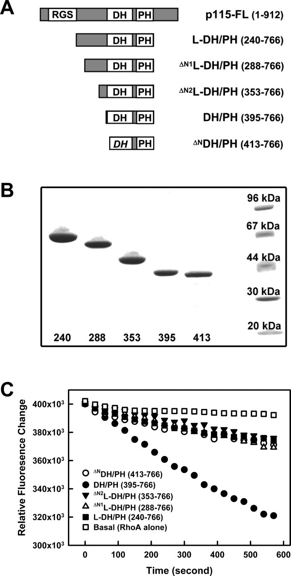
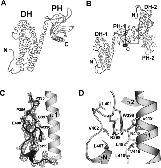
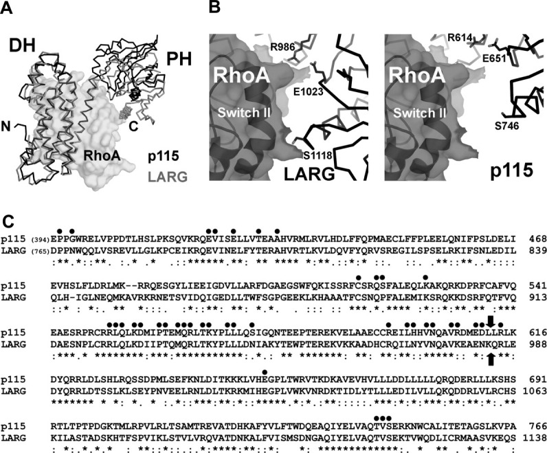
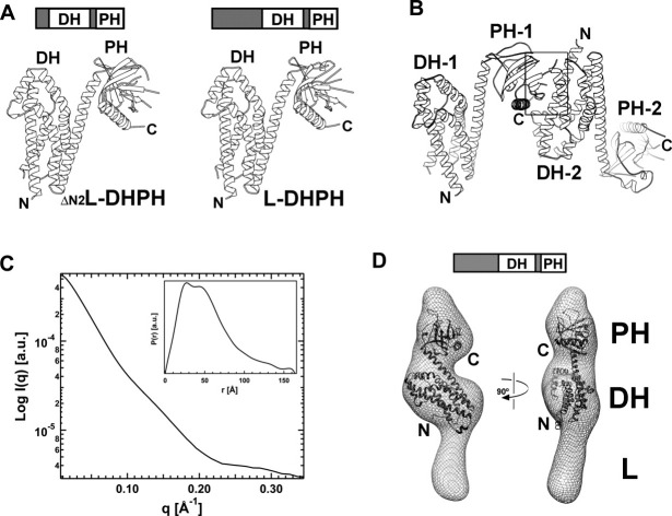
p115-RhoGEF (p115) belongs to the family of RGS-containing guanine nucleotide exchange factors for Rho GTPases (RGS-RhoGEFs) that are activated by G12 class heterotrimeric G protein α subunits. All RGS-RhoGEFs possess tandemly linked Dbl-homology (DH) and plekstrin-homology (PH) domains, which bind and catalyze the exchange of GDP for GTP on RhoA. We have identified that the linker region connecting the N-terminal RGS-homology (RH) domain and the DH domain inhibits the intrinsic guanine nucleotide exchange (GEF) activity of p115, and determined the crystal structures of the DH/PH domains in the presence or absence of the inhibitory linker region. An N-terminal extension of the canonical DH domain (the GEF switch), which is critical to GEF activity, is well folded in the crystal structure of DH/PH alone, but becomes disordered in the presence of the linker region. The linker region is completely disordered in the crystal structure and partially disordered in the molecular envelope calculated from measurements of small angle x-ray scattering (SAXS). It is possible that Gα subunits activate p115 in part by relieving autoinhibition imposed by the linker region.


Figures

Figure 1
Figure 1
The linker region between the RH and DH domains inhibits GEF activity of DH/PH. (a) Schematic representation of truncated forms of p115 used; included amino acids are listed in parentheses. (b) SDS-PAGE gel showing purified p115 fragments as detailed in panel (a). Numbers underneath represent the first amino acid of the protein. The last lane on the right contains protein standard markers with molecular weight labeled. (c) Nucleotide exchange assays with p115 and RhoA. For each time course, 0.5 μM RhoA loaded with mant-GDP was mixed with 100 μM GTP and the exchange reaction started at room temperature by addition of buffer (Basal, open squares), or 30 nM of p115: L-DH/PH (solid squares), ΔN1L-DH/PH (open triangles), ΔN2L-DH/PH (solid triangles), DH/PH (solid circles), or ΔNDH/PH (open circles). The subsequent decrease in fluorescence (λex = 356 nm, λem = 445 nm) was measured for 10 minutes.
Figure 2
Figure 2
Crystal structure of the p115 DH/PH domains. (a) Ribbon diagrams depicting the tertiary structure of p115 DH/PH. (b) Ribbon diagrams depicting the noncrystallographic dimer of p115 DH/PH (labeled 1 and 2). The dimer interface (marked by a box) involves a layer of β-strands near the C-terminus of one PH domain and the α4 helix from the dyad related DH domain. (c) The GEF switch in the DH domain. Electron density (cages) for the GEF switch from a 2.9 Å σA-weighted 2Fo-Fc total omit map calculated in SFCHECK (CCP4i) is contoured at 1.5 standard deviations above the mean. Side chains of residues 395–400 are depicted as stick models. The rest of the DH/PH domain is depicted as ribbon diagrams. (d) Trp-398 from the GEF switch is buried in a hydrophobic pocket between the GEF switch and the canonical DH domain. Side chains of residues involved are depicted as stick models. The putative hydrogen bond between side chains of Arg-399 and Glu-419 is drawn as a dotted line.
Figure 3
Figure 3
(a) Structural comparison of the DH/PH domains from p115 (black lines) and LARG (gray lines, PDB access code 1X86, with the bound nucleotide-free RhoA depicted as transparent solvent accessible surface). The structural alignment was based on coordinates from the DH domains only. The PH domain in LARG moves closer toward its DH domain upon binding to RhoA. (b) The interface between the PH domain and RhoA. In the crystal structure of the LARG-DH/PH:RhoA complex (left), residues from PH (depicted as sticks) make direct contacts with RhoA (depicted as transparent solvent accessible surface with black ribbons underneath). In the modeled p115-DH/PH:RhoA complex (right), where the two DH domains from p115 and LARG are superimposed, the same set of residues in p115 PH domain can not form direct contacts with RhoA. (c) Sequence alignment of DH/PH domains from p115 and LARG. Residues in LARG that are involved in contacts with the nucleotide-free RhoA are marked with dots on top. Block arrows mark the domain boundary between DH and PH. The sequence alignment is carried out by the program Clustal W.
Figure 4
Figure 4
Crystal and solution structures of the p115 L-DH/PH domains. (a) Ribbon diagrams depicting tertiary structures of ΔN2L-DH/PH (left) and L-DH/PH (right). (b) Ribbon diagrams depicting the noncrystallographic dimer of p115 L-DH/PH (labeled 1 and 2). The dimer interface (marked by a box) involves a layer of β-strands near the C-terminus of one PH domain and the α1-α2 helices from the dyad related DH domain. (c) Solution x-ray scattering profile for p115 L-DH/PH. The distance distribution function, P(r), of L-DH/PH was computed from the x-ray scattering using the program GNOM. (d) Solution structure of L-DH/PH of p115. The solution structure (molecular envelope) is depicted as a mesh and overlapped onto the crystal structure of p115 DH/PH domains (ribbon). The crystal structure is relatively well accommodated in the molecular envelope. The large unoccupied region in the molecular envelope beneath the DH domain is likely to be the location for the linker region with limited ordered structure.

References

    1. Sternweis PC, Carter AM, Chen Z, Danesh SM, Hsiung YF, Singer WD. Regulation of Rho guanine nucleotide exchange factors by G proteins. Adv Protein Chem. 2007;74:189–228. - PubMed
    1. Aittaleb M, Boguth CA, Tesmer JJ. Structure and function of heterotrimeric G protein-regulated Rho guanine nucleotide exchange factors. Mol Pharmacol. 2010;77:111–125. - PMC - PubMed
    1. Etienne-Manneville S, Hall A. Rho GTPases in cell biology. Nature. 2002;420:629–635. - PubMed
    1. Rossman KL, Der CJ, Sondek J. GEF means go: turning on RHO GTPases with guanine nucleotide-exchange factors. Nat Rev Mol Cell Biol. 2005;6:167–180. - PubMed
    1. Schmidt A, Hall A. Guanine nucleotide exchange factors for Rho GTPases: turning on the switch. Genes Dev. 2002;16:1587–1609. - PubMed

Publication types

MeSH terms

Substances

LinkOut - more resources