Review
. 2010 Dec;24(4):216-33.
doi: 10.3109/01677063.2010.514369. Epub 2010 Nov 11.

The history of the Drosophila TRP channel: the birth of a new channel superfamily

Affiliations
Review

The history of the Drosophila TRP channel: the birth of a new channel superfamily

Baruch Minke. J Neurogenet. 2010 Dec.

Abstract

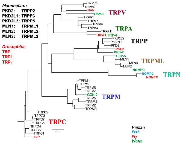
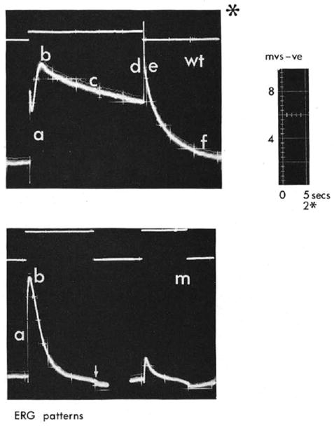
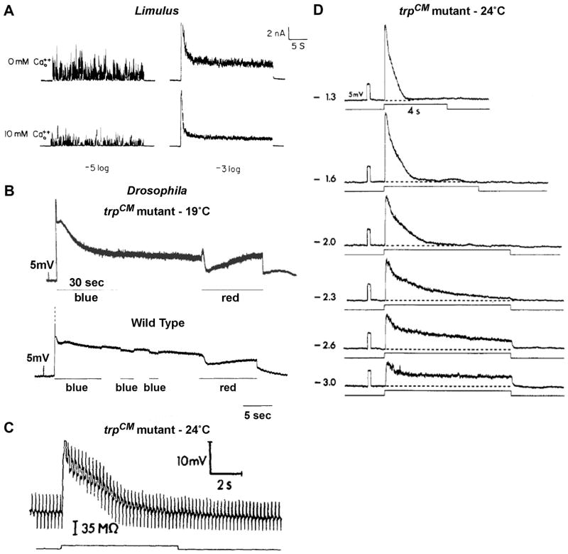
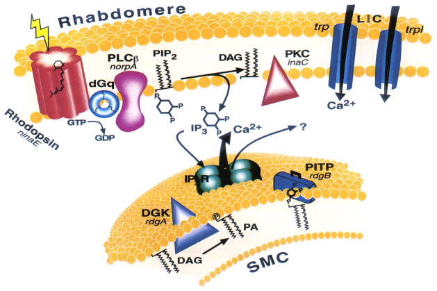
Transient receptor potential (TRP) channels are polymodal cellular sensors involved in a wide variety of cellular processes, mainly by changing membrane voltage and increasing cellular Ca(2+). This review outlines in detail the history of the founding member of the TRP family, the Drosophila TRP channel. The field began with a spontaneous mutation in the trp gene that led to a blind mutant during prolonged intense light. It was this mutant that allowed for the discovery of the first TRP channels. A combination of electrophysiological, biochemical, Ca(2+) measurements, and genetic studies in flies and in other invertebrates pointed to TRP as a novel phosphoinositide-regulated and Ca(2+)-permeable channel. The cloning and sequencing of the trp gene provided its molecular identity. These seminal findings led to the isolation of the first mammalian homologues of the Drosophila TRP channels. We now know that TRP channel proteins are conserved through evolution and are found in most organisms, tissues, and cell-types. The TRP channel superfamily is classified into seven related subfamilies: TRPC, TRPM, TRPV, TRPA, TRPP, TRPML, and TRPN. A great deal is known today about participation of TRP channels in many biological processes, including initiation of pain, thermoregulation, salivary fluid secretion, inflammation, cardiovascular regulation, smooth muscle tone, pressure regulation, Ca(2+) and Mg(2+) homeostasis, and lysosomal function. The native Drosophila photoreceptor cells, where the founding member of the TRP channels superfamily was found, is still a useful preparation to study basic features of this remarkable channel.


Figures

Figure 1
Figure 1. The phylogenetic tree of TRP channels superfamily
Depicted are the 7 subfamilies that constitute the TRP family. The 4 different species are indicated by different colors. Only some of the Drosophila and C. elegans (worm) members are included. For more details see (Venkatachalam & Montell, 2007). (From Nilius and Mahieu, (Nilius & Mahieu, 2006)).
Figure 2
Figure 2
The original electroretinogram (ERG) comparing the response to a light pulse between wild type Drosophila (wt) and a spontaneously formed mutant (m) that was designated later trp by Minke and colleagues. The traces were photographed from an oscilloscope and the different characters indicate various phases in the ERG response. Note that phases c,d,e,f are missing in the mutant. The upper traces represent the light monitor. The * at the upper panel indicates 2.5 faster timescale. Also note that all the traces are presented in an unconventional manner so that negative voltage (corneal negative) is presented upward (From (Cosens, 1971)).
Figure 3
Figure 3. The waveforms and light-induced bump noise of wild type and trp mutant during intense lights
A. The effect of lowering external Ca2+ on light responses of Limulus ventral photoreceptor in response to dim (−log I/I0=5) and intense (−log I/I0=3) lights, recorded by two electrode voltage clamp. Lowering external Ca2+ concentration increased the bump noise (left) and the steady state noise of the macroscopic response to light (right, from (Wong et al., 1982)). B, top. Intracellular recordings of the receptor potential and the following Prolonged Depolarizing Afterpotential (PDA) in response to intense blue lights in the trpCM mutant raised at 19°C (top) and in WT Drosophila (B bottom). In the trpCM mutant a large increase of bump noise is observed during the steady state phase of the light response and during the PDA of the mutant (top), but not in WT flies (bottom). Suppression of the PDA by red light resulted it a prompt suppression of the bump noise (B, top, red light) (from (Minke et al., 1975). C. The decay of the response to light of the trpCM mutant raised at 24°C is accompanied by a conductance decrease. The figure shows intracellular bridge measurement made in the trp mutant before, during and after green light stimulus. The bridge, which was balanced in the dark, shows during light an initial conductance increase that then decreases in parallele with the decrease in voltage, showing that the decay of the response is accompanied by closure of channels (from (Minke, 1982). D. The decay time of the intracellularly recorded response to light of the trpCM mutant raised at 24°C depends on light intensity. At very dim light the response does not decay to baseline (from (Minke, 1982).
Figure 4
Figure 4
The phosphoinositide cascade of vision. Cloned genes (for all of which mutants are available) are shown in italics, alongside their corresponding proteins. Upon absorption of light, rhodopsin (ninaE gene) is converted to the active metarhodopsin state, which activates a heterotrimeric G protein (dGq). This leads to activation of phospholipase C (PLCβ, norpA gene) and subsequent opening of two classes of light-sensitive channels encoded at by trp and trpl genes. PLC catalyzes the hydrolysis of phosphatidylinositol 4,5-bisphosphate (PIP2) into the soluble inositol 1,4,5-trisphosphate (IP3) and the membrane-bound diacylglycerol (DAG). DAG is recycled to PIP2 by the phosphatidylinositol (PI) cycle shown in an extension of the smooth endoplasmic reticulum called submicrovillar cisternae (SMC, shown on the bottom). DAG is converted to phosphatidic acid (PA) via DAG kinase (DGK, rdgA gene). After conversion to PI, PI is presumed to be transported back to the microvillar membrane by the PI transfer protein (PITP encoded by the rdgB gene). The InsP3 receptor (IP3R), which is an internal Ca2+ channel that opens and releases Ca2+ upon binding of InsP3. (From (Minke & Cook, 2002)).
Figure 5
Figure 5. Biochemical measurements of light-induced and G-protein-dependent hydrolysis of PIP2 and its absence by the norpA mutant which abolishes reversibly the response to light
A. Light-induced hydrolysis of phosphoinositides in Musca eye membrane preparation. Equivalents of 100 eyes were cut into halves and were incubated in the dark for 4 h at 30°C with [3H]inositol. Preparation of eye membranes and measurements of light dependent phosphoinositide hydrolysis were carried out as described previously (Devary et al., 1987). The upper panels show production of InsP2 and the lower panels show production of InsP3, which is a precursor of InsP2. The right column (top and bottom) shows the effect of the InsP3 phosphatase inhibitor (2,3 DPG) that causes an increase in InsP3 (right bottom) and concurrent reduction in InsP2 right top. The figure shows light dependent PLC activation leading to production of InsP3 and subsequent production of InsP2. B. Light-induced PIP2 hydrolysis is G-protein dependent. As in A, the figure shows enhancement of InsP2 production by GTPγS (10μM) and suppression of InsP2 production by GDPβS (100μM, right top as compared to left top) on inositol phosphate production. Due to the fast conversion of InsP3 to InsP2 only a small increase in InsP3 could be measured without 2,3 DPG. C. Light-induced PIP2 hydrolysis in eye membrane preparation of wild type (W.T., left panels) and its absence at elevated temperatures in the norp H52 temperature sensitive mutant of Drosophila. The experimental system was similar to that of panel A except that Drosophila heads were used. Systems depicted by dotted lines were pre-incubated for 4 min at the indicated temperatures in medium lacking ATP and ATP regenerating system which does not allow the biochemical reaction. The latter components were subsequently added (arrow) to initiate the reaction.
Figure 6
Figure 6. The trpCM mutant is not a null allele and expresses reduced amount of TRP
Western blot analysis of the homozygote trpCM mutant, raised at 24°C and at 19°C (as indicated) using momoclonal antibody against the TRP protein (Pollock et al., 1995). The TRP protein of WT and the mutant appears on the gel. (from (Yoon et al., 2000)).
Figure 7
Figure 7. Lanthanum (La3+) mimics the trp phenotype in wild type fly but has no effect on a trp homologue mutant
Intracellular recordings from single photoreptor cell of white-eyed Musca domestica (A) and from white-eyed nss mutant of Lucilia cuprina (Howard, 1984), which is a mutant homologue of the Drosophila trp (B). Responses to increasing intensities of orange lights are shown. The left columns show responses before application of La3+ (CONTROL). La3+ was applied by pressure injection into the extracellular space. Partial recovery of WT phenotype was observed 20 min after injection (A, right). Injection of La3+ to the extracellular space of the nss mutant had no effect on the rate of decline, but induced a small reduction in the amplitude of the initial peak response (from (Suss Toby et al., 1991)).
Figure 8
Figure 8. Model scheme that summarizes fly phototransduction according to the “conformational coupling” hypothesis
According to this model, TRP (trp) is a new type of channel/transporter, which is activated by light-induced depletion of the InsP3-sensitive Ca2+ stores (SMC), following production of InsP3 by G-protein (G) activated PLC and binding of InsP3 to the InsP3 Receptor. According to this model depletion of Ca2+ from the stores, couples TRP to the InsP3 receptor and opens the TRP Ca2+ channel (from (Minke & Selinger, 1991)).
Figure 9
Figure 9. Calcium indicator fluorescence reveals light-induced large elevation in intracellular Ca2+, which is smaller in the trp mutant relative to WT and it is totally abolished in a PLC null mutant
A and B: Calcium green-5N fluorescence (Kd< 30 μM) measured in WT at negative (−50 mV, A) and positive (+50 mV, B) membrane potentials. The reduced Ca2+ signal at positive membrane potential is due to reduced driving force for Ca2+ influx. The dotted lines indicate resting Ca2+ level in all panels. C and D: Measurements similar to A and B performed in the trpCM mutant (raised at 24°C) showing a large reduction of Ca2+ influx in the mutant. E: Both the light-induced current (lower trace in each pair) and increase in cellular Ca2+ are absent in the norpAP24, a virtually null PLC mutant. Fluo 3 (Kd< 1μM) fluorescence revealed only the resting Ca2+ level that was largely reduced by prolonged exposure to EGTA. (modified from (Peretz et al., 1994))
Figure 10
Figure 10. Current Voltage relationships of the native TRPL and TRP in WT and in the trp and trpl mutants
A. Current Voltage (I–V) relationships determined from voltage ramps in photoreceptors of wild type (WT), trpP301 and trpl302 mutants during whole cell recordings from ommatidia bathed in physiological Ringer’s solution containing nominal 0 Ca2+ and 4mM Mg2+. The current in trp shows simple exponential outward rectification (similar to that of panel A), however, in both WT and trpl flies there is a conspicuous S shape inward and outward rectification, which are markedly different than the I–V curve of TRP in pannel A (from (Reuss et al., 1997). B. Comparison of the I–V curves recorded by whole cell measurements from a photoreceptor of the trp mutant (expressing only TRPL) and the S2 cell expressing heterologously the TRPL channel. The top panel shows I–V curves derived from voltage ramps, while the bottom traces show voltage clamped currents in response to voltage steps between −100 mV and +80 mV in 20 mV increments (bottom traces). A striking similarity is observed between the data obtained from native and the heterologously expressed TRPL channels (from (Hardie et al., 1997).

Similar articles

Cited by

References

    1. Abramowitz J, Birnbaumer L. Physiology and pathophysiology of canonical transient receptor potential channels. FASEB J. 2009;23(2):297–328. - PMC - PubMed
    1. Acharya JK, Jalink K, Hardy RW, Hartenstein V, Zuker CS. InsP3 receptor is essential for growth and differentiation but not for vision in Drosophila. Neuron. 1997;18(6):881–887. - PubMed
    1. Berridge MJ. Capacitive calcium entry. Biochem J. 1995;312:1–11. - PMC - PubMed
    1. Berridge MJ, Irvine RF. Inositol trisphosphate, a novel second messenger in cellular signal transduction. Nature. 1984;312(5992):315–321. - PubMed
    1. Bloomquist BT, Shortridge RD, Schneuwly S, Perdew M, Montell C, Steller H, et al. Isolation of a putative phospholipase C gene of Drosophila, norpA, and its role in phototransduction. Cell. 1988;54:723–733. - PubMed

Publication types

Substances

LinkOut - more resources