. 2010 Nov 10:10:112.
doi: 10.1186/1471-213X-10-112.

Laminin-511 and integrin beta-1 in hair follicle development and basal cell carcinoma formation

Affiliations

Laminin-511 and integrin beta-1 in hair follicle development and basal cell carcinoma formation

Mindy C DeRouen et al. BMC Dev Biol. .

Abstract

Background: Initiation of the hair follicle placode and its subsequent growth, maturation and cycling in post-natal skin requires signaling interactions between epithelial cells and adjacent dermal cells and involves Shh signaling via the primary cilium. Previous reports have implicated laminins in hair follicle epithelial invagination.

Results: Here we use a human BCC model system and mouse mutants to re-evaluate the role of laminin-511 in epithelial invagination in the skin. Blocking laminin 511 and 332 in BCCs maintains primary cilia and Shh signalling, but prevents invagination. Similarly, in laminin-511 and dermal beta-1 integrin mutants, dermal papilla development and primary cilia formation are normal. Dermal beta-1 integrin mutants have normal hair follicle development.

Conclusions: Our data provides support for a primary role of laminin-511 promoting hair follicle epithelial downgrowth without affecting dermal primary cilia and Shh target gene induction.


Figures

Figure 1
Figure 1
LM511 and LM332 inhibition reduces tumor invasion in human BCC skin xenografts. A: Hematoxylin and eosin-stained sections of human skin xenografts carrying either control GFP or SHH retrovirus and treated with control IgG, 4c7 antibody against LM511 or BM165 antibody against LM332, as indicated. Images captured at 20x magnification. B: Indirect immunofluoresence for human-specific involucrin (red) confirms the human origin of graft tissue. C: Indirect immunofluoresence for keratin 10 (green) indicates all non-basal cells in graft epidermis. D: DIG RNA in-situ with anti-sense probe to PTCH1 transcript in indicated xenografts. Anti-sense probe signal is seen in all SHH-expressing grafts. Black dotted lines indicate epidermal/dermal boundary. Red arrows indicate DIG signal. D': DIG RNA in-situ with sense probe to PTCH1 transcript in indicated xenografts. Images are 20x magnification. E: Indirect immunofluoresence for Ki67 (red) in indicated graft tissues marks proliferating cells. F: TUNEL-TMR Red staining indicates apoptotic cells in graft tissues. E-F: White dashed lines indicate the epidermal/dermal boundary. Scale bars indicate 100 μm.
Figure 2
Figure 2
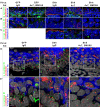
Xenograft tissues contain primary cilia. A-C: Indirect immunofluoresence for cilia components. Adenylyl cyclase III (green) indicates cilia shafts, rootletin (red) indicates the basal body at the base of the cilia and Hoechst dye (blue) indicates cell nuclei. A: Cilia are present in both epithelial and dermal cells. B: Zoom of epithelial cells in A. C: Zoom of dermal cells in A. D: Indirect immunofluoresence for cilia components and suprabasal cells. A cyclase III (green) and rootletin (red) mark cilia. Keratin10 (blue) marks suprabasal cells. Hoechst dye (white) indicates cell nuclei. Robust cilia are present only in cells excluding keratin 10. D': Zoom of boxed areas in D. All dashed lines indicate epidermal/dermal boundaries.
Figure 3
Figure 3
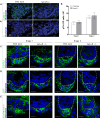
Laminin alpha 5 null hair follicles normally express dermal papilla markers. A: Images of indirect immunofluoresence for mouse LM511 (green) in E16.5 wild type and lamα5-/- mice (as indicated) showing a lack of antigen in null animals A': The same images as (A) indicating the Stage 2 hair follicles present in each view. B: Quantitation of p75-positive cells in dermal condensate of Stage 1 and Stage 2 hair follicles of E16.5 wild type and lama5-/- mice. C-E: Confocal images of indirect immunofluoresence for dermal markers CD133 (C), p75 (D) and syndecan-1 (E) in wild type and lamα5-/- Stage 1 and Stage 2 follicles within E16.5 skin. Hoechst (blue) labels cell nuclei. Dashed lines indicate epidermal/dermal boundary of hair follicles. Scale bars represent 10 μm. Abbreviations: DP, dermal papilla; HF, hair follicle.
Figure 4
Figure 4
Hair follicles in laminin alpha 5 null mice have primary cilia. A-D: Confocal z-stacks of primary cilia and dermal papilla in hair follicles of E14.5 (A-B) and E16.5 (C-D) skin of wild type (A, C) and lamα5-/- (B, D) animals. Adenylyl cyclase III (green) indicates the primary cilia, CD133 (red) marks the dermal papilla cells and Hoechst dye (blue) indicates cell nuclei. A'-D': Zoom of boxed areas in A-D. E: Confocal z-stacks of primary cilia in dermal papilla of wild type E16.5 skin treated overnight with either control IgM or Ha2/5 antibody to Itβ1 (as indicated). Immunofluoresence same as in A-D. F: H&E stained frozen sections of 9-day allografts of wild type E16.5 skin treated overnight with either control IgM or Ha2/5 antibody (as indicated). IgM treated allografts show significant hair growth and normal hair-free skin. Ha2/5 treated allografts show both blistered and intact epidermis with little to no hair growth, indicating the ability of this antibody to block Itβ1 function and hair follicle development. G: Percent dermal papilla cells with primary cilia in z-stacks of dermal papilla from E16.5 skin treated with either IgM or Ha2/5 antibody (as in E). All scale bars indicate 10 μm. Abbreviations: DPC, dermal papilla cells; PC, primary cilia; Ab, antibody.
Figure 5
Figure 5
Prx1-Cre; Itβ1flox/flox mice exhibit normal hair follicle morphogenesis and post-natal cycling. A: Mixed littermates (Ctrl) and Prx1-cre; Itβ1flox/flox (Itβ1 KO) mice at 1 day post-natal (P1). Conditional knockouts display slightly reduced size and a reddened appearance on ventral and lateral surfaces. B: Representative H&E stained skin sections from the ventral skin over the sternum of P1 control and conditional knockout mice. C: Control and conditional knockout mice at 5 days post-natal (P5) showing the reduced size and reddened ventral skin of mutant mice. D: H&E stained skin sections from the ventral skin of P5 control and conditional knockout mice showing hair follicles in advanced stages. E: Quantitation of hair follicle stages in P1 and P5 control and conditional knockout mice showing similar patterns of hair development. F: Control and conditional knockout mice at 82 days showing the normal appearance of coat on the ventral and lateral regions. G: Immunofluoresence for p75 (green) and Itβ1 (red) in sections of ventral skin from P1 control and conditional knockout mice at P1 (as indicated). Brackets indicate p75+ dermal papilla. Note lack of Itβ1 signal in the dermal papilla of a Stage 4 hair follicle (arrow). H: Immunofluoresence for mouse LM511 (green) shows normal incorporation of this laminin in P1 control and conditional knockout mice. Scale bars represent 20 μm. Abbreviations: Ctrl, control; Itβ1 KO, dermal Itβ1 knockout; HFs, hair follilces.
Figure 6
Figure 6
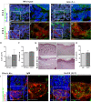
Dermal condensates in Prx1-Cre; Itβ1flox/flox mice appear normal and contain primary cilia. A: Immunofluoresence for loricrin (green) in P1 control and conditional knockout mice, showing similar staining of the statrum granulosum. B: Immunofluoresence for keratin 10 (green) in P1 control and conditional knockout mce, showing similar staining of the straturm spinosum Itα6 staining in A-B shows normal basal cell layers. C: Immunofluoresence for Itβ4 (red) and p75 (green), showing normal levels of Itβ4 staining in the epithlelia. D: Immunofluoresence for p75 (green) in P1 control and Prx1-Cre; Itβ1flox/flox mice showing similar dermal papilla expression patterns. A-D: Scale bars indicate 20 μm E: Number of dermal papilla cells in condensates of Stage 1 and 2 hair follicles of P1 control and Prx1-Cre; Itβ1flox/flox mice. F: Confocal z-stacks of immunofluoresence for primary cilia and dermal papilla in Stage 1 (top panels) and Stage 2 (bottom panels) hair follicles of P1 control and Prx1-Cre; Itβ1flox/flox mice. Adenylyl cyclase III (green) marks the primary cilium shaft, syndecan I (red) indicates dermal papilla cells and Hoechst dye (blue) indicates cell nuclei. Scale bars indicate 10 μm G: Percent dermal papilla cells with a primary cilium in Stage 1 and 2 hair follicles of P1 control and conditional knockout mice. All dotted lines indicate the epidermal/dermal border. Abbreviations: Ctrl, control; Itβ1 KO, dermal Itβ1 knockout; DP, dermal papilla; HF, hair follicle.

References

    1. Stamp GW, Quaba A, Braithwaite A, Wright NA. Basal cell carcinoma xenografts in nude mice: studies on epithelial differentiation and stromal relationships. J Pathol. 1988;156:213–25. doi: 10.1002/path.1711560306. - DOI - PubMed
    1. Van Scott EJ, Reinertson RP. The modulating influence of stromal environment on epithelial cells studied in human autotransplants. J Invest Dermatol. 1961;36:109–31. - PubMed
    1. Aszterbaum M, Rothman A, Johnson RL, Fisher M, Xie J, Bonifas JM, Zhang X, Scott MP, Epstein EH Jr. Identification of mutations in the human PATCHED gene in sporadic basal cell carcinomas and in patients with the basal cell nevus syndrome. J Invest Dermatol. 1998;110:885–8. doi: 10.1046/j.1523-1747.1998.00222.x. - DOI - PubMed
    1. Satir P. Cilia biology: stop overeating now! Curr Biol. 2007;17:R963–5. doi: 10.1016/j.cub.2007.09.006. - DOI - PubMed
    1. Michaud EJ, Yoder BK. The primary cilium in cell signaling and cancer. Cancer Res. 2006;66:6463–7. doi: 10.1158/0008-5472.CAN-06-0462. - DOI - PubMed

Publication types

MeSH terms

LinkOut - more resources