Modulating spontaneous brain activity using repetitive transcranial magnetic stimulation
- PMID: 21067612
- PMCID: PMC2993720
- DOI: 10.1186/1471-2202-11-145
Modulating spontaneous brain activity using repetitive transcranial magnetic stimulation
Abstract
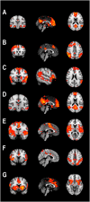
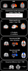
Background: When no specific stimulus or task is presented, spontaneous fluctuations in brain activity occur. Brain regions showing such coherent fluctuations are thought to form organized networks known as 'resting-state' networks, a main representation of which is the default mode network. Spontaneous brain activity shows abnormalities in several neurological and psychiatric diseases that may reflect disturbances of ongoing thought processes. Information about the degree to which such spontaneous brain activity can be modulated may prove helpful in the development of treatment options. We investigated the effect of offline low-frequency rTMS on spontaneous neural activity, as measured with fMRI, using a sequential independent-component-analysis and regression approach to investigate local changes within the default mode network.
Results: We show that rTMS applied over the left dorsolateral prefrontal cortex results in distal changes of neural activity, relative to the site of stimulation, and that these changes depend on the patterns of brain network activity during 'resting-state'.
Conclusions: Whereas the proximal changes may reflect the off-line effect of direct stimulation of neural elements, the distal changes likely reflect modulation of functional connectivity.
Figures


Similar articles
-
Effect of Repetitive Transcranial Magnetic Stimulation on fMRI Resting-State Connectivity in Multiple System Atrophy.Brain Connect. 2015 Sep;5(7):451-9. doi: 10.1089/brain.2014.0325. Epub 2015 Apr 22. Brain Connect. 2015. PMID: 25786196 Free PMC article.
-
Multimodal Imaging of Repetitive Transcranial Magnetic Stimulation Effect on Brain Network: A Combined Electroencephalogram and Functional Magnetic Resonance Imaging Study.Brain Connect. 2019 May;9(4):311-321. doi: 10.1089/brain.2018.0647. Epub 2019 Apr 8. Brain Connect. 2019. PMID: 30803271 Free PMC article.
-
Intranetwork and Internetwork Effects of Navigated Transcranial Magnetic Stimulation Using Low- and High-Frequency Pulse Application to the Dorsolateral Prefrontal Cortex: A Combined rTMS-fMRI Approach.J Clin Neurophysiol. 2020 Mar;37(2):131-139. doi: 10.1097/WNP.0000000000000528. J Clin Neurophysiol. 2020. PMID: 30335664
-
Effects of repetitive transcranial magnetic stimulation on resting-state connectivity: A systematic review.Neuroimage. 2020 May 1;211:116596. doi: 10.1016/j.neuroimage.2020.116596. Epub 2020 Jan 31. Neuroimage. 2020. PMID: 32014552 Free PMC article.
-
Mapping cognitive and emotional networks in neurosurgical patients using resting-state functional magnetic resonance imaging.Neurosurg Focus. 2020 Feb 1;48(2):E9. doi: 10.3171/2019.11.FOCUS19773. Neurosurg Focus. 2020. PMID: 32006946 Free PMC article. Review.
Cited by
-
Decreased neural activity and neural connectivity while performing a set-shifting task after inhibiting repetitive transcranial magnetic stimulation on the left dorsal prefrontal cortex.BMC Neurosci. 2015 Jul 22;16:45. doi: 10.1186/s12868-015-0181-3. BMC Neurosci. 2015. PMID: 26199083 Free PMC article. Clinical Trial.
-
Combined rTMS/fMRI Studies: An Overlooked Resource in Animal Models.Front Neurosci. 2018 Mar 23;12:180. doi: 10.3389/fnins.2018.00180. eCollection 2018. Front Neurosci. 2018. PMID: 29628873 Free PMC article. Review.
-
Dose-dependent effects of theta burst rTMS on cortical excitability and resting-state connectivity of the human motor system.J Neurosci. 2014 May 14;34(20):6849-59. doi: 10.1523/JNEUROSCI.4993-13.2014. J Neurosci. 2014. PMID: 24828639 Free PMC article.
-
cTBS delivered to the left somatosensory cortex changes its functional connectivity during rest.Neuroimage. 2015 Jul 1;114:386-397. doi: 10.1016/j.neuroimage.2015.04.017. Epub 2015 Apr 13. Neuroimage. 2015. PMID: 25882754 Free PMC article.
-
Resting-state and task-based centrality of dorsolateral prefrontal cortex predict resilience to 1 Hz repetitive transcranial magnetic stimulation.Hum Brain Mapp. 2020 Aug 1;41(11):3161-3171. doi: 10.1002/hbm.25005. Epub 2020 May 12. Hum Brain Mapp. 2020. PMID: 32395892 Free PMC article.
References
Publication types
MeSH terms
LinkOut - more resources
Full Text Sources
Medical

