

Acute sun damage and photoprotective responses in whales
- PMID: 21068035
- PMCID: PMC3081749
- DOI: 10.1098/rspb.2010.1903
Acute sun damage and photoprotective responses in whales
Abstract
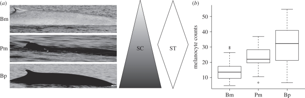
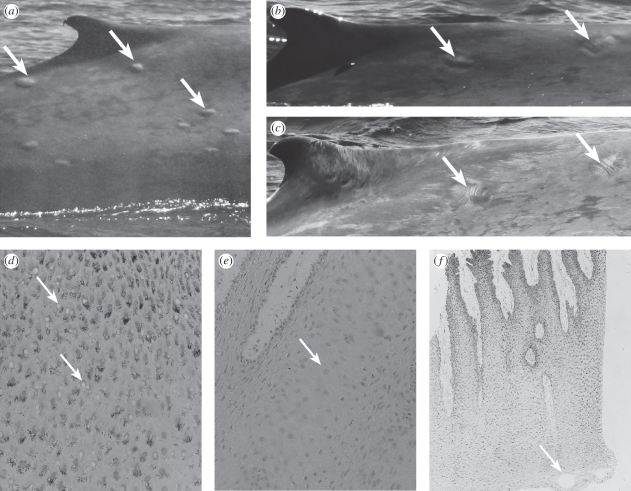
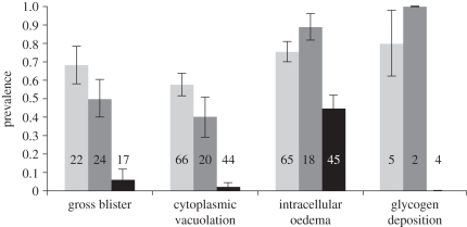
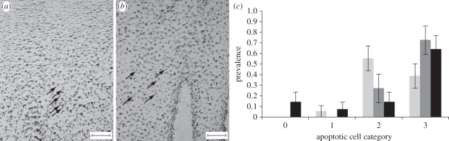
Rising levels of ultraviolet radiation (UVR) secondary to ozone depletion are an issue of concern for public health. Skin cancers and intraepidermal dysplasia are increasingly observed in individuals that undergo chronic or excessive sun exposure. Such alterations of skin integrity and function are well established for humans and laboratory animals, but remain unexplored for mammalian wildlife. However, effects are unlikely to be negligible, particularly for species such as whales, whose anatomical or life-history traits force them to experience continuous sun exposure. We conducted photographic and histological surveys of three seasonally sympatric whale species to investigate sunburn and photoprotection. We find that lesions commonly associated with acute severe sun damage in humans are widespread and that individuals with fewer melanocytes have more lesions and less apoptotic cells. This suggests that the pathways used to limit and resolve UVR-induced damage in humans are shared by whales and that darker pigmentation is advantageous to them. Furthermore, lesions increased significantly in time, as would be expected under increasing UV irradiance. Apoptosis and melanocyte proliferation mirror this trend, suggesting that whales are capable of quick photoprotective responses. We conclude that the thinning ozone layer may pose a risk to the health of whales and other vulnerable wildlife.
Figures
References
-
- McKenzie R. L., Aucamp P. J., Bais A. F., Bjorn L. O., Ilyas M. 2007. Changes in biologically-active ultraviolet radiation reaching the Earth's surface. Photochem. Photobiol. Sci. 6, 218–231 10.1039/B700017K (doi:10.1039/B700017K) - DOI - PubMed
-
- Li F., Stolarski R. S., Newman P. A. 2009. Stratospheric ozone in the post-CFC era. Atmos. Chem. Phys. 9, 2207–2213 10.5194/acp-9-2207-2009 (doi:10.5194/acp-9-2207-2009) - DOI
-
- Dameris M. 2009. Depletion of the ozone layer in the 21st century. Angew. Chem. Int. Ed. 49, 489–491 10.1002/anie.200906334 (doi:10.1002/anie.200906334) - DOI - PubMed
-
- De Gruijl F. R., Longstreth J., Norval M., Cullen A. P., Slaper H., Kripke M. L., Takizawa Y., Van der Leun J. C. 2003. Health effects from stratospheric ozone depletion and interactions with climate change. Photochem. Photobiol. Sci. 2, 16–28 10.1039/b211156j (doi:10.1039/b211156j) - DOI - PubMed
-
- Gallagher R. P., Lee T. K. 2006. Adverse effects of ultraviolet radiation: a brief review. Prog. Biophys. Mol. Biol. 92, 119–131 10.1016/j.pbiomolbio.2006.02.011 (doi:10.1016/j.pbiomolbio.2006.02.011) - DOI - PubMed
MeSH terms
LinkOut - more resources
Full Text Sources
Other Literature Sources
Medical