. 2011 Mar 22;278(1707):940-51.
doi: 10.1098/rspb.2010.2161. Epub 2010 Nov 10.

Parasitoidism, not sociality, is associated with the evolution of elaborate mushroom bodies in the brains of hymenopteran insects

Affiliations

Parasitoidism, not sociality, is associated with the evolution of elaborate mushroom bodies in the brains of hymenopteran insects

Sarah M Farris et al. Proc Biol Sci. .

Abstract

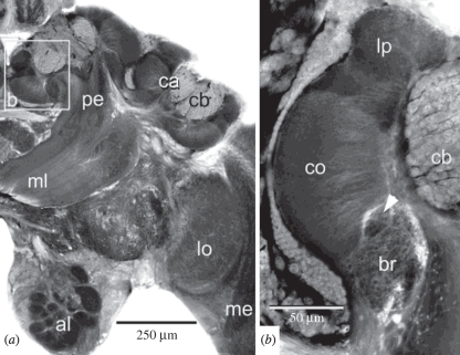
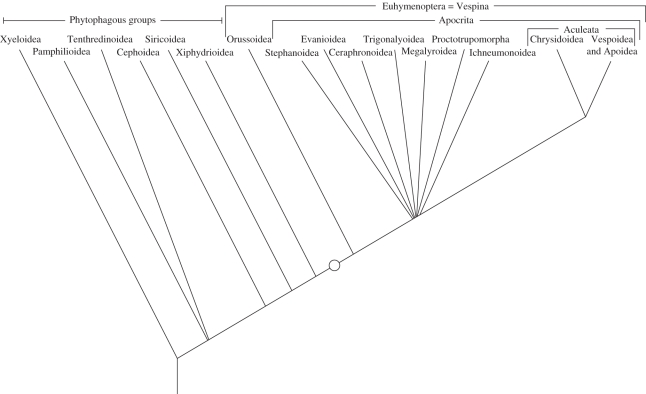
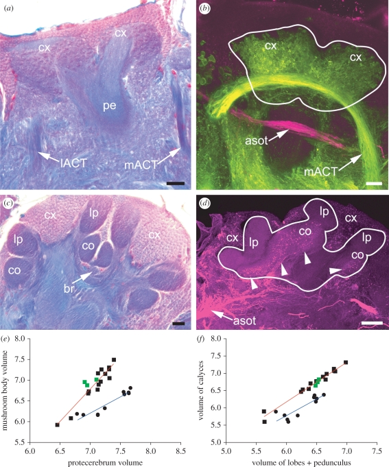
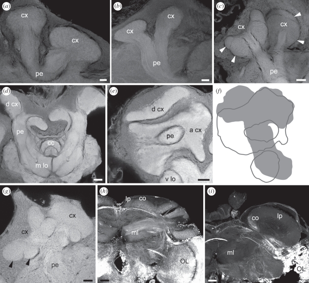
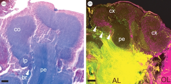
The social brain hypothesis posits that the cognitive demands of social behaviour have driven evolutionary expansions in brain size in some vertebrate lineages. In insects, higher brain centres called mushroom bodies are enlarged and morphologically elaborate (having doubled, invaginated and subcompartmentalized calyces that receive visual input) in social species such as the ants, bees and wasps of the aculeate Hymenoptera, suggesting that the social brain hypothesis may also apply to invertebrate animals. In a quantitative and qualitative survey of mushroom body morphology across the Hymenoptera, we demonstrate that large, elaborate mushroom bodies arose concurrent with the acquisition of a parasitoid mode of life at the base of the Euhymenopteran (Orussioidea + Apocrita) lineage, approximately 90 Myr before the evolution of sociality in the Aculeata. Thus, sociality could not have driven mushroom body elaboration in the Hymenoptera. Rather, we propose that the cognitive demands of host-finding behaviour in parasitoids, particularly the capacity for associative and spatial learning, drove the acquisition of this evolutionarily novel mushroom body architecture. These neurobehavioural modifications may have served as pre-adaptations for central place foraging, a spatial learning-intensive behaviour that is widespread across the Aculeata and may have contributed to the multiple acquisitions of sociality in this taxon.


Figures

Figure 1.
Figure 1.
Mushroom body architecture in aculeate hymenopterans, as illustrated by the honeybee Apis mellifera (Apidae, Apoidea). (a) The mushroom body neuropil consisting of the calyces (ca), pedunculus (pe) and lobes (medial lobe (ml) visible in this plane of section). The calyces are elaborated into deeply invaginated cups, and the cell bodies of mushroom body intrinsic neurons, called Kenyon cells (cb) fill and surround the calyces. (b) High-magnification view of boxed area in (a) showing functional subdivisions of the calyx. Olfactory projection neurons target the lip (lp), visual projection neurons target the collar (co) and the basal ring (br) receives collaterals from both. Figures borrowed with permission from Ehmer & Gronenberg [37].
Figure 2.
Figure 2.
Cautious estimate of the phylogeny of Hymenoptera based on [41,56,120,121] and recent molecular analyses by the HymAToL project (not yet published). Only relationships that are supported independently by morphological and molecular analyses are shown. The present study examined brains of species representing all groups indicated in the tree with the exception of the phytophagous Pamphilioidea. Proctotrupomorpha includes Chalcidoidea, Proctotrupoidea, Cynipoidea, Platygastroidea, Diaprioidea and Mymarommatoidea. Circle indicates the appearance of large, elaborate mushroom bodies at the base of the Euhymenoptera. Eusocial species are found only within the Vespoidea and Apoidea.
Figure 3.
Figure 3.
Comparisons of mushroom body morphology in phytophagous versus parasitoid Hymenoptera. (a) Mushroom bodies of the phytophagous Dolerus sp. (Tenthredinidae) showing simple, ovoid mushroom body calyces (cx) with no obvious compartmentalization into lip, collar or basal ring regions. (b) Dextran fills of the antennal lobes (green) reveal arborizations of olfactory projection neurons from the mACT throughout both calyces. Dextran fills of the optic lobes (magenta) label projection neuron axons in the anterior superior optic tract (asot), which passes beneath the mushroom bodies without providing collaterals to the calyces. (c) Mushroom bodies of the euhymenopteran parasitoid Ophion sp. (Ichneumonidae) have elaborate, deeply invaginated calyces divided into subcompartments like those observed in aculeate hymenopterans (lip (lp), collar (co) and basal ring (br), labelled in one calyx). (d) Dextran fills of the optic lobe of an ichneumonid reveal innervation of the calyx collar by optic lobe projection neurons (arrowheads). (e) Linear regression of log-transformed mushroom body volumes relative to the volume of the remaining protocerebrum in phytophagous (blue line) versus euhymenopteran (red line) species. Green points indicate measurements for the social aculeate Tetramorium caespitum (Formicidae, Vespoidea). (f) Linear regression of log-transformed calyx volumes versus lobe volumes (n = 10 for phytophagous species, n = 17 for parasitoid species). lACT, mACT; lateral and medial antennocerebral tracts. Scale bars (ac) 20 µm; (d) 50 µm.
Figure 4.
Figure 4.
Mushroom body morphology in phytophagous and parasitoid Hymenoptera. Simple ovoid calyces (cx) without subcompartments were observed in the phytophagous (a) Tremex columba (Siricidae) and (b) Xiphydria maculata (Xiphydriidae). (c) The calyces of stem-boring sawflies such as Cephus spinipes (Cephidae) appeared to possess small subcompartments, although they were not otherwise elaborated like those of euhymenopterans (di). (d,e) The elaborate calyces of Orussus abietinus (Orussidae) were greatly enlarged, with one calyx oriented dorsally (d cx) and one anteriorly (a cx). (f) Outline of the Orussus mushroom bodies illustrating the great size of the calyces. (g) Subcompartments (arrowheads) in the elaborate calyces of the basal parasitoid Stephanus serrator (Stephanidae) are structurally similar to the lip and collar of the higher Euhymenoptera, suggesting that they are similarly subdivided by olfactory and visual inputs. (h) Optic lobe dextran fills reveal visual input to collar (co) subcompartments of the elaborate calyces of the parasitoid Gasteruption sp. (Gasteruptiidae, Evanoidea). Unlabelled dorsal compartments probably correspond to the lip (lp). (i) The mushroom bodies of Leucospis sp. (Leucospidae, Chalcidoidea) possess a single flask-shaped calyx that receives dextran-labelled optic lobe projection neurons to a small ventral collar (co). Again, the dorsal unfilled compartment probably represents an olfactory input-receiving lip. Medial lobe (m lo); optic lobe (OL); pedunculus (pe); vertical lobe (v lo). Scale bars (ac,g) 20 µm; (d,e,h,i) 50 µm.
Figure 5.
Figure 5.
Mushroom body calyces of a solitary aculeate wasp (Chrysididae, Chrysidoidea). (a) Chrysidids have elaborate and deeply invaginated calyces (one visible here) with a clear lip, collar and basal ring (arrow). (b) Dextran fills of the antennal lobes (AL, green) and optic lobes (OL, magenta) show segregation of inputs into the calyx lip, collar and basal ring (arrows). The position of the collar and lip are inverted relative to what is observed in aculeates so that the greatly enlarged collar lies on top of the much smaller lip. Scale bars (a) 20 µm; (b) 50 µm.

References

    1. McGuire S. E., Le P. T., Davis R. L. 2001. The role of Drosophila mushroom body signaling in olfactory memory. Science 293, 1330–133310.1126/science.1062622 (doi:10.1126/science.1062622) - DOI - DOI - PubMed
    1. Perez-Orive J., Mazor O., Turner G. C., Cassenaer S., Wilson R. I., Laurent G. 2002. Oscillations and sparsening of odor representation in the mushroom body. Science 297, 359–36510.1126/science.1070502 (doi:10.1126/science.1070502) - DOI - DOI - PubMed
    1. Blum A. L., Li W., Cressy M., Dubnau J. 2009. Short- and long-term memory in Drosophila require cAMP signaling in distinct cell types. Curr. Biol. 19, 1341–135010.1016/j.cub.2009.07.016 (doi:10.1016/j.cub.2009.07.016) - DOI - DOI - PMC - PubMed
    1. Mizunami M., Weibrecht J. M., Strausfeld N. J. 1993. A new role for the insect mushroom bodies: place memory and motor control. In Biological neural networks in invertebrate neuroethology and robotics (eds Beer R. D., Ritzman R. E., McKenna T.), pp. 199–225 New York, NY: Academic Press
    1. Mizunami M., Weibrecht J. M., Strausfeld N. J. 1998. Mushroom bodies of the cockroach: their participation in place memory. J. Comp. Neurol. 402, 520–53710.1002/(SICI)1096-9861(19981228)402:4<520::AID-CNE6>3.0.CO;2-K (doi:10.1002/(SICI)1096-9861(19981228)402:4<520::AID-CNE6>3.0.CO;2-K) - DOI - DOI - PubMed

Publication types