. 2010 Dec;137(24):4201-9.
doi: 10.1242/dev.055046. Epub 2010 Nov 10.

dFMRP and Caprin, translational regulators of synaptic plasticity, control the cell cycle at the Drosophila mid-blastula transition

Affiliations

dFMRP and Caprin, translational regulators of synaptic plasticity, control the cell cycle at the Drosophila mid-blastula transition

Ophelia Papoulas et al. Development. 2010 Dec.

Abstract

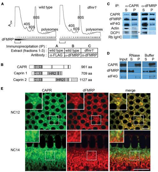
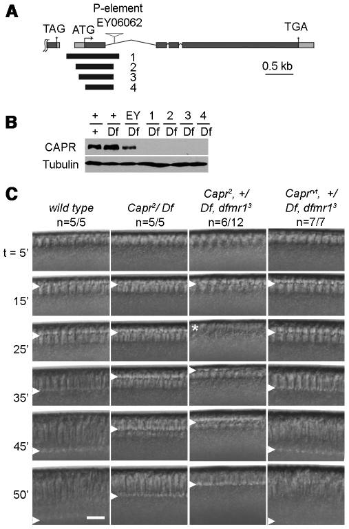
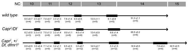
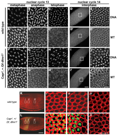
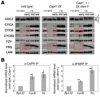
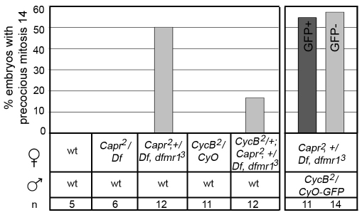
The molecular mechanisms driving the conserved metazoan developmental shift referred to as the mid-blastula transition (MBT) remain mysterious. Typically, cleavage divisions give way to longer asynchronous cell cycles with the acquisition of a gap phase. In Drosophila, rapid synchronous nuclear divisions must pause at the MBT to allow the formation of a cellular blastoderm through a special form of cytokinesis termed cellularization. Drosophila Fragile X mental retardation protein (dFMRP; FMR1), a transcript-specific translational regulator, is required for cellularization. The role of FMRP has been most extensively studied in the nervous system because the loss of FMRP activity in neurons causes the misexpression of specific mRNAs required for synaptic plasticity, resulting in mental retardation and autism in humans. Here, we show that in the early embryo dFMRP associates specifically with Caprin, another transcript-specific translational regulator implicated in synaptic plasticity, and with eIF4G, a key regulator of translational initiation. dFMRP and Caprin collaborate to control the cell cycle at the MBT by directly mediating the normal repression of maternal Cyclin B mRNA and the activation of zygotic frühstart mRNA. These findings identify two new targets of dFMRP regulation and implicate conserved translational regulatory mechanisms in processes as diverse as learning, memory and early embryonic development.


Figures

Fig. 1.
Fig. 1.
dFMRP associates with Caprin. (A) Extracts from Drosophila embryos laid by wild-type or dfmr mutant [dfmr13/Df(3R)Exel6265] females were fractionated on sucrose gradients. UV traces (A254) show positions of ribosomal material (40S, 60S, 80S, polysomes). Fractions (1-22) were analyzed by immunoblotting for dFMRP. Fractions 1-3 (brackets) from each gradient were separately pooled for immunoprecipitations using anti-FLAG (sample A) or anti-dFMRP (samples B and C) antibodies. (B) Alignment of Drosophila Caprin (CAPR) (CG18811) with human CAPRIN1 and 2 showing Homology Region 1 and 2 (shaded boxes HR1 and HR2) (Grill et al., 2004), G3BP-binding motif (hatched boxes) (Solomon et al., 2007), Caprin 2-specific C1q-related domain (stippled box) (Aerbajinai et al., 2004; Grill et al., 2004), RNA-binding RGG motifs (thick black bars) (Shiina et al., 2005; Wang et al., 2005) and lengths (aa, amino acid). For Drosophila CAPR, the percentage identity/similarity to human CAPRIN1 HR1 is 32%/52% and to human CAPRIN2 HR1 is 51%/73%. (C) Immunoblot of supernatants (S) and pellets (P) from anti-CAPR and anti-dFMRP immunoprecipitations from wild-type nuclear cycle (NC) 13 to early NC14 embryo extracts probed with antibodies to the proteins listed on the left. DCP1, an mRNA decapping protein, and Actin served as controls and were not present in either pellet. Asterisk marks a non-specific signal (Rb IgHC) detected by goat anti-rabbit HRP secondary antibody alone (compare with bottom panel). (D) Immunoblot of anti-CAPR immunoprecipitate from wild-type embryo extract (Input) and equal percentages of supernatants (S) and pellets (P) from Input incubated with RNase A or buffer. Probing was with antibodies to the proteins listed on the left. (E) Immunofluorescence analysis of fixed wild-type embryos reveals partial colocalization between CAPR (green) and dFMRP (red). Optical sections are through the apical cytoplasm of an NC12 embryo and the sagittal plane of an NC14 embryo. The boxed regions are shown at higher magnification in the insets. Scale bars: 10 μm.
Fig. 2.
Fig. 2.
Maternal expression of Capr is required for the MBT. (A) Structure of Drosophila Capr showing start (ATG) and stop (TGA) codons, introns (thin lines), exons (gray boxes) and untranslated regions (light gray boxes). The P element transposon (EY06062) was excised to generate four deletion alleles. Black bars (1-4) indicate the deleted regions, none of which removes the 3′ end of the upstream gene (TAG stop codon). (B) Immunoblots with equal amounts of adult extracts were probed for CAPR or Tubulin. +, wild type; Df, Df(3L)Cat (which removes Capr); EY, transposon insertion EY06062; 1-4, Capr alleles. (C) Frames from representative DIC movies of single embryos from females of the genotypes indicated. Caprrvt is a perfect excision of EY06062, with no detectable phenotype. Frames show a sagittal portion of each embryo at times (in minutes) relative to NC14 onset; the number of embryos displaying the phenotype over the total period analyzed is indicated (n). Arrowheads indicate the furrow front. The asterisk indicates premature mitosis 14 with furrow disassembly.
Fig. 3.
Fig. 3.
Reduction of CAPR and dFMRP function specifically disrupts timing of the MBT. The length of interphase (black line) and mitosis (black box) for nuclear cycles (NC) 10-14 for Drosophila embryos derived from females of the indicated genotypes. Elapsed times (minutes ± s.d.) and number of embryos analyzed (n) are indicated beneath the line that represents each genotype. Note the precocious mitosis of embryos from Capr2, +/Df, dfmr13 females at 18.9 minutes into interphase of NC14. Minor differences noted in NC12 and NC13 interphase and mitosis lengths produced no net change in overall cycle length.
Fig. 4.
Fig. 4.
Mitosis appears normal in embryos with a disrupted MBT. (A-J′) Immunofluorescence analysis of Drosophila embryos laid by females of the indicated genotypes (left) during the indicated cell cycle phase (top), stained with TO-PRO-3 iodide (DNA, A-E,F-J) and with antibodies to α-Tubulin 67C to visualize microtubules (MT, A′-E′,F′-J′). The boxed regions of D, D′, I and I′ are enlarged as E, E′, J and J′, respectively. (K,L) Embryos of the indicated genotypes were imaged for F-actin (red) and phospho-Histone H3-Ser10, a marker for mitotic chromosomes (green). The boxed regions are enlarged as labeled (i-iii). The wild-type embryo (K) is in interphase of NC14, whereas a portion of the mutant embryo is in interphase of NC14 (iii) and another portion is undergoing precocious mitosis (i,ii). Scale bars: 50 μm in D,D′,I,I′,K,L; 10 μm in all other panels.
Fig. 5.
Fig. 5.
dFMRP and CAPR associate with CycB and frs mRNAs in wild-type embryos and specifically alter CYCB and FRS protein expression at the MBT. (A) An immunoblot of ten Drosophila embryos per lane of the indicated genotypes (top) was probed for the indicated proteins (left), which included Lamin (LAM) as a loading control. Hand-sorted embryos were of the indicated stages: NC13 interphase (I13) and mitosis (M13), or NC14 early, middle and late interphase (I14). Phospho-isoforms of CDC2 (Edgar et al., 1994) are labeled (1-4). Red boxes highlight differences in protein expression between genotypes. (B) mRNA specifically immunoprecipitating from NC13 to early NC14 embryo extracts with CAPR or dFMRP as determined by quantitative PCR. Levels are presented as ratios (wild type/mutant) of RpL32 (control), CycB and frs mRNAs in immunoprecipitates. Error bars indicate s.d. Significance was determined for normalized mRNA values using a two-tailed Student's t-test. *, P=0.022 for CycB and P=0.020 for frs; **, P=0.003 for CycB and P=0.00005 for frs.
Fig. 6.
Fig. 6.
Elevated levels of maternally derived CYCB promote premature mitosis at the MBT. The percentage of embryos (n, number of embryos analyzed) derived from the indicated crosses (beneath) that display a premature mitosis 14 phenotype. Progeny derived from Capr2, +/Df, dfmr13 females receiving either a CycB2 (light-gray bar, GFP) or GFP-marked (dark-gray bar, GFP+) paternal chromosome displayed the premature mitosis 14 phenotype at similar frequencies. The reduction of maternal CycB mRNA partially rescues the precocious mitosis 14 phenotype (left panel, far right-hand bar), whereas the reduction of zygotic CycB mRNA does not (right panel, GFP).

Similar articles

Cited by

References

    1. Aerbajinai W., Lee Y. T., Wojda U., Barr V. A., Miller J. L. (2004). Cloning and characterization of a gene expressed during terminal differentiation that encodes a novel inhibitor of growth. J. Biol. Chem. 279, 1916-1921 - PubMed
    1. Anderson P., Kedersha N. (2009). RNA granules: post-transcriptional and epigenetic modulators of gene expression. Nat. Rev. Mol. Cell Biol. 10, 430-436 - PubMed
    1. Arbeitman M. N., Furlong E. E., Imam F., Johnson E., Null B. H., Baker B. S., Krasnow M. A., Scott M. P., Davis R. W., White K. P. (2002). Gene expression during the life cycle of Drosophila melanogaster. Science 297, 2270-2275 - PubMed
    1. Barbee S. A., Estes P. S., Cziko A. M., Hillebrand J., Luedeman R. A., Coller J. M., Johnson N., Howlett I. C., Geng C., Ueda R., et al. (2006). Staufen- and FMRP-containing neuronal RNPs are structurally and functionally related to somatic P bodies. Neuron 52, 997-1009 - PMC - PubMed
    1. Bechara E. G., Didiot M. C., Melko M., Davidovic L., Bensaid M., Martin P., Castets M., Pognonec P., Khandjian E. W., Moine H., et al. (2009). A novel function for fragile X mental retardation protein in translational activation. PLoS Biol. 7, e16 - PMC - PubMed

Publication types

MeSH terms