Review
. 2010 Dec;2(12):a006734.
doi: 10.1101/cshperspect.a006734. Epub 2010 Nov 10.

Integration of clearance mechanisms: the proteasome and autophagy

Affiliations
Review

Integration of clearance mechanisms: the proteasome and autophagy

Esther Wong et al. Cold Spring Harb Perspect Biol. 2010 Dec.

Abstract

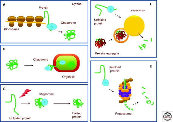
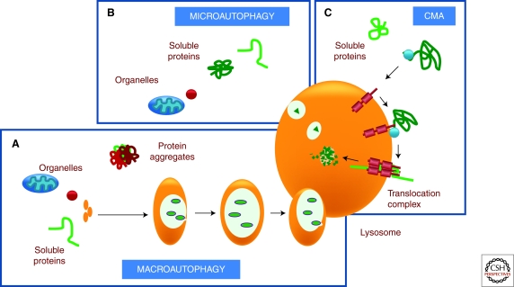
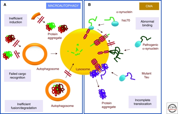
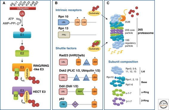
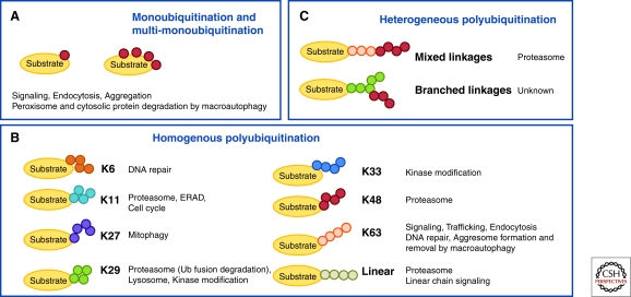
Cells maintain a healthy proteome through continuous evaluation of the quality of each of their proteins. Quality control requires the coordinated action of chaperones and proteolytic systems. Chaperones identify abnormal or unstable conformations in proteins and often assist them to regain stability. However, if repair is not possible, the aberrant protein is eliminated from the cellular cytosol to prevent undesired interactions with other proteins or its organization into toxic multimeric complexes. Autophagy and the ubiquitin/proteasome system mediate the complete degradation of abnormal protein products. In this article, we describe each of these proteolytic systems and their contribution to cellular quality control. We also comment on the cellular consequences resulting from the dysfunction of these systems in common human protein conformational disorders and provide an overview on current therapeutic interventions based on the modulation of the proteolytic systems.


Figures

Figure 1.
Figure 1.
Coordinate action of chaperones and the proteolytic systems in quality control. Chaperones assist in the folding of de novo synthesized proteins (A), unfolding and refolding of proteins as they traffic into cellular compartments (B), and in the refolding of proteins when damaged by cellular aggressors (C). Proteins that fail to fold can be eliminated from the cell by two proteolytic systems: autophagy (D) and the ubiquitin/proteasome system (E).
Figure 2.
Figure 2.
Autophagic pathways. Cytosolic proteins can reach the lysosomal lumen for degradation via autophagy through three different mechanisms. (A) In macroautophagy, a whole region of the cytosol is sequestered into a double membrane vesicle that fuses with lysosomes for cargo delivery. (B) In microautophagy, the lysosomal membrane invaginates to trap regions of the cytosol that are internalized into the lysosomal lumen as single membrane vesicles. (C) In chaperone-mediated autophagy, a targeting motif in the substrate proteins is recognized by a cytosolic chaperone that delivers it to the surface of the lysosome. Once there, the substrate protein binds to a lysosomal receptor that multimerizes to form a translocation complex. A luminal chaperone mediates the translocation of the substrate protein into the lumen where it is rapidly degraded.
Figure 3.
Figure 3.
The autophagic system in quality control. Autophagy contributes to the removal of both soluble cytosolic proteins and proteins organized into irreversible complexes or aggregates. Impairment of the autophagic system leads to the accumulation of damaged proteins in the form of protein inclusions. Failure of both macroautophagy (A) and chaperone-mediated autophagy (B) has been described to contribute to pathogenesis in different protein conformational disorders. Some of the steps described to be affected in each of the autophagic pathways are illustrated here.
Figure 4.
Figure 4.
Components of the ubiquitin/proteasome system. Substrates destined for proteasomal elimination are tagged with polymers of ubiquitin (Ub) through repeated sequential reactions catalyzed by ubiquitin activating (E1), conjugated (E2), and ligating (E3) enzymes (A) RING/RING-like E3 catalyzes the transfer of Ub directly from E2 to substrate whereas HECT E3 accepts activated Ub from E2 before transferring it to the substrate. Ubiquitinated substrates either bind directly to the ubiquitin receptors in the proteasome regulatory particle or shuttle to the proteasome by shuttle factors (B) * indicates domain binding polyubiquitin chain. Binding of substrate is followed by protein unfolding by the six ATPases forming the base of 19S regulatory particle, removal of polyubiquitin chain by deubiquitinating enzymes (DUBs) to regenerate free Ub, and translocation of the unfolded protein into the core proteolytic chamber, where it is cleaved into short peptides (C).
Figure 5.
Figure 5.
The ubiquitin code. The ubiquitin molecule can be attached to a single site (A) or multisites (B-C) on a substrate to yield mono- and multi-ubiquitination respectively. In addition, the ubiquitin sequence contains seven lysine residues that can support the assembly of polyubiquitin of different chain topologies. The plethora of ubiquitin linkages makes ubiquitination a highly versatile modification that serves diverse roles in the cell. Heterogeneous polyubiquitination (C) occurs when a ubiquitin chain has alternating linkage types (mixed linkages) or when a single ubiquitin is extended at two or more lysine residues (branched linkages).

References

    1. Adams J 2004. The development of proteasome inhibitors as anticancer drugs. Cancer Cell 5: 417–421 - PubMed
    1. Ahlberg J, Glaumann H 1985. Uptake–microautophagy–and degradation of exogenous proteins by isolated rat liver lysosomes. Effects of pH, ATP, and inhibitors of proteolysis. Exp Mol Pathol 42: 78–88 - PubMed
    1. Alves G, Forsaa EB, Pedersen KF, Dreetz Gjerstad M, Larsen JP 2008. Epidemiology of Parkinson's disease. J Neurol 255: 18–32 - PubMed
    1. Axe EL, Walker SA, Manifava M, Chandra P, Roderick HL, Habermann A, Griffiths G, Ktistakis NT 2008. Autophagosome formation from membrane compartments enriched in phosphatidylinositol 3-phosphate and dynamically connected to the endoplasmic reticulum. J Cell Biol 182: 685–701 - PMC - PubMed
    1. Baboshina OV, Haas AL 1996. Novel multiubiquitin chain linkages catalyzed by the conjugating enzymes E2EPF and RAD6 are recognized by 26S proteasome subunit 5. J Biol Chem 271: 2823–2831 - PubMed

Publication types

MeSH terms

LinkOut - more resources