. 2011 Jan;17(1):64-73.
doi: 10.1261/rna.2395211. Epub 2010 Nov 10.

Origin of the 1918 pandemic H1N1 influenza A virus as studied by codon usage patterns and phylogenetic analysis

Affiliations

Origin of the 1918 pandemic H1N1 influenza A virus as studied by codon usage patterns and phylogenetic analysis

Darisuren Anhlan et al. RNA. 2011 Jan.

Abstract

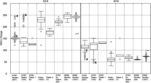
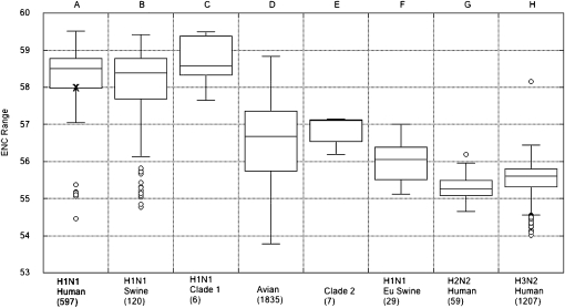
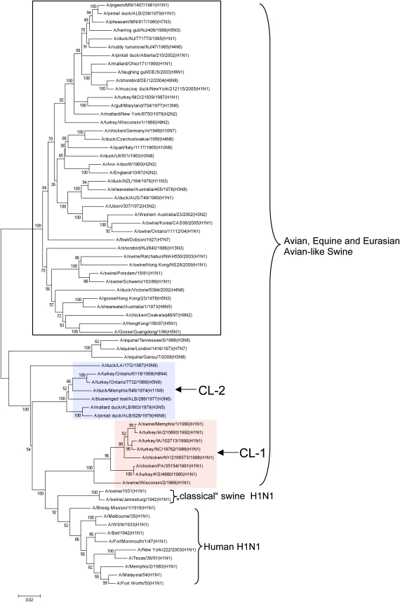
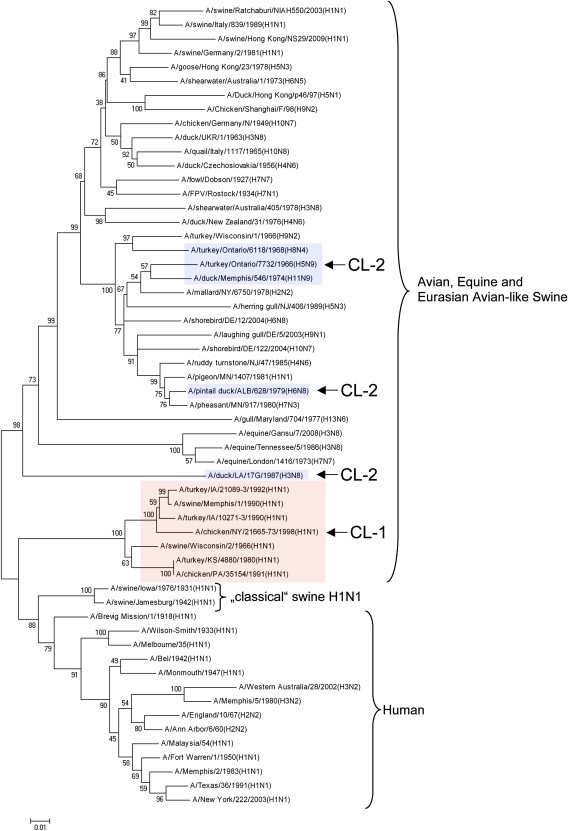
The pandemic of 1918 was caused by an H1N1 influenza A virus, which is a negative strand RNA virus; however, little is known about the nature of its direct ancestral strains. Here we applied a broad genetic and phylogenetic analysis of a wide range of influenza virus genes, in particular the PB1 gene, to gain information about the phylogenetic relatedness of the 1918 H1N1 virus. We compared the RNA genome of the 1918 strain to many other influenza strains of different origin by several means, including relative synonymous codon usage (RSCU), effective number of codons (ENC), and phylogenetic relationship. We found that the PB1 gene of the 1918 pandemic virus had ENC values similar to the H1N1 classical swine and human viruses, but different ENC values from avian as well as H2N2 and H3N2 human viruses. Also, according to the RSCU of the PB1 gene, the 1918 virus grouped with all human isolates and "classical" swine H1N1 viruses. The phylogenetic studies of all eight RNA gene segments of influenza A viruses may indicate that the 1918 pandemic strain originated from a H1N1 swine virus, which itself might be derived from a H1N1 avian precursor, which was separated from the bulk of other avian viruses in toto a long time ago. The high stability of the RSCU pattern of the PB1 gene indicated that the integrity of RNA structure is more important for influenza virus evolution than previously thought.


Figures

FIGURE 1.
FIGURE 1.
RSCU of threonine of the PB1 gene of influenza A viruses. The pattern is divided into human H1N1, H2N2, and H3N2, “classical” and European (Eu) swine H1N1 isolates, and avian influenza viruses, including clade 1 and 2 in respect to two synonymous codons of threonine: ACA and ACU, respectively. The numbers in the chamber shows estimated PB1 gene sequences. Only the full open reading frame (ORF) sequenced PB1 genes of all human H1N1, H2N2, H3N2, swine H1N1, and avian viruses of all subtypes found in GenBank were analyzed. The cross (x) indicates the RSCU value of the 1918 virus PB1 gene. The crosshatch (#) represents human and new swine-origin (SO) 2009 H1N1 reassortants, which had acquired their PB1 gene from human H3N2 viruses (for review, see Scholtissek 1998; Garten et al. 2009). The ampersand (&) shows outliers of Euroasian swine H1N1 viruses (Smith et al. 2009b) and H1N1 reassortant viruses isolated from pigs in the USA (Vincent et al. 2009) and six swine H1N1 reassortants from North America (Accession numbers [Acc. No]: EU409959, EU692905, EU692906, EU692907, EU409945, and GQ150326), bearing the PB1 gene of avian origin, respectively (see also, outliers in Fig. 2B). Clade 1 and clade 2 viruses were isolated from birds, but they are outliers of bird viruses. They have a distinct RSCU pattern. Phylogenetically, their PB1 gene is closely related to human H1N1 viruses (Table 1; Fig. 3). The H1N1 European swine viruses (Eu) are “avian-like” swine viruses (Fig. 3; Schultz et al. 1991). The asterisk (*) presents the A/Victoria/1968 (H3N2), a reassortant strain with a human H1N1 PB1 gene. The RSCU has been calculated according to Sharp et al. (1986). Each box encloses 50% of the data with the median value of the variable displayed as a line. The top and bottom of the box mark the data value located halfway between the median and the largest or the smallest data value and they define the limits of ±25% of the variable population. The lines extending from the top and bottom of each box mark the minimum and maximum values within the data set that fall within an acceptable range. Any value outside of this range, called an outlier, is displayed as an individual point. The outliers are defined as values, which are located outside the box by >150% of its size.
FIGURE 2.
FIGURE 2.
The ENC value of the PB1 gene of influenza A viruses. The cross (x) indicates the ENC value of the 1918 PB1 gene. The bulk of the PB1 gene of human H2N2 and H3N2 viruses as well as the H1N1 European swine viruses is of avian origin. The exceptional swine or human H1N1 viruses are “avian-like” swine viruses like those of the Euroasian lineage (Fig. 3), and eight human H1N1 reassortants (Accession numbers: DQ889683, CY019745, CY026417, CY021723, M38376, CY028730, CY021915, and AF342823), which contain an avian PB1 gene, respectively. The numbers in the chamber shows estimated PB1 gene sequences with full-length ORF. The ENC value is calculated according to Novembre (2002).
FIGURE 3.
FIGURE 3.
Phylogenetic tree of the PB1 genes of representative influenza viruses. Nucleotide sequences were aligned by using MUSCLE multiple sequence alignment program with default parameters (Edgar 2004) and constructed for the dendrogram using the neighbor-joining method (MEGA 4.0) (Tamura et al. 2007). Bootstrap values were estimated based on 1000 replicates and were given for all presented branches. A distance bar scale was shown under the tree. Clades (CL) are identified with different color backgrounds and with large letters as well as arrows, respectively.
FIGURE 4.
FIGURE 4.
Phylogenetic tree of the NP genes of representative influenza viruses. Nucleotide sequences were aligned and analyzed as described in the legend for Figure 3.

Similar articles

Cited by

References

    1. Antonovics J, Hood ME, Baker CH 2006. Molecular virology: Was the 1918 flu avian in origin? Nature 440: E9 doi: 10.1038/nature04824 - PubMed
    1. Bao Y, Bolotov P, Dernovoy D, Kiryutin B, Zaslavsky L, Tatusova T, Ostell J, Lipman D 2008. The influenza virus resource at the National Center for Biotechnology Information. J Virol 82: 596–601 - PMC - PubMed
    1. Benson DA, Karsch-Mizrachi I, Lipman DJ, Ostell J, Sayers EW 2009. GenBank. Nucleic Acids Res 37: D26–D31 doi: 10.1093/nar/gkp1024 - PMC - PubMed
    1. Chun J 1919. Influenza including its infection among pigs. Nat Med J China (Peking) 5: 34–44
    1. Cortazzo P, Cervenansky C, Marin M, Reiss C, Ehrlich R, Deana A 2002. Silent mutations affect in vivo protein folding in Escherichia coli. Biochem Biophys Res Commun 293: 537–541 - PubMed