. 2011 Feb;300(2):R330-9.
doi: 10.1152/ajpregu.00487.2010. Epub 2010 Nov 10.

Pathogen-induced heart rate changes associated with cholinergic nervous system activation

Affiliations

Pathogen-induced heart rate changes associated with cholinergic nervous system activation

Karen D Fairchild et al. Am J Physiol Regul Integr Comp Physiol. 2011 Feb.

Abstract

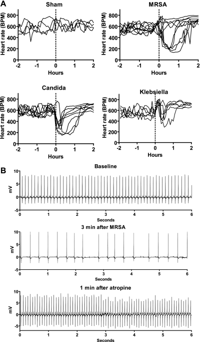
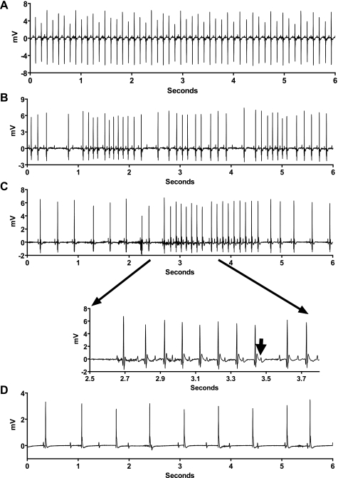
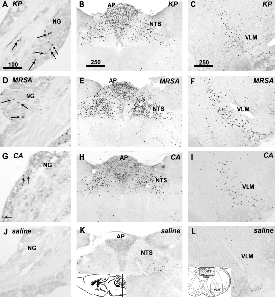
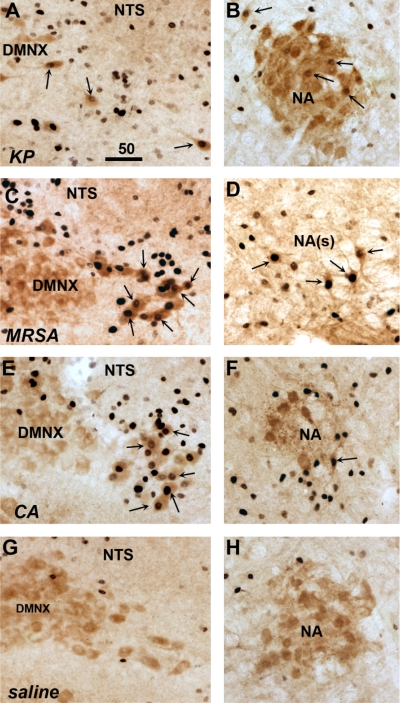
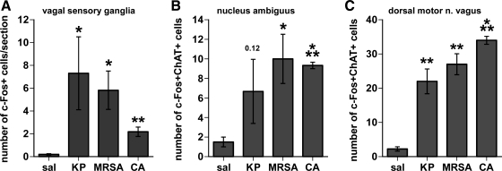
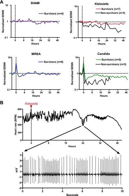
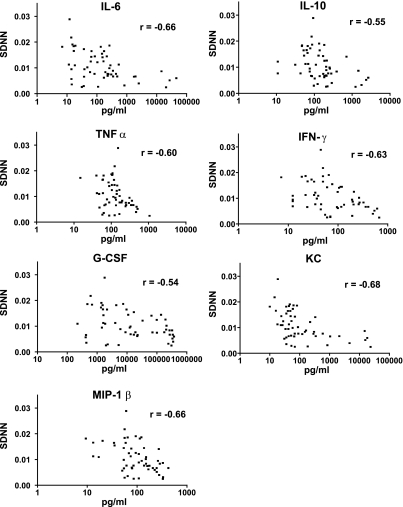
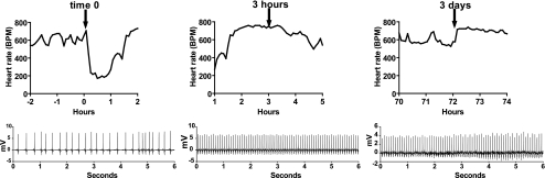
The autonomic nervous system plays a central role in regulation of host defense and in physiological responses to sepsis, including changes in heart rate and heart rate variability. The cholinergic anti-inflammatory response, whereby infection triggers vagal efferent signals that dampen production of proinflammatory cytokines, would be predicted to result in increased vagal signaling to the heart and increased heart rate variability. In fact, decreased heart rate variability is widely described in humans with sepsis. Our studies elucidate this apparent paradox by showing that mice injected with pathogens demonstrate transient bradyarrhythmias of vagal origin in a background of decreased heart rate variability (HRV). Intraperitoneal injection of a large inoculum of Gram-positive or Gram-negative bacteria or Candida albicans rapidly induced bradyarrhythmias of sinus and AV nodal block, characteristic of cardiac vagal firing and dramatically increased short-term HRV. These pathogen-induced bradycardias were immediately terminated by atropine, an antagonist of muscarinic cholinergic receptors, demonstrating the role of vagal efferent signaling in this response. Vagal afferent signaling following pathogen injection was demonstrated by intense nuclear c-Fos activity in neurons of the vagal sensory ganglia and brain stem. Surprisingly, pathogen-induced bradycardia demonstrated rapid and prolonged desensitization and did not recur on repeat injection of the same organism 3 h or 3 days after the initial exposure. After recovery from the initial bradycardia, depressed heart rate variability developed in some mice and was correlated with elevated plasma cytokine levels and mortality. Our findings of decreased HRV and transient heart rate decelerations in infected mice are similar to heart rate changes described by our group in preterm neonates with sepsis. Pathogen sensing and signaling via the vagus nerve, and the desensitization of this response, may account for periods of both increased and decreased heart rate variability in sepsis.


Figures

Fig. 1.
Fig. 1.
Pathogens induce bradycardia, which is terminated by atropine. A: heart rate (BPM, beats per min) of each mouse from 2 h before to 2 h after intraperitoneal injection of saline (sham n = 4) or high-inoculum pathogens at time 0, indicated by the dotted line (n = 6–9). B: atropine terminates pathogen-induced bradycardia. Six-second ECG tracings from a representative mouse at baseline, 3 min after intraperitoneal injection of methicillin-resistant Staphylococcus aureus (MRSA), and 1 min after injection of atropine. Similar results were obtained from mice injected with Klebsiella pneumoniae (KP), and Candida albicans (CA).
Fig. 2.
Fig. 2.
Electrocardiographic characteristics of pathogen-induced bradycardias. Representative ECG tracings within several minutes of injection of saline (A) or high-inoculum pathogens (BD). Second-degree sinus exit block (B) and type 1 second-degree AV node block (Wenckebach) (C) show sinus bradycardia or exit block and AV node Wenckebach (inset, lengthening PR intervals followed by nonconducted P wave indicated by the arrow). D: short epoch of complete AV node block. These arrhythmias occurred in all mice injected with CA, 7 of 9 mice injected with MRSA, and 4 of 6 mice injected with KP.
Fig. 3.
Fig. 3.
Pathogens rapidly activate vagal sensory neurons in the nodose ganglia and viscerosensory neurons in the caudal brain stem. Immunohistochemical staining for c-Fos was performed 90 min after inoculation with KP (AC), MRSA (DF), CA (GI) or saline (JL). c-Fos protein is seen as black staining in cell nuclei. All three pathogens induced c-Fos in vagal sensory neurons of the nodose ganglia (NG; A, D, G, J). Strong increases in c-Fos expression were also seen in caudal brain stem viscerosensory regions, including the area postrema (AP) and nucleus of the solitary tract (NTS) (B, E, H, K). The ventrolateral medulla also exhibited c-Fos staining following inoculation with each pathogen compared with sham (ventrolateral medulla, VLM; C, F, I, L). Sections shown are representative of findings in 3 mice in each group. Insets: level of brain stem section (K) and the regions of interest (L). Scale bars in AC indicate micrometers.
Fig. 4.
Fig. 4.
Pathogens activate cholinergic visceromotor neurons of the dorsal motor nucleus of the vagus (DMNX) and nucleus ambiguus (NA). Dual immunohistochemical staining was performed for choline acetyltransferase (ChAT, reddish-brown cytoplasm staining) and c-Fos protein (black nuclear staining) 90 min after inoculation of mice with KP (A and B), methicillin-resistant SA (MRSA; C and D), CA (E and F), or saline (G and H). Double-labeled neurons indicated by arrows are found in the dorsal motor nucleus of the vagus (DMNX; A, C, E, G) and the nucleus ambiguus (NA; B, D, F, H), both within compact and more caudal semicompact portions [NA(s)]. Saline injection resulted in few or no double-labeled neurons (G and H). Sections shown are representative of findings in 3 mice in each group. Scale bar in A indicates micrometers.
Fig. 5.
Fig. 5.
Quantitation of pathogen-induced c-Fos expression in vagal sensory and motor neurons. Graphs summarize the immunohistochemistry results from mice 90 min after intraperitoneal injection of saline (sal; n = 4) or pathogens (KP, MRSA, or CA, n = 3 each). c-Fos-positive cells were counted in vagal sensory neurons (A) and dual c-Fos and choline acetyltransferase (ChAT)-positive cells in cholinergic neurons in the nucleus ambiguus and the dorsal motor nucleus of the vagus (B, C). (*P < 0.05; **P < 0.005, ***P < 0.0005 vs. saline control).
Fig. 6.
Fig. 6.
Pathogens decrease and increase heart rate variability. A: decreased heart rate variability (HRV) is associated with mortality in sepsis. At time 0 (vertical dashed line), mice were injected with saline (SHAM), KP, MRSA, or CA. HRV (SDNN, standard deviation of normal R-R intervals) was normalized to the 4-h baseline prior to injection, denoted as “1” (horizontal dashed line). Average normalized SDNN from −4 h to +42 h relative to injection for surviving (colored lines) and nonsurviving mice (black lines) is shown (n = 8–10 per pathogen). B: sepsis induces both decreased HRV and transient HR decelerations. Heart rate of a mouse injected with Klebsiella at time 0 (red arrow) is shown. Note absence of the early bradycardic response with 106 colony-forming units of inoculum. At 18 h after injection, HRV was depressed, and at 24 h, there was a transient episode of recurrent heart rate decelerations (ECG tracing shown in inset) resulting in increased HRV. Subsequently, HR returned to normal but variability was decreased, and 40 h after injection, there was sinus bradycardia followed by death.
Fig. 7.
Fig. 7.
Cytokines are inversely correlated with HRV. Seven cytokines were assayed in plasma 18 h and 42 h after injection of saline (n = 4) or KP, MRSA, or CA (n = 8–10 each). Cytokine level (pg/ml, note logarithmic scale) is plotted against HRV (SDNN, ms). Spearman correlation coefficients “r” ranged from −0.54 to −0.68, as indicated.
Fig. 8.
Fig. 8.
Desensitization to pathogen-induced bradycardia. Heart rate responses to identical pathogen injections (arrows) at time 0, 3 h, and 3 days. Representative 6-s ECG tracings within 15 min of each injection are shown. Data are from a single mouse injected with MRSA and are representative of results from 6 mice injected with MRSA or CA (n = 3 each).

References

    1. American Physiological Society Guiding principles for research involving animals, and human beings. Am J Physiol Regul Integr Comp Physiol 283: R281–R283, 2002 - PubMed
    1. Akiho H, Khan WI, Al-Kaabi A, Blennerhassett P, Deng Y, Collins SM. Cytokine modulation of muscarinic receptors in the murine intestine. Am J Physiol Gastrointest Liver Physiol 293: G250–G255, 2007 - PubMed
    1. Barnes PJ, Haddad EB, Rousell J. Regulation of muscarinic M2 receptors. Life Sci 60: 1015–1021, 1997 - PubMed
    1. Borovikova LV, Ivanova S, Zhang M, Yang H, Botchkina GI, Watkins LR, Wang H, Abumrad N, Eaton JW, Tracey KJ. Vagus nerve stimulation attenuates the systemic inflammatory response to endotoxin. Nature 405: 458–462, 2000 - PubMed
    1. Cao H, Lake DE, Griffin MP, Moorman JR. Increased nonstationarity of neonatal heart rate before the clinical diagnosis of sepsis. Ann Biomed Eng 32: 233–244, 2004 - PubMed

Publication types

MeSH terms

LinkOut - more resources