. 2011 Jan;85(2):753-64.
doi: 10.1128/JVI.01835-10. Epub 2010 Nov 10.

Norovirus P particle, a novel platform for vaccine development and antibody production

Affiliations

Norovirus P particle, a novel platform for vaccine development and antibody production

Ming Tan et al. J Virol. 2011 Jan.

Abstract

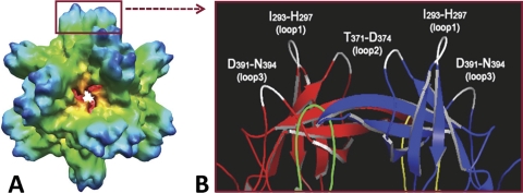
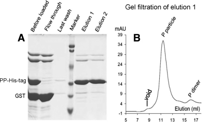
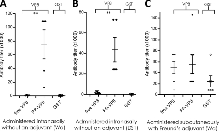
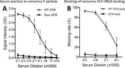
The norovirus P particle is an octahedral nanoparticle formed by 24 copies of the protrusion (P) domain of the norovirus capsid protein. This P particle is easily produced in Escherichia coli, extremely stable, and highly immunogenic. There are three surface loops per P domain, making a total of 72 loops per particle, and these are potential sites for foreign antigen presentation for immune enhancement. To prove this concept, a small peptide (His tag, 7 amino acids [aa]) and a large antigen (rotavirus VP8, 159 aa) were inserted into one of the loops. Neither insertion affects P particle formation, while both antigens were presented well on the P particle surface. The immune-enhancement effect of the P particle was demonstrated by significantly increased antibody titers induced by the P particle-presented antigens compared to the titers induced by free antigens. In addition, the measured neutralization antibody titers and levels of protection against rotavirus shedding in mice immunized with the VP8 chimeric P particles were significantly higher than those of mice immunized with the free VP8 antigen. Sera from P particle-VP8 chimera-vaccinated animals also blocked norovirus virus-like particle (VLP) binding to the histo-blood group antigen (HBGA) receptors. From these data, the P particle appears to be an excellent vaccine platform for antigen presentation. The readily available three surface loops and the great capacity for foreign antigen insertion make this platform attractive for wide application in vaccine development and antibody production. The P particle-VP8 chimeras may serve as a dual vaccine against both rotavirus and norovirus.


Figures

FIG. 1.
FIG. 1.
Norovirus P particle and its surface loops. (A) The structure of norovirus P particle of VA387 (GII.4) reconstructed by cryo-EM. (B) The distal end of a protrusion of the P particle is illustrated in a crystal structure (cartoon model), with the surface loops indicated. Red (P2 domain) and green (P1 domain) indicate one P protomer, while blue (P2 domain) and yellow (P1 domain) indicate the other protomer of a P dimer. Three surface loops of each P monomer that may be suitable sites for antigen insertion are shown in white.
FIG. 2.
FIG. 2.
Production and analysis of the P particle-His tag chimera. (A) Expression construct of the P particle-His tag chimera. The His tag was inserted in loops 2 between the N372 and D374 of the P domain. pGEX-4T-1 is an expression vector of the GST gene fusion system. The circled C represents a cysteine-containing peptide (CDCRGDCFC) at the C terminus of the P domain, which is to stabilize P particle formation (34). (B) The distal end of a protrusion of the P particle in the crystal structure (cartoon model) indicates the location of two residues N373 in loop 2 (dot models), where the inserted His tags are expected to be located. (C) Expression and purification of the P domain-His tag chimera. SDS-PAGE analysis revealed that the GST-P domain-His tag fusion protein (GST fusion) is ∼52 kDa. Digestion of the fusion protein in solution by thrombin resulted in GST (∼27 kDa) and the P domain-His tag chimera (PD-His-tag) (∼35 kDa) (left panel). The P domain-His tag chimera can also be released from the purification resin by thrombin digestion (right panel). M represents a prestained protein marker, with bands from top to bottom representing 113, 92, 50, 35, 29, and 21 kDa. (D) The elution curve of a gel filtration chromatography of the thrombin-released P-domain-His tag protein through the Superdex 200 size exclusion column. Three major peaks representing void, P particle-His tag, and P dimer-His tag were indicated, respectively. The sizes of these three peaks were calibrated with blue dextran 2000 (∼2,000 kDa; void), wild-type P particle (∼830 kDa), and wild-type P dimer (∼70 kDa), respectively. mAU, milli-absorbance units. (E) The fractions from gel filtration chromatography (see panel D) were analyzed by SDS-PAGE, and the fractions representing the three peaks are indicated.
FIG. 3.
FIG. 3.
Exposure of the His tag on the P particle (P particle-His tag binding on Talon resin). (A) The GST-P domain-His tag fusion protein was digested by thrombin, resulting in a mixture of the P particle-His tag chimera (PP-His-tag), GST, and other copurified proteins. These mixed proteins (“before loaded”) were loaded onto the Talon resin, and the flowthrough contained all proteins except the P particle-His tag chimera. After being washed (last wash), the P particle-His tag chimera was eluted (elutions 1 and 2) by 250 mM imidazole. “Marker” represents a prestained protein standard, with bands from top to bottom representing 107, 81, 49, 33, 27, and 20 kDa. (B) The elution curve of gel filtration chromatography of the eluted protein (elution 1 of panel A) through the Superdex 200 size exclusion column, indicating that vast majority (>98%) of the eluted protein formed chimeric P particles as shown by a defined peak at ∼840 kDa. The gel filtration column was calibrated as described in the legend to Fig. 2.
FIG. 4.
FIG. 4.
Antibody responses of mice to the P particle-presented His tag. (A) Immune reactivity of mouse sera (5 mice) after immunization with equal molar amounts of the P particle-His tag chimera (PP-His), P dimer-His tag chimera (PD-His), free 7×His peptide, and wild-type P particle (WT PP) to recombinant His-tagged-α-fucosidase of T. maritima (His-tag) in EIAs. (B) Antibody titers of the sera described for panel A were determined by an end-point dilution approach. Microtiter plates were coated with antigens at 5 ng/μl for EIAs. Sera at indicated dilutions were used to measure immune reactivity. *, P < 0.05.
FIG. 5.
FIG. 5.
Production and analysis of the P particle-VP8 chimera. (A) The P particle-VP8 chimera expression construct based on plasmid pGEX-4T-1 containing the P domain-encoding cDNA sequences. The rotavirus (Wa) VP8 antigen was inserted in loops 2 of the P domain between T368 and L375 through a cloning cassette with enzyme sites SpeI and ClaI/EcoRV. The circled C represents the cysteine-containing peptide (CDCRGDCFC). (B) Expression and purification of the P particle-VP8 chimera. An SDS-PAGE analysis revealed that the GST-P-VP8 fusion protein (GST fusion) is ∼80 kDa (left panel). Digestion of the fusion protein by thrombin results in GST (∼27 kDa) and the P-VP8 chimera (∼52 kDa) (middle panel). The free P-VP8 chimera can also be released from the purification resin by a thrombin digestion (right panel). (C) The elution curve of the gel filtration chromatography of the thrombin-released P-VP8 protein from panel B through the Superdex 200 size exclusion column. An SDS-PAGE analysis of the fractions of the peaks is shown at the top. The single major peak near the void volume indicated that almost all P-VP8 protein formed chimeric P particles. The column was calibrated with blue dextran 2000 (∼2,000 kDa; void), wild-type P particles (∼830 kDa), and wild-type P dimer (∼70 kDa). M, marker. (D) The P-VP8 protein (left panel) reacted to antibodies against rotavirus VP8 (middle panel) and norovirus VLPs (right panel) in a Western blot analysis. In panels B and D, lanes M represent prestained protein markers, with bands from top to bottom representing 113, 92, 50, 35, 29, and 21 kDa.
FIG. 6.
FIG. 6.
Cryo-EM structure of the P particle-VP8 chimera. (A) A wild-type P particle. (B) A P particle-VP8 chimera. Compared to the wild-type P particle, the chimera shows extended protrusions with nicks in the middle, suggesting the boundary between the P2 subdomain and the inserted VP8 antigen. The radii of the particles in panels A and B are indicated by the same color schemes. (C) Fitting of two copies of the crystal structures (green and blue in the cartoon model) of the rotavirus (Wa) VP8 antigen into the density map of the extended protrusion of the chimera (transparent gray), confirming the exposure of the VP8 antigen on the chimeric P particle. Enlarged side (D) and top (E) views of a protrusion of the P particle-VP8 chimera are shown.
FIG. 7.
FIG. 7.
Immune responses of mice to P particle-presented VP8s. Equal molar amounts of the P particle-VP8 chimera and free VP8 were used to immunize mice, either intranasally without an adjuvant (5 to 7 mice; panels A and B) or subcutaneously with Freund's adjuvant (6 or 7 mice; panel C). Free VP8 and GST were used as antigens for antibody titer determination in EIAs. (A and B) Anti-VP8 and -GST antibody titers of mouse sera after immunization with free VP8 antigen (free VP8) and the chimeric P particles (PP-VP8) containing VP8s of Wa (A) and DS1 (B). (C) Anti-VP8 antibody titers after immunization with free VP8 antigen (free VP8) and the chimeric P particle (PP-VP8) containing VP8 of the Wa strain. In all experiments, an equal molar amount of GST in the immunogens served as an internal control. **, P < 0.01.
FIG. 8.
FIG. 8.
Neutralization of rotavirus by mouse sera after immunization with P particle-VP8 chimeras. (A) Mouse sera after intranasal immunization with a P particle-VP8 (Wa, P[8]) chimera without an adjuvant show strong neutralization of the same Wa strain (PP-VP8 sera), while sera after immunization with free VP8 show a significantly lower level of neutralization (VP8 sera). Sera from mice receiving no antigen served as negative controls (control sera). (B) Mouse sera after immunization with the P particle-VP8 (DS1, P[4]) chimera show weak cross-neutralization of Wa (PP-VP8 sera), whereas sera after immunization with free VP8 of DS1 (VP8 sera) and the negative-control sera did not show this cross-neutralization. (C) Immune reactivity against VP8 of sera from mice immunized with free Wa VP8 (VP8 sera) and P particle-VP8 (Wa) chimera (PP-VP8 sera) subcutaneously with Freund's adjuvant. The two antigens induced similar antibody responses. Sera from mice receiving no antigen served as negative controls (control sera). (D) The sera from panel C after immunization with the P particle-VP8 chimera showed significantly higher rotavirus (Wa) neutralization titers (PP-VP8 sera) than sera induced by immunization with free VP8 (VP8 sera). Control sera from mice receiving no antigen served as a negative control (control sera). Asterisks indicate the P values between the neutralization levels of the sera after immunization with the two forms of VP8. *, P < 0.05; **, P < 0.01; ***, P < 0.001.
FIG. 9.
FIG. 9.
Protection after immunization of mice with P particle-VP8 chimeric vaccine against a mouse rotavirus infection. (A) Antibody responses of mice to free murine rotavirus VP8 after vaccination with free (free mVP8) and P particle-presented (PP-mVP8) murine rotavirus VP8. The wild-type P particle (WT PP) served as a vector control. **, P < 0.01. (B) Rotavirus shedding (μg/ml) by mice was measured after vaccination with four different vaccines and challenged by murine rotavirus (EDIM). WT PP, mice vaccinated with the wild-type norovirus P particle (vector control; n = 7); PP-hVP8, mice vaccinated with the P particle-VP8 (Wa) chimera (n = 5); free mVP8, mice vaccinated with free murine VP8 (EDIM) antigen (n = 5); PP-mVP8, mice vaccinated with the P particle-VP8 (EDIM) chimera (n = 5). Results of data calculation and statistic analysis are shown in Table 1.
FIG. 10.
FIG. 10.
Mouse sera after immunization with P particle-VP8 chimeras block binding of norovirus VLPs to HBGA receptors. (A) Mouse sera after immunization with the P particle-VP8 (Wa) chimeras reacted strongly to norovirus P particle (PP-VP8), while sera after immunization with the free VP8 did not show this reactivity (free VP8). (B) Mouse sera from panel A blocked binding of norovirus VLPs to HBGA receptors (type A saliva, PP-VP8 sera), while sera after immunization with free VP8 did not show this blockade (VP8 sera).

Similar articles

Cited by

References

    1. Bertolotti-Ciarlet, A., L. J. White, R. Chen, B. V. Prasad, and M. K. Estes. 2002. Structural requirements for the assembly of Norwalk virus-like particles. J. Virol. 76:4044-4055. - PMC - PubMed
    1. Blanchard, H., X. Yu, B. S. Coulson, and M. von Itzstein. 2007. Insight into host cell carbohydrate-recognition by human and porcine rotavirus from crystal structures of the virion spike associated carbohydrate-binding domain (VP8*). J. Mol. Biol. 367:1215-1226. - PubMed
    1. Cao, S., Z. Lou, M. Tan, Y. Chen, Y. Liu, Z. Zhang, X. C. Zhang, X. Jiang, X. Li, and Z. Rao. 2007. Structural basis for the recognition of blood group trisaccharides by norovirus. J. Virol. 81:5949-5957. - PMC - PubMed
    1. Chatterji, A., L. L. Burns, S. S. Taylor, G. P. Lomonossoff, J. E. Johnson, T. Lin, and C. Porta. 2002. Cowpea mosaic virus: from the presentation of antigenic peptides to the display of active biomaterials. Intervirology 45:362-370. - PubMed
    1. Chatterji, A., W. Ochoa, L. Shamieh, S. P. Salakian, S. M. Wong, G. Clinton, P. Ghosh, T. Lin, and J. E. Johnson. 2004. Chemical conjugation of heterologous proteins on the surface of Cowpea mosaic virus. Bioconjug. Chem. 15:807-813. - PubMed

Publication types