. 2011 Jan;85(2):715-24.
doi: 10.1128/JVI.01742-10. Epub 2010 Nov 10.

Oxidative stress induces reactivation of Kaposi's sarcoma-associated herpesvirus and death of primary effusion lymphoma cells

Affiliations

Oxidative stress induces reactivation of Kaposi's sarcoma-associated herpesvirus and death of primary effusion lymphoma cells

Xudong Li et al. J Virol. 2011 Jan.

Abstract

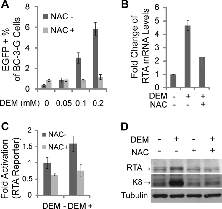
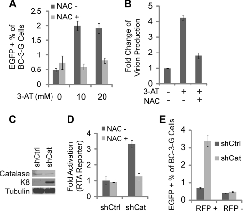
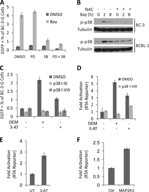
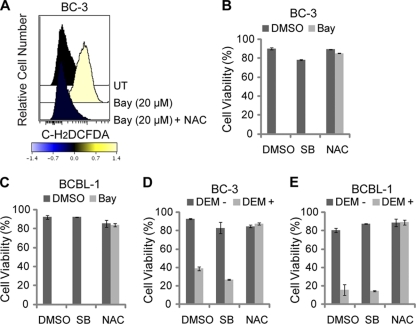
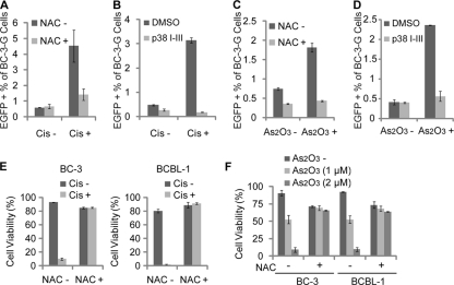
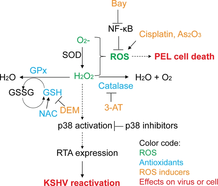
Kaposi's sarcoma (KS) and primary effusion lymphoma (PEL) cells are predominantly infected with latent Kaposi's sarcoma-associated herpesvirus (KSHV), presenting a barrier to the destruction of tumor cells. Latent KSHV can be reactivated to undergo lytic replication. Here we report that in PEL cells, oxidative stress induced by upregulated reactive oxygen species (ROS) can lead to KSHV reactivation or cell death. ROS are upregulated by NF-κB inhibition and are required for subsequent KSHV reactivation. Disruption of the intracellular redox balance through depletion of the antioxidant glutathione or inhibition of the antioxidant enzyme catalase also induces KSHV reactivation, suggesting that hydrogen peroxide induces reactivation. In addition, p38 signaling is required for KSHV reactivation induced by ROS. Furthermore, treatment of PEL cells with a higher concentration of the NF-κB inhibitor than that used for inducing KSHV reactivation further upregulates ROS and induces massive cell death. ROS, but not p38 signaling, are required for PEL cell death induced by NF-κB inhibition as well as by glutathione depletion. Importantly, anticancer drugs, such as cisplatin and arsenic trioxide, also induce KSHV reactivation and PEL cell death in a ROS-dependent manner. Our study thus establishes a critical role for ROS and oxidative stress in the regulation of KSHV reactivation and PEL cell death. Disrupting the cellular redox balance may be a potential strategy for treating KSHV-associated lymphoma.


Figures

FIG. 1.
FIG. 1.
Inhibition of NF-κB induces ROS-dependent KSHV reactivation. (A) Fluorescence-activated cell sorter (FACS) analysis of c-H2DCFDA staining, showing ROS levels in BC-3 and BCBL-1 cells untreated (UT) or treated with 5 μM Bay 11-7082 (Bay) for 2 h in the presence or absence of 10 mM NAC added 1 h prior to Bay treatment. (B) Cell viability measured by trypan blue exclusion assay of BC-3 cells treated with 0, 5, or 10 μM Bay for 24 h in the presence of conditioned medium or fresh medium. (C) KSHV reactivation measured by FACS analysis of EGFP expression in BC-3-G cells untreated or treated with 5 μM Bay for 48 h in the presence or absence of 10 mM NAC added 1 h prior to Bay treatment. (D) KSHV reactivation measured by FACS analysis of EGFP expression in BC-3-G cells untreated or treated with 5 ng/ml of TPA or 0.2 mM NaB for 48 h in the presence or absence of 10 mM NAC added 1 h prior to Bay treatment. (E) KSHV RTA promoter activity measured by a luciferase reporter assay in BC-3 cells treated with 5 μM Bay or its vehicle dimethyl sulfoxide (DMSO) for 48 h in the presence or absence of 10 mM NAC added 1 h prior to Bay treatment. (F) Western blotting with anti-K8 and anti-α-tubulin antibodies of cell lysates of BC-3 and BCBL-1 cells treated with 5 μM Bay or its vehicle DMSO for 48 h in the presence or absence of 10 mM NAC added 1 h prior to Bay treatment. (G) Relative virion production by BCBL-1 cells treated with 5 μM Bay or its vehicle DMSO for 72 h in the presence or absence of 10 mM NAC added 1 h prior to Bay treatment. For panels A and F, data are representative of three independent experiments. For panels B, C, D, E, and G, data are the means ± standard deviations from three independent experiments.
FIG. 2.
FIG. 2.
Glutathione depletion induces ROS-dependent reactivation of KSHV. (A) KSHV reactivation measured by FACS analysis of EGFP expression in BC-3-G cells untreated or treated with 0.05, 0.1, or 0.2 mM DEM for 48 h in the presence or absence of 10 mM NAC added 1 h prior to DEM treatment. (B) RT-Q-PCR analysis of RTA transcripts in BC-3 cells untreated or treated with 0.1 mM DEM for 48 h in the presence or absence of 10 mM NAC pretreatment. (C) KSHV RTA promoter activity measured by a luciferase reporter assay in BC-3 cells untreated or treated with 0.1 mM DEM for 48 h in the presence or absence of 10 mM NAC pretreatment. (D) Western blotting with anti-RTA, anti-K8, and anti-α-tubulin antibodies of cell lysates of BCBL-1 cells untreated or treated 0.1 mM DEM for 48 h in the presence or absence of 10 mM NAC pretreatment. For panel D, data are representative of three independent experiments. For panels A to C, data are the mean ± standard deviations from three independent experiments.
FIG. 3.
FIG. 3.
Inhibition or depletion of catalase induces ROS-dependent reactivation of KSHV. (A) KSHV reactivation measured by FACS analysis of EGFP expression in BC-3-G cells untreated or treated with 10 or 20 mM 3-AT for 48 h in the presence or absence of 10 mM NAC pretreatment. (B) Relative virion production by BC-3 cells untreated or treated with 10 mM 3-AT for 72 h in the presence or absence of 10 mM NAC pretreatment. (C) Western blotting with anti-catalase, anti-K8, and anti-α-tubulin antibodies of cell lysates of BC-3 cells transfected with the shRNA plasmid targeting catalase (shCat) or the control shRNA plasmid (shCtrl). Transfected cells were selected with 3 μg/ml of puromycin at 24 h posttransfection and collected for analysis at 72 h posttransfection. (D) KSHV RTA promoter activity measured by a luciferase reporter assay in BC-3 cells transfected with shCat or shCtrl. Cells were untreated or treated with 10 mM NAC at 12 h posttransfection and collected for analysis at 60 h posttransfection. (E) KSHV reactivation measured by FACS analysis of EGFP expression in RFP-positive and RFP-negative BC-3-G cells cotransfected with an RFP expression plasmid and shCat or shCtrl at 72 h posttransfection. For panel C, data are representative of three independent experiments. For panels A, B, D, and E, data are the means ± standard deviations from three independent experiments.
FIG. 4.
FIG. 4.
Requirement of p38 signaling for KSHV reactivation induced by ROS. (A) KSHV reactivation measured by FACS analysis of EGFP expression in BC-3-G cells treated with 5 μM Bay or DMSO vehicle control for 48 h in the presence or absence of pretreatment with 20 μM MEK inhibitor PD 98059 (PD), 20 μM p38 inhibitor SB 203580 (SB), PD and SB, or DMSO vehicle control. (B) Western blotting with anti-phospho-p38 (Thr180/Tyr182) and anti-α-tubulin antibodies of cell lysates of BC-3 and BCBL-1 cells untreated or treated with 5 μM Bay for 2 or 8 h in the presence or absence of 10 mM NAC pretreatment. (C) KSHV reactivation measured by FACS analysis of EGFP expression in BC-3-G cells untreated or treated with 0.1 mM DEM or 10 mM 3-AT for 48 h in the presence or absence of pretreatment with 20 μM p38 inhibitor III (p38 I-III), 40 μM p38 inhibitor VIII (p38 I-VIII), or DMSO vehicle control. (D) KSHV RTA promoter activity measured by a luciferase reporter assay in BC-3 cells untreated or treated with 0.1 mM DEM or 10 mM 3-AT for 48 h in the presence of 40 μM p38 I-VIII or DMSO pretreatment. (E) KSHV RTA promoter activity measured by a luciferase reporter assay in 293T cells untreated or treated with 20 mM 3-AT for 24 h. (F) KSHV RTA promoter activity measured by a luciferase reporter assay in 293T cells transfected with a MAP2K3 expression plasmid or a control plasmid. For panel B, data are representative of two independent experiments. For panels A, C, D, E, and F, data are the means ± standard deviations from three independent experiments.
FIG. 5.
FIG. 5.
ROS induce PEL cell death. (A) FACS analysis of c-H2DCFDA staining, showing ROS levels in BC-3 cells untreated (UT) or treated with 20 μM Bay for 2 h in the presence or absence of 10 mM NAC added 1 h prior to Bay treatment. Data are representative of three independent experiments. (B and C) Cell viability measured by trypan blue exclusion assay of BC-3 cells (B) and BCBL-1 cells (C) treated with 20 μM Bay or DMSO vehicle control for 48 h in the presence of pretreatment with 10 mM NAC, 20 μM SB, or DMSO vehicle control. (D and E) Cell viability measured by trypan blue exclusion assay of BC-3 cells (D) and BCBL-1 cells (E) untreated or treated with 0.25 mM DEM for 72 h in the presence of pretreatment with 10 mM NAC, 20 μM SB, or DMSO vehicle control. For panels B to E, data are the means ± standard deviations from three independent experiments.
FIG. 6.
FIG. 6.
Cisplatin and arsenic trioxide induce KSHV reactivation and PEL cell death in a ROS-dependent manner. (A) KSHV reactivation measured by FACS analysis of EGFP expression in BC-3-G cells untreated or treated with 4.1 μg/ml of cisplatin for 48 h in the presence or absence of 10 mM NAC pretreatment. (B) KSHV reactivation measured by FACS analysis of EGFP expression in BC-3-G cells untreated or treated with 4.1 μg/ml of cisplatin for 48 h in the presence or absence of pretreatment with 20 μM p38 inhibitor III or DMSO vehicle control. (C) KSHV reactivation measured by FACS analysis of EGFP expression in BC-3-G cells untreated or treated with 1 μM arsenic trioxide for 48 h in the presence or absence of 10 mM NAC pretreatment. (D) KSHV reactivation measured by FACS analysis of EGFP expression in BC-3-G cells untreated or treated with 0.5 μM arsenic trioxide for 48 h in the presence or absence of pretreatment with 20 μM p38 inhibitor III or DMSO vehicle control. (E) Cell viability measured by trypan blue exclusion assay of BC-3 and BCBL-1 cells untreated or treated with 4.1 μg/ml of cisplatin for 72 h in the presence or absence of pretreatment with 10 mM NAC. (F) Cell viability measured by trypan blue exclusion assay of BC-3 and BCBL-1 cells untreated or treated with 1 μM or 2 μM arsenic trioxide for 48 h in the presence or absence of pretreatment with 10 mM NAC. Data are the means ± standard deviations from three independent experiments.
FIG. 7.
FIG. 7.
Model of the regulation of KSHV reactivation and PEL cell death by oxidative stress. Cellular antioxidant systems protect cells from oxidative stress by converting reactive oxygen species such as superoxide (O2) and hydrogen peroxide (H2O2) into H2O and O2. These antioxidant systems include superoxide dismutase (SOD), glutathione peroxidase (GPx), and catalase (CAT). SOD converts O2 into H2O2, which can be further converted into H2O and O2 by catalase or into H2O by GPx. Glutathione (GSH) provides the reducing potential for GPx to reduce H2O2 to H2O, while being oxidized into an oxidized form (GSSG). NAC functions as an antioxidant by increasing intracellular concentrations of GSH. Oxidative stress resulting from inhibition of NF-κB or inhibition of various other intracellular antioxidant systems leads to p38 activation, which enhances RTA expression and ultimately reactivation of KSHV. Oxidative stress can also lead to death of primary effusion lymphoma cells.

Similar articles

Cited by

References

    1. Ambinder, R. F., K. D. Robertson, S. M. Moore, and J. Yang. 1996. Epstein-Barr virus as a therapeutic target in Hodgkin's disease and nasopharyngeal carcinoma. Semin. Cancer Biol. 7:217-226. - PubMed
    1. Bellare, P., and D. Ganem. 2009. Regulation of KSHV lytic switch protein expression by a virus-encoded microRNA: an evolutionary adaptation that fine-tunes lytic reactivation. Cell Host Microbe 6:570-575. - PMC - PubMed
    1. Brander, C., T. Suscovich, Y. Lee, P. T. Nguyen, P. O'Connor, J. Seebach, N. G. Jones, M. van Gorder, B. D. Walker, and D. T. Scadden. 2000. Impaired CTL recognition of cells latently infected with Kaposi's sarcoma-associated herpes virus. J. Immunol. 165:2077-2083. - PubMed
    1. Brinkmann, M. M., M. Glenn, L. Rainbow, A. Kieser, C. Henke-Gendo, and T. F. Schulz. 2003. Activation of mitogen-activated protein kinase and NF-κB pathways by a Kaposi's sarcoma-associated herpesvirus K15 membrane protein. J. Virol. 77:9346-9358. - PMC - PubMed
    1. Brown, H. J., M. J. Song, H. Deng, T. T. Wu, G. Cheng, and R. Sun. 2003. NF-κB inhibits gammaherpesvirus lytic replication. J. Virol. 77:8532-8540. - PMC - PubMed

Publication types

MeSH terms

LinkOut - more resources