. 2011 Jan;85(2):853-64.
doi: 10.1128/JVI.01873-10. Epub 2010 Nov 10.

Noncytotoxic inhibition of viral infection through eIF4F-independent suppression of translation by 4EGi-1

Affiliations

Noncytotoxic inhibition of viral infection through eIF4F-independent suppression of translation by 4EGi-1

Robert McMahon et al. J Virol. 2011 Jan.

Abstract

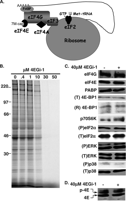
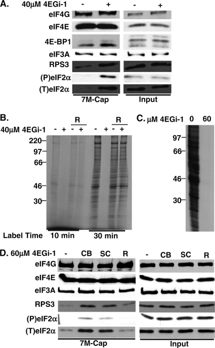
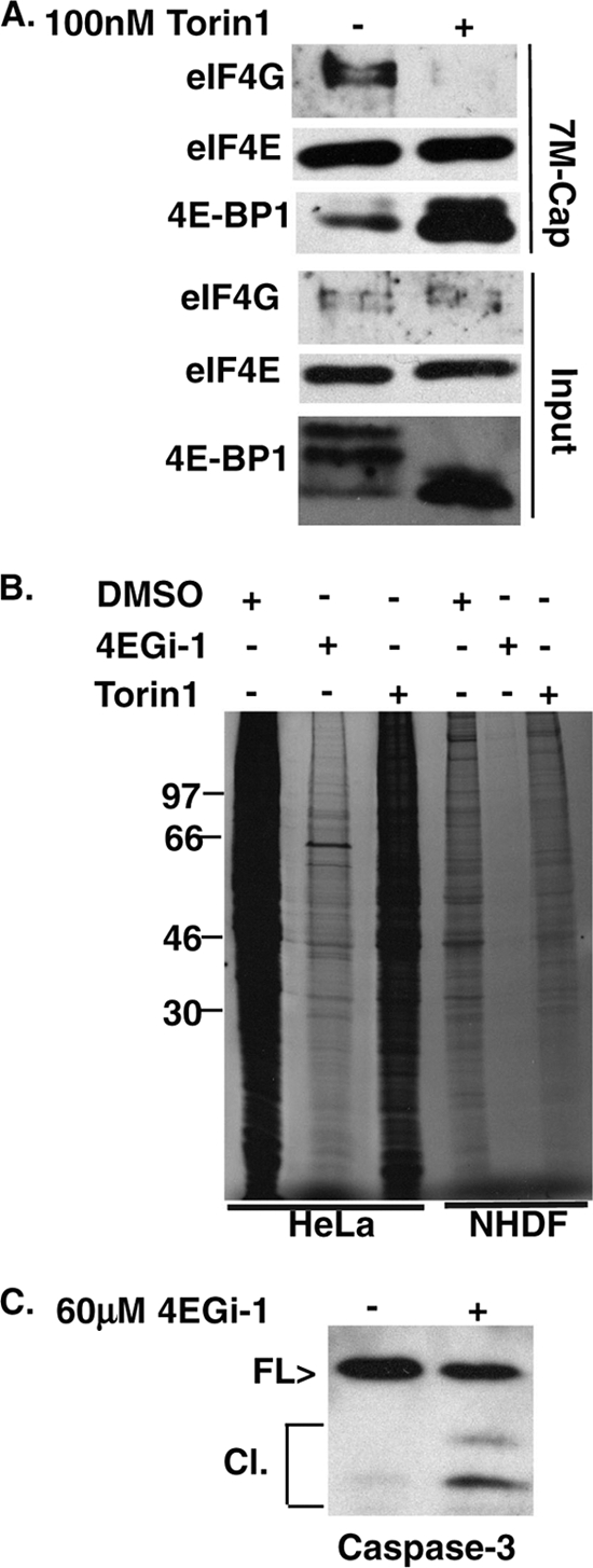
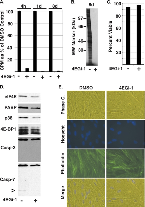
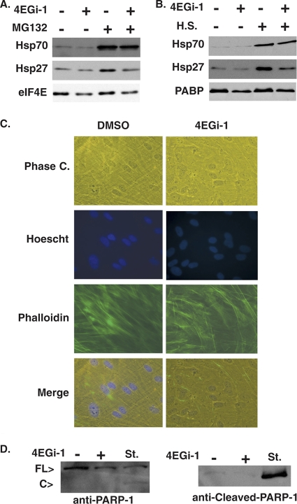
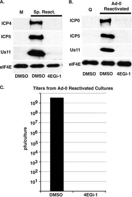
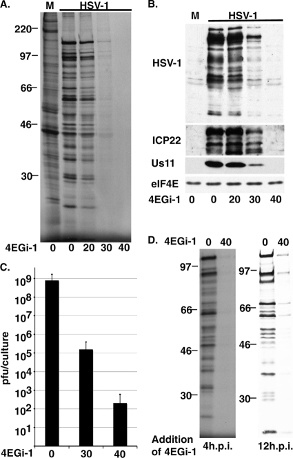
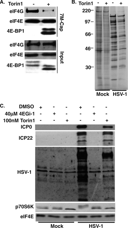
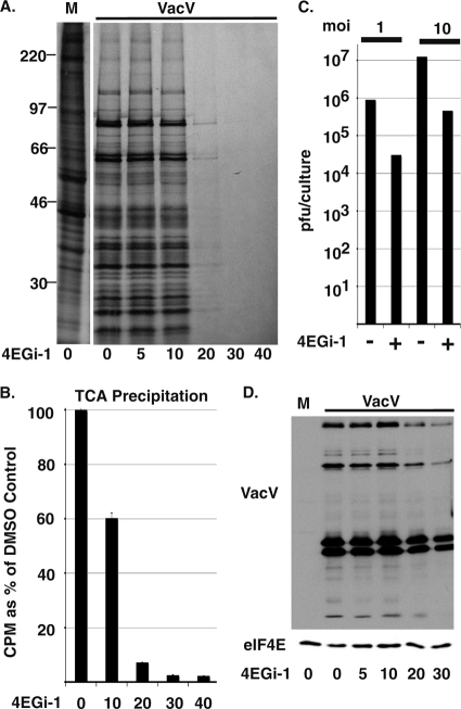
The eukaryotic initiation factor eIF4F recruits ribosomes to capped mRNAs while eIF2 mediates start codon recognition to initiate protein synthesis. Increasing interest in targeting translation to suppress tumor growth has led to the development of new classes of inhibitors, including 4EGi-1, which disrupts eIF4F complexes. However, the full effects of this inhibitor and its potential uses in the treatment of other disease states remain unclear. Here, we show that overall rates of protein synthesis in primary human cells were affected only modestly by eIF4F disruption using the mTOR inhibitor Torin1, yet were highly sensitive to 4EGi-1. Translational suppression occurred even at concentrations of 4EGi-1 that were below those required to significantly alter eIF4F levels but were instead found to increase the association of ribosomal complexes containing inactive eIF2α. Although highly stable in culture, the effects of 4EGi-1 on both cellular protein synthesis and ribosome association were readily reversible upon inhibitor removal. In addition, despite potently inhibiting translation, prolonged exposure to 4EGi-1 had only modest effects on cell morphology and protein abundance without affecting viability or stress tolerance to any significant degree, although differential effects on heat shock protein (hsp) expression highlighted distinct 4EGi-1-sensitive modes of hsp induction. In contrast, 4EGi-1 potently suppressed poxvirus replication as well as both reactivation and lytic phases of herpesvirus infection. These findings identify a novel way in which 4EGi-1 affects the host cell's protein synthesis machinery and demonstrate its potential as a noncytotoxic inhibitor of diverse forms of viral infection.


Figures

FIG. 1.
FIG. 1.
Inhibition of translation in primary human fibroblasts by 4EGi-1. (A) Regulation of ribosome recruitment by cellular translation initiation factors. The core eIF4F complex consists of eIF4E, which binds the 7-methyl-GTP (7M-cap) present at the 5′ end of the mRNA; eIF4G, the central scaffolding protein; and eIF4A, an RNA helicase. eIF4G also associates with the multifactor complex eIF3, which bridges the cap-binding complex and the 43S ribosome to form the 48S initiation complex. The 43S ribosome is formed through the interaction of the 40S ribosome and a ternary complex consisting of the multisubunit initiation factor, eIF2, together with GTP and the initiator Met tRNA (fork symbol). Finally, additional interactions between eIF4G, PABP, and the poly(A) tail are thought to mediate circularization of the mRNA. (B) Effects of 4EGi-1 on rates of translation in primary cells. NHDFs were treated for 3 h with increasing micromolar concentrations of 4EGi-1 and then metabolically labeled with [35S]Met/Cys for 1 h. Whole-cell extracts were resolved by SDS-PAGE, and then fixed, dried gels were exposed to X-ray film. Migration of molecular weight (MW) standards is indicated to the left of the autoradiogram. (C) NHDFs were treated for 4 h with DMSO or 40 μM 4EGi-1, and then whole-cell extracts were analyzed by Western blotting with the indicated antibodies. Total levels of 4E-BP1 (T) were examined using nonresolving 7.5% gels, while phosphorylated forms of 4E-BP1 were resolved (R) in 17.5% gels. Phosphorylation of p70S6K is evident as retarded mobility in 12.5% gels. (D) Samples described for panel C were fractionated by isoelectric focusing, and membranes were probed with anti-eIF4E antibody. Migration of the phosphorylated (p-4E) and hypophosphorylated (4E) forms of eIF4E are indicated to the left.
FIG. 2.
FIG. 2.
Reversible effects of 4EGi-1 on both the composition of translation initiation complexes and rates of translation. (A) NHDFs were treated for 4 h with DMSO or 40 μM 4EGi-1, and then soluble cell extracts were subjected to 7-methyl-GTP chromatography. Cap-bound and input samples were analyzed by Western blotting with the indicated antibodies. (P), phosphorylated; (T), total. 4E-BP1 was examined using 17.5% gels to resolve phosphorylated species. (B) NHDFs were treated with DMSO (−) or 40 μM 4EGi-1 (+) for 4 h. Cells were then rinsed in growth medium containing DMSO or 4EGi-1 and metabolically labeled for 10 min or 30 min in the continued presence of inhibitor (lanes 1 to 2 and 5 to 6) or rinsed in medium containing DMSO to remove (R) inhibitor and labeled in the presence of DMSO alone (lanes 3 to 4 and 7 to 8). Whole-cell extracts were resolved by SDS-PAGE and then fixed, dried gels were exposed to X-ray film. Migration of molecular weight (MW) standards is indicated to the left of the autoradiogram. (C) HeLa cells were treated with DMSO solvent control (0) or 60 μM 4EGi-1 for 3 h and then labeled with [35S]Met/Cys for 1 h. Whole-cell extracts were prepared and resolved by SDS-PAGE and fixed, dried gels were exposed to X-ray film. Migration of MW markers is indicated to the left. (D) HeLa cells were treated with DMSO (−) or 60 μM 4EGi-1 for 4 h. Independent sources of 4EGi-1 from Calbiochem (CB) and Santa Cruz Biotechnology (SC) were compared. An additional sample was prepared from cells treated with 60 μM 4EGi-1, but the inhibitor was removed (R) by omitting it from wash buffers at the end of the assay to examine reversibility. Samples were resolved by SDS-PAGE and analyzed by Western blotting with the indicated antibodies.
FIG. 3.
FIG. 3.
4EGi-1 and the mTORC inhibitor Torin1 have distinct effects on rates of translation. (A) NHDFs were treated with DMSO (−) or 100 nM Torin1 (+) for 1 day, and then eIF4E and associated proteins were recovered from soluble cell extracts on 7-methyl-GTP-Sepharose and analyzed by Western blotting with the indicated antibodies. 4E-BP1 was resolved in 17.5% SDS-PAGE gels to separate phosphorylated species. (B) NHDFs or HeLa cells were treated with DMSO, 40 μM (NHDFs) or 60 μM (HeLa cells) 4EGi-1, or 100 nM Torin1 for 1 day. Cultures were metabolically labeled for 1 h, and then whole-cell extracts were resolved by SDS-PAGE and fixed, dried gels were exposed to X-ray film. MW standards are indicated to the left. (C) HeLa cells were treated with DMSO or 60 μM 4EGi-1 for 1 day, and then whole-cell extracts were analyzed by Western blotting against caspase-3. Migration of full-length (FL) and cleaved (Cl) forms is indicated to the left.
FIG. 4.
FIG. 4.
The effects of extended exposure to 4EGi-1 in NHDFs. (A) NHDFs were treated with DMSO (−) or 40 μM 4EGi-1 (+) and then 1 h prior to the indicated time points cultures were metabolically labeled and whole-cell extracts prepared. 35S incorporation was quantified by TCA precipitation and counts per minute (CPM) are presented as a percentage of DMSO controls, arbitrarily set at 100%. (B) An autoradiograph of samples from cultures treated with DMSO (−) or 40 μM 4EGi-1 (+) for 8 days. MW standards are indicated to the left. (C) NHDFs were treated with DMSO (−) or 40 μM 4EGi-1 (+) for 8 days, and then cells were trypsinized and incubated with trypan blue. Percentage viability represents the number of dye-excluding cells as a percentage of the total cell number. (D) Whole-cell extracts from NHDFs treated for 8 days with DMSO or 40 μM 4EGi-1 were analyzed by Western blotting with the indicated antibodies. Phosphorylated species of 4E-BP1 were resolved in 17.5% gels. > indicates a low-level caspase-7 cleavage product evident in the DMSO-treated sample. (E) NHDFs were treated with DMSO or 40 μM 4EGi-1 for 8 days and then washed in PBS and fixed in formaldehyde. Samples were permeabilized and stained with FITC-conjugated phalloidin (actin; green) and Hoechst (DNA; blue). Phase contrast and fluorescent images were captured on a Leica DFC 500 microscope.
FIG. 5.
FIG. 5.
4EGi-1-treated NHDFs remain tolerant of and responsive to distinct stresses. (A) NHDFs were treated with DMSO (−) or 40 μM 4EGi-1 (+) for 7 days and then treated with fresh DMSO (−) or 40 μM 4EGi-1 (+) combined with either DMSO (−) or 10 μM MG132 (+) for an additional 24 h. Whole-cell extracts were prepared and analyzed by Western blotting with the indicated antibodies. (B) NHDFs were treated for 7 days with DMSO (−) or 40 μM 4EGi-1 (+), followed by either continued incubation at 37°C (−) or heat shock (H.S.) at 41°C (+) for 24 h. Whole-cell extracts were analyzed by Western blotting with the indicated antibodies. (C) NHDFs were treated for 7 days with DMSO or 40 μM 4EGi-1 and then heat shocked at 41°C for a further 24 h. Cultures were washed in PBS, fixed in formaldehyde, and incubated with FITC-conjugated phalloidin (actin; green) and Hoechst (DNA; blue). Images were captured on a Leica DFC 500 microscope. (D) Whole-cell extracts from cultures treated with DMSO (−) or 40 μM 4EGi-1 (+) for 7 days and then cultured at 41°C for 24 h were analyzed by Western blotting with antibodies against full-length or cleaved forms of PARP-1. Whole-cell extracts from NHDFs treated for 4 h with 1 μM staurosporine (St.) were used as a control for apoptosis. Full-length (FL) and cleaved (C) forms of the protein detected using anti-PARP-1 antibody are indicated to the left.
FIG. 6.
FIG. 6.
4EGi-1 inhibits reactivation of HSV-1. (A) NHDFs were mock infected (M) or infected with HSV-1 at an elevated temperature to establish quiescent infection, which was maintained for 6 days. Cultures were then returned to 37°C and quiescent virus was allowed to spontaneously reactivate (Sp. React.) in the presence of DMSO or 40 μM 4EGi-1 for 5 days. Whole-cell extracts were prepared and analyzed by Western blotting with the indicated antibodies. (B) NHDFs were quiescently infected with HSV-1 for 6 days and then returned to 37°C. Cultures were transduced with medium, maintaining quiescence (Q), or an adenovirus encoding HSV-1 ICP0 (Ad-0 Reactivated) in the presence of DMSO or 40 μM 4EGi-1 for 2 days. Whole-cell extracts were prepared and analyzed by Western blotting using the indicated antibodies. (C) NHDFs were quiescently infected for 6 days and then reactivated by transduction with an adenovirus encoding HSV-1 ICP0 in the presence of DMSO or 40 μM 4EGi-1 for 2 days. Infectious virus in cultures was determined by titration on permissive Vero cells and presented as PFU/culture.
FIG. 7.
FIG. 7.
4EGi-1 potently suppresses lytic HSV-1 replication. (A) NHDFs were pretreated for 4 h and then mock infected (M) or infected with HSV-1 at an MOI of 5 in the presence of DMSO (0) or increasing micromolar concentrations of 4EGi-1. Ten hours postinfection, cultures were metabolically labeled for 1 h and whole-cell extracts were resolved by SDS-PAGE. Fixed, dried gels were exposed to X-ray film. MW standards are indicated to the left. (B) Samples as described for panel A were analyzed by Western blotting using the indicated antibodies. M, mock infection. (C) NHDFs were infected with HSV-1 at an MOI of 5 in the presence of increasing micromolar concentrations of 4EGi-1 for 11 h. Infectious virus production was determined by titration on permissive Vero cells and represented as PFU/culture. (D) NHDFs were infected with HSV-1 at an MOI of 5 for 4 h (left) or 12 h (right), and then DMSO or 40 μM 4EGi-1 was added for 4 h, followed by metabolic labeling for 1 h. Whole-cell extracts were resolved by SDS-PAGE and fixed, dried gels were exposed to X-ray film. Migration of MW markers is indicated to the left.
FIG. 8.
FIG. 8.
The mTORC inhibitor Torin1 disrupts eIF4F but does not suppress HSV-1 protein production as potently as 4EGi-1. (A) NHDFs were pretreated for 4 h with DMSO (−) or 100 nM Torin1 (+) and then infected with HSV-1 at an MOI of 5 for 11 h. eIF4E and associated proteins were recovered from soluble cell extracts on 7-methyl-GTP-Sepharose and analyzed by Western blotting with the indicated antibodies. Phosphorylated forms of 4E-BP1 were resolved using 17.5% gels. (B) NHDFs were pretreated for 4 h with DMSO (−) or 100 nM Torin1 (+) and then mock infected (mock) or infected (HSV-1) at an MOI of 5 for 11 h. Cultures were metabolically labeled from 10 to 11 hpi and then whole-cell extracts were resolved by SDS-PAGE and fixed, dried gels exposed to X-ray film. Migration of MW markers is indicated to the left of autoradiograms. (C) NHDFs were pretreated for 4 h with DMSO, 40 μM 4EGi-1, or 100 nM Torin1 as indicated in the panels above, then mock infected or infected with HSV-1 at an MOI of 5 for 11 h. Whole-cell extracts were analyzed by Western blotting with the indicated antibodies.
FIG. 9.
FIG. 9.
4EGi-1 suppresses poxvirus protein synthesis and replication. (A) NHDFs were pretreated for 4 h with DMSO (0) or increasing micromolar concentrations of 4EGi-1 and then mock infected (M) or infected with VacV at an MOI of 10 for 15 h, followed by metabolic labeling for 1 h. Whole-cell extracts were resolved by SDS-PAGE and then gels were fixed, dried, and exposed to X-ray film. Migration of MW standards is indicated to the left. (B) 35S-labeled protein in VacV samples described in panel A was quantified by TCA precipitation, and counts per minute (CPM) are presented as a percentage of DMSO controls, arbitrarily set at 100%. (C) NHDFs were pretreated with DMSO (−) or 30 μm 4EGi-1 (+) and infected with VacV at an MOI of 1 or 10 for 16 h. Infectious virus was quantified by titration on permissive BSC40 cells and presented as PFU/culture. (D) NHDFs were treated with DMSO (0) or increasing micromolar concentrations of 4EGi-1 and then mock infected (M) or infected with VacV at an MOI of 10 for 16 h. Whole-cell extracts were analyzed by Western blotting using antibodies against VacV or eIF4E as a loading control.

Similar articles

Cited by

References

    1. Arias, C., D. Walsh, J. Harbell, A. C. Wilson, and I. Mohr. 2009. Activation of host translational control pathways by a viral developmental switch. PLoS Pathog. 5:e1000334. - PMC - PubMed
    1. Beere, H. M., D. B. Wolf, K. Cain, D. D. Mosser, A. Mahboubi, T. Kuwana, P. Tailor, R. I. Morimoto, G. M. Cohen, and D. R. Green. 2000. Heat-shock protein 70 inhibits apoptosis by preventing recruitment of procaspase-9 to the Apaf-1 apoptosome. Nat. Cell Biol. 2:469-475. - PubMed
    1. Castelló, A., A. Quintas, E. G. Sánchez, P. Sabina, M. Nogal, L. Carrasco, and Y. Revilla. 2009. Regulation of host translational machinery by African swine fever virus. PLoS Pathog. 5:e1000562. - PMC - PubMed
    1. Coldwell, M. J., and S. J. Morley. 2006. Specific isoforms of translation initiation factor 4GI show differences in translational activity. Mol. Cell. Biol. 26:8448-8460. - PMC - PubMed
    1. De Benedetti, A., and R. E. Rhoads. 1990. Overexpression of eukaryotic protein synthesis initiation factor 4E in HeLa cells results in aberrant growth and morphology. Proc. Natl. Acad. Sci. U. S. A. 87:8212-8216. - PMC - PubMed

Publication types

MeSH terms

LinkOut - more resources