. 2011 Jan;85(2):1011-24.
doi: 10.1128/JVI.01092-10. Epub 2010 Nov 10.

The bovine herpesvirus 4 Bo10 gene encodes a nonessential viral envelope protein that regulates viral tropism through both positive and negative effects

Affiliations

The bovine herpesvirus 4 Bo10 gene encodes a nonessential viral envelope protein that regulates viral tropism through both positive and negative effects

Bénédicte Machiels et al. J Virol. 2011 Jan.

Abstract

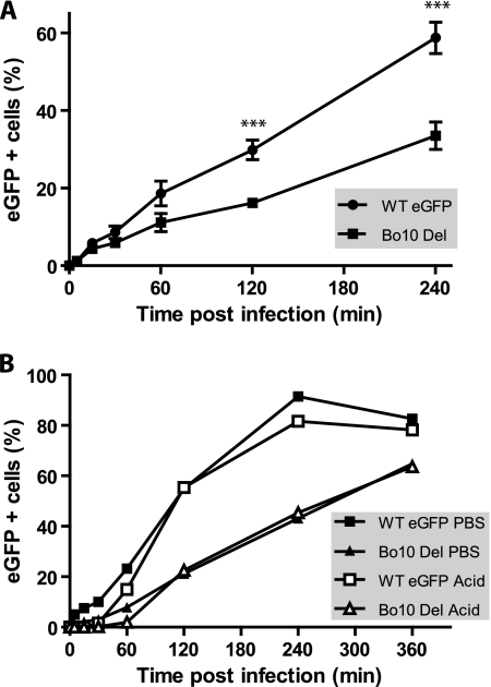
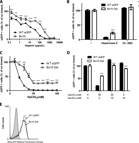
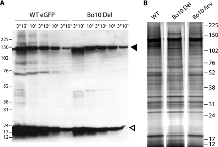
All gammaherpesviruses encode a glycoprotein positionally homologous to the Epstein-Barr virus gp350 and the Kaposi's sarcoma-associated herpesvirus (KSHV) K8.1. In this study, we characterized the positional homologous glycoprotein of bovine herpesvirus 4 (BoHV-4), encoded by the Bo10 gene. We identified a 180-kDa gene product, gp180, that was incorporated into the virion envelope. A Bo10 deletion virus was viable but showed a growth deficit associated with reduced binding to epithelial cells. This seemed to reflect an interaction of gp180 with glycosaminoglycans (GAGs), since compared to the wild-type virus, the Bo10 mutant virus was both less infectious for GAG-positive (GAG(+)) cells and more infectious for GAG-negative (GAG(-)) cells. However, we could not identify a direct interaction between gp180 and GAGs, implying that any direct interaction must be of low affinity. This function of gp180 was very similar to that previously identified for the murid herpesvirus 4 gp150 and also to that of the Epstein-Barr virus gp350 that promotes CD21(+) cell infection and inhibits CD21(-) cell infection. We propose that such proteins generally regulate virion attachment both by binding to cells and by covering another receptor-binding protein until they are displaced. Thus, they regulate viral tropism both positively and negatively depending upon the presence or absence of their receptor.


Figures

FIG. 1.
FIG. 1.
The Bo10 gene is expressed as a late gene and encodes a 180-kDa protein. (A) Determination of Bo10 kinetic class of transcription. MDBK cells were mock infected or infected with BoHV-4 V. test strain in the absence (/) or presence of CHX or PAA. Twenty-four hours postinfection, expression of immediate-early Bo5, early ORF21, and late ORF22 and Bo10 was studied using an RT-PCR approach as described in Materials and Methods. mRNA and cDNA represent PCR products generated when reverse transcriptase was omitted from the reaction mixtures and RT-PCR products, respectively. Amplification of viral genomic DNA is provided as control. UI and Neg. represent uninfected cell samples and PCR negative controls, respectively. The locations of the marker sizes (in base pairs) are indicated to the right of the gel. (B) Predicted product of expression of BoHV-4 Bo10 gene. Signal peptide and transmembrane (TM) regions were identified with SignalP and TMHMM software programs, respectively (http://www.expasy.ch/tools/). The sequence of the peptide used for rabbit immunization is shown below the map. Cyt., intracytoplasmic region. (C) Immunization with Bo10-c15 peptide elicited antibody response against the peptide. The antipeptide antibody responses were determined for two immunized rabbits by indirect immunofluorescence staining of permeabilized cells and FACS analysis as described in Materials and Methods. (D) Detection of specific BoHV-4 protein by the anti-Bo10-c15 serum. MDBK cells were left uninfected or infected with the WT V. test strain of BoHV-4 (1 PFU/cell). Forty-eight hours later, cells were scraped off the plate, and Western blotting was carried out as described in Materials and Methods. The positions of molecular mass (MM) standards (in kilodaltons) are shown to the left of the gel. The open and filled triangles indicate the specific 180-kDa protein and a background band, respectively.
FIG. 2.
FIG. 2.
Production of a recombinant BoHV-4 strain deleted for Bo10. Using the BoHV-4 V. test strain as the parental strain, a recombinant BoHV-4 strain deleted for Bo10 and a derived revertant strain were produced by homologous recombination. (A) Recombination cassettes were constructed. The HindIII restriction map of the entire BoHV-4 V. test strain is shown at the top. Letters were assigned to the different restriction fragments according to their lengths, as described by Bublot et al. (4a). Most of Bo10 exons 1 and 2 were replaced by an eGFP expression cassette as described in Materials and Methods. prDNA, polyrepetitive DNA; CMV IE promoter, cytomegalovirus immediate-early promoter; SV40 pA, simian virus 40 pA. (B) Bo10 Del and Bo10 Rev strains were then characterized by a combined restriction endonuclease and Southern blotting approach. The DNA of the parental strain V. test and the derived recombinant strains Bo10 Del and Bo10 Rev were analyzed by HindIII restriction and further tested by Southern blotting using probes corresponding to the entire V. test Bo10 ORF or to the eGFP ORF or to the deleted part of the V. test Bo10 ORF. The asterisk and arrows indicate the restriction fragments containing the entire Bo10 and eGFP ORFs, respectively. Marker sizes (in kilobase pairs) are indicated on the left.
FIG. 3.
FIG. 3.
gp180 detection in infected cells and virions. (A) MDBK cells were either mock infected or infected with the WT, Bo10 Del, or Bo10 Rev virus strain (1 PFU/cell). At 24 h postinfection, the cells were fixed and stained with either preimmune serum or anti-Bo10-c15 serum for FACS analysis as described in Materials and Methods. Alexa 633, Alexa Fluor 633. (B and C) Detection of specific BoHV-4 protein by the anti-Bo10-c15 serum. Infected MDBK cells (48 h, 1 PFU/cell) (B) or purified virions (5 × 10⁵ virions per lane) (C) were subjected to Western blotting with anti-Bo10-c15 serum as described in Materials and Methods. The position of a MM standard is shown. Open and filled triangles indicate the specific 180-kDa protein and a background band, respectively. (D) BoHV-4 WT purified virions (5 × 10⁵ virions per lane) were treated with proteinase K (pK) in the presence (+) or absence (−) of Triton X-100 (TX-100) (0.1% [vol/vol]) and then subjected to Western blotting with anti-Bo10-c15 serum as described in Materials and Methods. The open and filled triangles indicate the specific 180-kDa protein and the protected C-terminal end of gp180, respectively. The positions of MM standards (in kilodaltons) are shown to the left of the gels in panels B to D.
FIG. 4.
FIG. 4.
Effect of Bo10 deletion on BoHV-4 replication in vitro. (A and B) MDBK cells grown in 6-well cluster dishes were infected at an MOI of 0.05 in a multistep assay (A) or at an MOI of 5 in a one-step assay (B) as described in Materials and Methods with BoHV-4 wild-type V. test, Bo10 Del, and Bo10 Rev. Supernatant of infected cultures and infected cells were harvested at different times (days) postinfection (p.i.), and the amount of infectious virus was determined by plaque assay on MDBK cells. For supernatants, time zero p.i. is retitration of the inoculums to ensure that similar amounts of virus were put on the cells. Plaques were visualized by immunofluorescence staining as described in Materials and Methods. The data presented are the averages ± standard errors of the means (SEMs) (error bars) for triplicate measurements. The data were analyzed by two-way analysis of variance (ANOVA) and Bonferroni posttests. Statistical significance is indicated as follows: *, P < 0.05; **, P < 0.01; ***, P < 0.001. (C) Bo10 deletion has no effect on BoHV-4 plaque size. MDBK cells grown on coverslips were infected with BoHV-4 wild type V. test, Bo10 Del, and Bo10 Rev strains and then overlaid with MEM containing CMC as described in Materials and Methods. At successive intervals after infection, plaques were revealed by indirect immunofluorescence staining using MAb 35 (recognizing gB) and Alexa Fluor 568-conjugated goat anti-mouse IgG as the primary and secondary antibodies, respectively. The pictures of plaques were captured with a CCD camera and analyzed with AnalySIS 3.2 software (Soft Imaging System) in order to determine plaque area. Each value presented is the average ± SEM (error bars) for the measurement of 30 plaques. The data were analyzed by two-way ANOVA and Bonferroni posttests, and no significant difference was observed.
FIG. 5.
FIG. 5.
The Bo10 deletion phenotype is not associated with a virion release deficit. (A) Electron microscopy of virus-infected cells. MDBK cells were infected (24 or 48 h; 1 PFU/cell) with WT or Bo10 Del virus as indicated. The pictures shown are representative of at least 10 sections per sample. For each time point, two different pictures at different magnifications are provided. (B) Fluid-phase virus spread. A total of 2 × 10⁵ infected MDBK cells (MOI of 0.5) or 2 × 10⁵ uninfected MDBK cells were seeded either on a 6-cm-diameter dish or in a 3.5-cm-diameter dish placed inside the first one. The cells were therefore separated by a physical barrier but connected by medium so that virus could not spread directly from the infected to the uninfected population but only via their common supernatant. After 48 h, the cells were harvested and analyzed for eGFP expression (eGFP + cells, eGFP-expressing cells). The data presented are the averages plus SEMs (error bars) for triplicate measurements and were analyzed by t test. The values for the WT eGFP and Bo10 Del virus strains were not significantly different.
FIG. 6.
FIG. 6.
Infectivity assays with Bo10 Del BoHV-4 mutant. (A) MDBK cells were exposed to eGFP-expressing (eGFP +) wild-type virus (WT eGFP) or Bo10 Del BoHV-4 strain (0.5 PFU/cell) for the times indicated and then washed with PBS and cultured overnight. Viral infection was assayed 18 h later by flow cytometry for eGFP expression. The data presented are the average ± SEMs for triplicate measurements. The data were analyzed by two-way ANOVA and Bonferroni posttests. The values for WT eGFP-infected cells were significantly different (P < 0.001) from the values for the Bo10 Del strain as indicated (***). (B) MDBK cells were exposed to WT eGFP or Bo10 Del BoHV-4 strain (0.5 PFU/cell) for the times indicated and then washed either with PBS (pH 7.4) or with isotonic (pH 3) buffer (acid wash). Viral infection was assayed by measuring eGFP expression 18 h later as for panel A.
FIG. 7.
FIG. 7.
gp180 is one of the BoHV-4 proteins interacting with heparan sulfate (HS). (A) WT eGFP or Bo10 Del BoHV-4 virions were preincubated with increasing amounts of soluble heparin for 2 h at 37°C. Viruses and heparin were then added to MDBK cells (MOI of 0.5 PFU/cell). Eighteen hours later, viral eGFP expression was assayed by flow cytometry. Each value is expressed as a percentage of the eGFP expression with untreated virus (no treatment [no ttment]). The data presented are the averages ± SEMs (error bars) for triplicate measurements and were analyzed by two-way ANOVA and Bonferroni posttests. Statistical significance was indicated as follows: **, P < 0.01; ***, P < 0.001. (B) Cell surface HS or chondroitin sulfate (CS) was enzymatically removed from MDBK cells after treatment with heparinase II or chondroitinase ABC (Ch. ABC), respectively. WT eGFP or Bo10 Del BoHV-4 was then added for 2 h on ice before extensive PBS washing to remove unbound virions. Eighteen hours later, viral eGFP expression was assayed by flow cytometry. Each value is expressed as a percentage of eGFP expression with untreated cells shown in the leftmost pair of bars. The data presented are the averages ± SEMs (error bars) for six measurements and were analyzed by two-way ANOVA and Bonferroni posttests. Heparinase II treatment of the cells resulted in a significantly lower level of infection by the B010 Del virus than by the WTeGFP virus (P < 0.001), as indicated (***). (C and D) Sulfation inhibition causes less entry of Bo10 Del virions than of WT virions. MDBK cells were cultured overnight in the presence of indicated amounts of sodium chlorate (NaClO3). The cells were then infected with WT eGFP or Bo10 Del BoHV-4 strain (MOI of 0.5 PFU/cell). Eighteen hours later, viral eGFP expression was assayed by flow cytometry. Each value is expressed as a percentage of the eGFP expression with untreated cells. The data presented are the averages ± SEMs (error bars) for four measurements and were analyzed by two-way ANOVA and Bonferroni posttests. Statistical significance is indicated as follows: ***, P < 0.001. (C) Addition of sodium sulfate (10 mM) restores WT eGFP and Bo10 Del infectivity. MDBK cells were cultured overnight with the indicated concentrations of sodium chlorate (NaClO3) and/or sodium sulfate (Na2SO4). The cells were then infected with WT eGFP or Bo10 Del BoHV-4 strain (MOI of 0.5 PFU/cell). Eighteen hours later, viral eGFP expression was assayed by flow cytometry. Each value is expressed as a percentage of the eGFP expression with untreated cells. The data presented are the averages ± SEMs for triplicate measurements and were analyzed by two-way ANOVA and Bonferroni posttests, Statistical significance is indicated as follows: ***, P < 0.001. (E) MDBK cells were left uninfected (filled histogram) or infected (1 PFU/cell) with either the WT eGFP or Bo10 Del strain of BoHV-4. Each population was then analyzed by flow cytometry for BoHV-4 gp8 (MAb 103) expression as described in Materials and Methods.
FIG. 8.
FIG. 8.
Bo10 deletion increases BoHV-4 entry in GAG-deficient (GAG−) cells. (A) CHO-K1 cells (CHO GAG+) and the GAG-deficient derivative CHO-pgsA-745 (CHO GAG−) were infected at the indicated MOI with the WT eGFP or Bo10 Del strain of BoHV-4. Twenty-four hours later, viral eGFP expression was assayed by flow cytometry. (B) CHO GAG+ and CHO GAG− cells were exposed to WT eGFP or Bo10 Del BoHV-4 strain (3 PFU/cell) for the times indicated and then washed either with PBS (pH 7.4) or with isotonic (pH 3) buffer (acid wash). Viral infection was assayed by measuring eGFP expression 18 h later as for panel A. The data presented are the averages ± SEMs (error bars) for three measurements. For the same treatment, WT eGFP and Bo10 del BoHV-4 strains were compared by two-way ANOVA and Bonferroni posttests. Statistical significance is indicated as follows: *, P < 0.05; ***, P < 0.001. (C and D) Bovine PBMC were isolated on Ficoll gradient and then left uninfected or infected with the WT eGFP or Bo10 Del strain of BoHV-4 (1 PFU/cell). (C) Twenty-four hours later, the cells were analyzed by flow cytometry for CD14 and viral eGFP expression as described in Materials and Methods. The data presented in panel D are the averages ± SEMs for six measurements and were analyzed by one-way ANOVA and Bonferroni posttests. Statistical significance is indicated as follows: ***, P < 0.001.
FIG. 9.
FIG. 9.
Comparison of the structural proteins of the different strains. (A) WT eGFP and Bo10 Del concentrated stocks were compared for protein content by immunoblotting with anti-BoHV-4 rabbit polyserum. The black and white triangles indicate bands corresponding to VP7 and VP24 proteins, respectively; the two bands probably correspond to ORF25 and ORF65 encoding major capsid protein and capsid protein, respectively. 3*105, 3 × 10⁵. (B) Tartrate-purified virions were loaded onto 4 to 12% Bis-Tris polyacrylamide gels and silver stained. The positions of molecular mass markers (in kilodaltons) are indicated to the left of the gel in panel A and to the right of the gel in panel B.

Similar articles

Cited by

References

    1. Baeuerle, P. A., and W. B. Huttner. 1986. Chlorate-a potent inhibitor of protein sulfation in intact cells. Biochem. Biophys. Res. Commun. 141:870-877. - PubMed
    1. Birkmann, A., K. Mahr, A. Ensser, S. Yaguboglu, F. Titgemeyer, B. Fleckenstein, and F. Neipel. 2001. Cell surface heparan sulfate is a receptor for human herpesvirus 8 and interacts with envelope glycoprotein K8.1. J. Virol. 75:11583-11593. - PMC - PubMed
    1. Boerner, B., W. Weigelt, H. J. Buhk, G. Castrucci, and H. Ludwig. 1999. A sensitive and specific PCR/Southern blot assay for detection of bovine herpesvirus 4 in calves infected experimentally. J. Virol. Methods 83:169-180. - PubMed
    1. Borza, C. M., and L. M. Hutt-Fletcher. 2002. Alternate replication in B cells and epithelial cells switches tropism of Epstein-Barr virus. Nat. Med. 8:594-599. - PubMed
    1. Bublot, M., M. F. Van Bressem, E. Thiry, J. Dubuisson, and P. P. Pastoret. 1990. Bovine herpesvirus 4 genome: cloning, mapping and strain variation analysis. J. Gen. Virol. 71:133-142. - PubMed

Publication types