. 2011 Feb;85(3):1348-59.
doi: 10.1128/JVI.01716-10. Epub 2010 Nov 10.

Characterization of Bafinivirus main protease autoprocessing activities

Affiliations

Characterization of Bafinivirus main protease autoprocessing activities

Rachel Ulferts et al. J Virol. 2011 Feb.

Abstract

The production of functional nidovirus replication-transcription complexes involves extensive proteolytic processing by virus-encoded proteases. In this study, we characterized the viral main protease (M(pro)) of the type species, White bream virus (WBV), of the newly established genus Bafinivirus (order Nidovirales, family Coronaviridae, subfamily Torovirinae). Comparative sequence analysis and mutagenesis data confirmed that the WBV M(pro) is a picornavirus 3C-like serine protease that uses a Ser-His-Asp catalytic triad embedded in a predicted two-β-barrel fold, which is extended by a third domain at its C terminus. Bacterially expressed WBV M(pro) autocatalytically released itself from flanking sequences and was able to mediate proteolytic processing in trans. Using N-terminal sequencing of autoproteolytic processing products we tentatively identified Gln↓(Ala, Thr) as a substrate consensus sequence. Mutagenesis data provided evidence to suggest that two conserved His and Thr residues are part of the S1 subsite of the enzyme's substrate-binding pocket. Interestingly, we observed two N-proximal and two C-proximal autoprocessing sites in the bacterial expression system. The detection of two major forms of M(pro), resulting from processing at two different N-proximal and one C-proximal site, in WBV-infected epithelioma papulosum cyprini cells confirmed the biological relevance of the biochemical data obtained in heterologous expression systems. To our knowledge, the use of alternative M(pro) autoprocessing sites has not been described previously for other nidovirus M(pro) domains. The data presented in this study lend further support to our previous conclusion that bafiniviruses represent a distinct group of viruses that significantly diverged from other phylogenetic clusters of the order Nidovirales.


Figures

FIG. 1.
FIG. 1.
(A to D) Proteins expressed in the present study. (A) Schematic showing the location of the putative Mpro domain (dark gray) within the pp1a/1ab replicase polyproteins. Also shown are the locations of the putative transmembrane domains in pp1a/1ab (light gray) as predicted by TMHMM (38). The putative WBV Mpro core domain was expressed with flanking regions (Gln-3424 to Met-3725) as a fusion with the maltose-binding protein (MBP) at its N-terminal and glutathione S-transferase (GST) at its C-terminal (MBP-pp1a-3424-3725-GST). This construct was presumed to contain both upstream and downstream flanking cleavage sites (indicated by arrows). (B) Expression of WBV pp1a/pp1ab residues Gln-3424 to Thr-3707 as an N-terminal fusion with MBP and containing a C-terminal hexahistidine (His6) tag. This construct only contained the N-terminal autoprocessing site, thus allowing a single-step purification (by IMAC) of the Mpro domain (Mpro-CHis) following its intracellular autocatalytic release from the fusion protein precursor. (C) Intermolecular (trans) cleavage reaction of the proteolytically inactive MBP-pp1a-3424-3725_S3589A-GST fusion protein by Mpro-CHis. (D) To obtain molecular mass markers for the four theoretical forms of the Mpro domain resulting from cleavage at two alternative N- and C-terminal cleavage sites, respectively, the respective parts of the polyprotein (see also Fig. 3A) were expressed as N-terminal fusions with MBP. Cleavage of the purified fusion proteins with factor Xa yielded size markers with authentic N termini. To prevent autoprocessing at cleavage sites present in some of the proteins, the active-site Ser residue (Ser-3589) was substituted with Ala in all constructs. (E to G) Purification of processing products. (E) The MBP-pp1a-3424-3725-GST fusion protein was expressed in E. coli TB1 (for details, see Materials and Methods) and the C-terminal, GST-containing processing product was purified using GST-Bind resin. The cleared lysate (lane 2) and the purified protein (lane 3) were separated by SDS-PAGE and stained with Coomassie brilliant blue R-250. Lane 1: protein ladder. The filled arrowhead indicates the purified C-terminal processing product. (F) MBP-pp1a-3424-3725_Q3716A-GST was expressed in E. coli TB1 and the C-terminal processing product was affinity purified using GST-Bind resin. The cleared lysate (lane 2) and the purified protein (lane 3) were analyzed by SDS-PAGE and Coomassie blue staining. The C-terminal processing product of the MBP-pp1a-3424-3725-GST fusion protein purified in panel E was electrophoresed on the same gel for size comparison (lane 4). Lane 1, protein ladder. The open arrowhead indicates the purified C-terminal autoprocessing product of the MBP-pp1a-3424-3725_Q3716A-GST fusion protein; the filled arrowhead indicates the C-terminal autoprocessing product of the MBP-pp1a-3424-3725-GST fusion protein. (G) Purification of Mpro-CHis. The MBP-Mpro-CHis fusion protein was expressed in E. coli TB1 and the C-terminal, His-tagged processing product was purified by IMAC. Samples taken at different steps of the purification protocol were analyzed by SDS-PAGE and Coomassie blue staining. Lane 1, protein ladder; lane 2, total lysate; lane 3, insoluble protein fraction; lane 4, soluble supernatant fraction; lane 5, eluate fraction. Mpro-CHis is indicated by a filled arrowhead. Molecular masses (in kilodaltons) of the protein ladder proteins are given to the left for each of the gels shown.
FIG. 2.
FIG. 2.
In trans proteolytic processing of the MBP-pp1a-3424-3725_S3589A-GST fusion protein by Mpro-CHis. MBP-pp1a-3424-3725_S3589A-GST was expressed in E. coli TB1 and affinity purified using amylose resin. The MBP-pp1a-3424-3725_S3589A-GST fusion protein was then incubated with (lane 3) or without (lane 2) Mpro-CHis. Processing products were separated through SDS-PAGE and stained with Coomassie brilliant blue R-250. Lane 4, the amount of Mpro-CHis equal to that present in lane 3; lane 1, protein ladder. The molecular masses (in kilodaltons) of the ladder proteins are indicated to the left of the gel.
FIG. 3.
FIG. 3.
Western blot analysis of Mpro forms expressed in WBV-infected cells. (A) Schematic representation of the four putative forms of the Mpro domain. The location of the two upstream (N1 and N2) and the two downstream (C1 and C2) cleavage sites identified after expression of the putative Mpro and flanking sequences in E. coli are depicted. Numbers refer to the amino acid position in the WBV pp1a/pp1ab. Depending on the cleavage sites used, four forms of the Mpro domain would be possible (N1-C1, N1-C2, N2-C1, and N2-C2). The theoretical molecular mass of each form is indicated. (B) EPC cells were infected with WBV at an MOI of 10 TCID50 per cell or mock infected. At 24 h postinfection, lysates were prepared. These were separated in a 15% discontinuous SDS-polyacrylamide gel alongside heterologously expressed molecular mass markers that conform to the four theoretical forms of the Mpro domain: N1-C1, N1-C2, N2-C1, and N2-C2. Mpro present in infected cells and the Mpro marker proteins were detected by immunoblotting with rabbit anti-WBV-Mpro antiserum. The molecular masses of the protein ladder proteins (in kilodaltons) are indicated to the left. Two forms of Mpro were detected in WBV-infected cells (indicated by arrowheads).
FIG. 4.
FIG. 4.
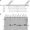
Putative WBV Mpro active-site residues. (A) Multiple sequence alignment of astro-, arteri-, toro-, and bafinivirus serine proteases. Partial protease sequences were aligned by using ClustalX 2.0 (30) and rendered with ESPript 2.2 (http://espript.ibcp.fr/ESPript/cgi-bin/ESPript.cgi) (22). Abbreviations of virus names and accession numbers are as follows: HAstV-1, human astrovirus 1 (AAW51880); EToV, equine torovirus (X52374); BToV, bovine torovirus (AY427798); PRRSV, porcine reproductive and respiratory syndrome virus (Q04561); LDV, lactate dehydrogenase-elevating virus (NC_02534); EAV, equine arteritis virus (NP_127506). Secondary structure elements of the astrovirus Mpro are shown above the sequence (PDB code: 2W5E). These were extracted from the PDB file and added to the alignment using ESPript 2.2 and manually adjusted to conform to the numbering used by Speroni et al. (42). Conserved residues that were identified as possible catalytic or S1 subsite residues based on data available for the corresponding residues in astrovirus and nidovirus homologs (see the text for details) are marked with closed or open arrowheads, respectively. The gray arrowhead marks the (nonconserved) Asp residue that was used as a negative control in this mutagenesis study. Residues were numbered according to their position in the respective viral polyprotein sequence. (B to D) Autoprocessing activities of mutant forms of the WBV Mpro with substitutions of putative active-site residues. MBP-pp1a-3424-3725-GST and mutant proteins were expressed in E. coli TB1. After induction of expression for 3 h at 25°C, total cell lysates were prepared and separated in a 15% discontinuous SDS-polyacrylamide gel and transferred onto nitrocellulose. GST-containing unprocessed precursor proteins, processing intermediates, and processing end products were detected by Western blot analysis with rabbit anti-GST antiserum (D) as the primary antibody and IRDye800CW anti-rabbit IgG as the secondary antibody. After imaging, bound antibodies were removed. MBP- and Mpro-containing proteins were subsequently detected by using mouse anti-MBP monoclonal antibody (B) and rabbit anti-WBV Mpro antiserum (C), followed by labeling with IRDye680 anti-mouse IgG and IRDye 800CW anti-rabbit IgG, respectively. Images were acquired by using an LI-COR Odyssey system and software. Wild-type and mutant proteins analyzed in this experiment are indicated above the lanes. MBP-LacZα, E. coli TB1 transformed with pMal-c2X and induced with 1 mM IPTG; control, untransformed and mock induced E. coli TB1; Mpro marker 1, Mpro markers N1-C1 and N1-C2; Mpro marker 2, Mpro markers N2-C1 and N2-C2. The molecular masses of the marker proteins (in kilodaltons) and the identities of the Mpro marker proteins (in panel C) are indicated to the left. Black arrowheads, processing end products; gray arrowheads, processing intermediates; open arrowhead, unprocessed precursor.
FIG. 5.
FIG. 5.
Purification and proteolytic activities of Mpro-CHis wild-type and mutant proteins. (A) Mpro-CHis wild-type and mutant proteins D3518A and T3584A were expressed in E. coli and purified by IMAC. One microgram of each of the indicated proteins was electrophoresed in an SDS-15% polyacrylamide gel and stained with Coomassie brilliant blue R-250. The molecular masses (in kilodaltons) of marker proteins are given to the left. (B to C) A synthetic peptide (0.5 mM final concentration) whose sequence corresponded to the previously determined N1 cleavage site (N1 peptide) was incubated with 0.3 μM Mpro-CHis wild-type or mutant protein or no protein (control) for 3 h (B) or 20 h (C). The reaction products were analyzed by reversed-phase chromatography. The y axis conforms to the control reaction, all other chromatograms are depicted as shifted up by 100 mAU (B) or 70 mAU (C) with respect to the one below.

Similar articles

Cited by

References

    1. Adams, M. J., J. F. Antoniw, and F. Beaudoin. 2005. Overview and analysis of the polyprotein cleavage sites in the family Potyviridae. Mol. Plant Pathol. 6:471-487. - PubMed
    1. Allaire, M., M. M. Chernaia, B. A. Malcolm, and M. N. James. 1994. Picornaviral 3C cysteine proteinases have a fold similar to chymotrypsin-like serine proteinases. Nature 369:72-76. - PubMed
    1. Anand, K., G. J. Palm, J. R. Mesters, S. G. Siddell, J. Ziebuhr, and R. Hilgenfeld. 2002. Structure of coronavirus main proteinase reveals combination of a chymotrypsin fold with an extra alpha-helical domain. EMBO J. 21:3213-3224. - PMC - PubMed
    1. Anand, K., J. Ziebuhr, P. Wadhwani, J. R. Mesters, and R. Hilgenfeld. 2003. Coronavirus main proteinase (3CLpro) structure: basis for design of anti-SARS drugs. Science 300:1763-1767. - PubMed
    1. Ash, E. L., J. L. Sudmeier, E. C. De Fabo, and W. W. Bachovchin. 1997. A low-barrier hydrogen bond in the catalytic triad of serine proteases? Theory versus experiment. Science 278:1128-1132. - PubMed

Publication types

LinkOut - more resources