. 2011 Jan;85(2):804-17.
doi: 10.1128/JVI.01173-10. Epub 2010 Nov 10.

Alternative splicing of the human cytomegalovirus major immediate-early genes affects infectious-virus replication and control of cellular cyclin-dependent kinase

Affiliations

Alternative splicing of the human cytomegalovirus major immediate-early genes affects infectious-virus replication and control of cellular cyclin-dependent kinase

Guixin Du et al. J Virol. 2011 Jan.

Abstract

The major immediate-early (MIE) gene locus of human cytomegalovirus (HCMV) is the master switch that determines the outcomes of both lytic and latent infections. Here, we provide evidence that alteration in the splicing of HCMV (Towne strain) MIE genes affects infectious-virus replication, movement through the cell cycle, and cyclin-dependent kinase activity. Mutation of a conserved 24-nucleotide region in MIE exon 4 increased the abundance of IE1-p38 mRNA and decreased the abundance of IE1-p72 and IE2-p86 mRNAs. An increase in IE1-p38 protein was accompanied by a slight decrease in IE1-p72 protein and a significant decrease in IE2-p86 protein. The mutant virus had growth defects, which could not be complemented by wild-type IE1-p72 protein in trans. The phenotype of the mutant virus could not be explained by an increase in IE1-p38 protein, but prevention of the alternate splice returned the recombinant virus to the wild-type phenotype. The lower levels of IE1-p72 and IE2-p86 proteins correlated with a delay in early and late viral gene expression and movement into the S phase of the cell cycle. Mutant virus-infected cells had significantly higher levels of cdk-1 expression and enzymatic activity than cells infected with wild-type virus. The mutant virus induced a round-cell phenotype that accumulated in the G(2)/M compartment of the cell cycle with condensation and fragmentation of the chromatin. An inhibitor of viral DNA synthesis increased the round-cell phenotype. The round cells were characteristic of an abortive viral infection.


Figures

FIG. 1.
FIG. 1.
Multiple sequence alignment of the primate CMV IE1-p72 protein homologs. Amino acid sequences for the proteins from human CMV Towne strain IE1 (AAR31448), chimpanzee CMV IE1 (AAM00752), rhesus CMV IE1 (AAB00487), and African green monkey (AGM) CMV IE1 (AAB16882) were aligned using MultAlin. Multiple sequence alignments are displayed using BoxShade. Identical residues appear shaded in black, while similar residues appear shaded in gray. A star indicates a residue that is identical in all four aligned sequences, while a dot indicates a residue that is conserved in at least half of the aligned sequences. The numbers appearing between the species and the amino acid sequence represent the amino acid position for that particular species. A hyphen designates a gap in the sequence that was inserted for optimal alignment. The black bar underlines amino acids 412 to 419.
FIG. 2.
FIG. 2.
IE1 exon 4 mutations altered MIE gene splicing and expression pattern. (A) Schematic diagram of the NheI-linearized DNA fragment from the shuttle vector carrying mutated IE1 exon 4 for constructing recombinant BACs. The plasmid pdlMCATdl-694/-583+Kan+IE1/IE2 carries the genomic sequence of the HCMV Towne strain from UL121 to UL128. The UL127 locus was replaced by the CAT reporter. A kanamycin resistance (KanR) cassette was inserted between UL127 and UL128 for selection of recombinant BACs. Twenty-four nucleotides of exon 4 that encode amino acid residues from 412 to 419 of IE1-p72 were either mutated or deleted to generate IE1 X412-419A, IE1 dl412-419, and IE1 Pu/Py412-419. The upstream 3′ splice site (3′ss) in exon 4 was mutated in IE1 WTSS and IE1 X412-419ASS recombinant viruses. MIEP, MIE promoter. HFF cells infected with recombinant viruses were harvested at 8 h p.i. (C) or 48 h p.i. (B and D) for MIE mRNA and protein expression analysis. (B) RT-PCR detecting IE1-p72 or IE1-p38 mRNAs as described in Materials and Methods. Mock, mock infected. (C) The levels of viral transcripts in WT and IE1 X412-419A recombinant virus-infected cells were determined by quantitative real-time RT-PCR as described in Materials and Methods using probes specific for MIE, IE1-p72, and IE2-p86 mRNA. A representative experiment of three is shown here. rel., relative. (D) Protein levels of MIE proteins IE1-p72, IE1-p38, and IE2-p86 were determined by Western blotting with monoclonal antibody MAB810, which recognizes an epitope in exon 2/exon 3 of IE1-p72, IE1-p38, and IE2-p86. Asterisk shows a degraded viral protein in all virus-infected samples.
FIG. 3.
FIG. 3.
Recombinant virus IE1 X412-419A has a growth defect. Cells were infected in triplicate with recombinant viruses at 0.05 PFU/cell. Cells and supernatant were harvested at each time point after infection, and the triplicate samples were pooled and stored. A plaque assay was performed in triplicate on ihfie1.3 cells. A representative experiment of at least three is shown here. (A) HFF cells infected with IE1 WT, IE1 X412-419A, or IE1 Rev. (B) HFF cells infected with IE1 WT, IE1 WT SS, IE1 X412-419A, IE1 X412-419A SS, or CR208. (C) The same as panel B except ihfie1.3 cells were used, which rescues recombinant virus CR208.
FIG. 4.
FIG. 4.
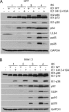
Viral protein expression is delayed with recombinant virus IE1 X412-419A in both HFF and ihfie1.3 cells. Cells were infected with IE1 WT or IE1 X412-419A recombinant viruses at an MOI of 0.2. Cells were harvested at the time points indicated. MIE viral proteins IE2-p86, IE1-p72, and IE1-p38, early viral proteins UL44 (p52) and UL84, early/late viral protein pp65 and late viral protein pp28, and cellular protein GAPDH were detected using specific antibodies as described in Materials and Methods. (A) HFF cells. (B) ihfie1.3 cells.
FIG. 5.
FIG. 5.
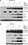
IE1-p38 protein does not suppress HCMV viral protein expression. HFF cells were transduced with 20 PFU/cell of either AdGFP or AdIE38 in the presence of AdTrans (20 PFU/cell). After 24 h, cells were infected with HCMV Towne strain at an MOI of 0.05 (A) or 2 (B), harvested at the indicated times postinfection, and analyzed by Western blotting for MIE viral proteins IE2-p86, IE1-p72, and IE1-p38, early viral protein UL44 (p52), late viral protein (UL99) pp28, and cellular protein β-tubulin as described in Materials and Methods.
FIG. 6.
FIG. 6.
Recombinant virus IE1 X412-419A induces a round-cell phenotype. HFF or ihfie1.3 cells were infected with recombinant viruses IE1 WT, IE1 X412-419A, or IE1 Rev at an MOI of 0.2 and analyzed at 5 days p.i. as described in Materials and Methods. (A) Fluorescence and phase-contrast microscopy; magnification, ×100. Florescence images of ihfie1.3 cells are not shown. (B) Percent round cells in the presence or absence of PFA (200 μg/ml).
FIG. 7.
FIG. 7.
Round cells infected with recombinant virus IE1 X412-419A have IE1 protein condensed on the chromatin and abnormal mitotic figures. HFF cells were infected with recombinant viruses IE1 WT or IE1 X412-419A at MOI of 0.1 and stained for an immunofluorescence assay with either MIE exon 4-specific antibody 6E1 (red) or DAPI (blue). In addition, round cells were enriched for an apoptotic assay or for mitotic spreads as described in Materials and Methods. (A) Immunofluorescence assay. Arrowheads indicate infected monolayer cells. Arrows indicate round cells; magnification ×400. (B) Genomic DNAs purified from 2 × 10⁶ cells and resolved on a 0.8% agarose gel. The positive control was U937 cells treated with 4 μg/ml of camptothecin for 3 h. (C) Mitotic spreads. (a) Interphase nuclei of mock-infected cells; (b) IE1 WT-infected cells; (c) mitotic nuclei of mock-infected cells; (d to f) IE1 X412-419A-infected round cells. Magnification, ×1,000.
FIG. 8.
FIG. 8.
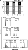
Cells infected with recombinant virus IE1 X412-419A move more into the S phase, and the round cells accumulate in the G2/M compartment of the cell cycle. (A) HFF cells were synchronized by contact inhibition and released to the cell cycle by culturing at a lower density. Cells were infected with recombinant virus IE1 WT or IE1 X412-419A at an MOI of 1. After 24 h, the cells were harvested. (B) Confluent HFF cells were mock infected or infected with recombinant virus IE1 WT or IE1 X412-419A at an MOI of 0.2 in the presence or absence of PFA (200 μg/ml). Monolayer cells or round cells were harvested at 5 days (5d) postinfection. All cells (A and B) were fixed in 70% ethanol, stained with anti-MIE antibody MAB810X and propidium iodide, and then subjected to FACS analysis as described in Materials and Methods. MIE-positive cells were sorted and counted according to DNA content. Cell cycle profiles were analyzed using FlowJo software to determine the percentage of infected cells in each phase of cell cycle for each sample. With the exception of samples for the bottom right panel (w/o PFA), all samples were treated with PFA during infection.
FIG. 9.
FIG. 9.
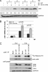
cdk-1 accumulates in cells infected with recombinant virus IE1 X412-419A. Cells were infected with IE1 WT or IE1 X412-419A at an MOI of 1 and analyzed by Western blotting, quantitative RT-PCR, and cdk-1 enzyme activity as described in Materials and Methods. (A) Western blot of cdk-1 protein in infected cells at various days after infection (dpi). (B) cdk-1 mRNA in infected cells. (C) cdk-1 enzyme activity in anti-cdk-1 immunoprecipitates of cell lysates from infected cells at 2 and 3 days p.i. Western blot of IE1-p72, IE2-p86, cdk-1, and GAPDH proteins in the cell lysates of infected cells at 2 and 3 days p.i. The data in the figures are representative of results of at least two independent experiments.
FIG. 10.
FIG. 10.
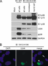
Early and late viral proteins were not detected in recombinant virus IE1 X412-419A-infected round cells. (A) HFF cells were infected with recombinant virus IE1 WT or IE1 X412-419A (MOI, 0.1). Cells were harvested or enriched for round cells and analyzed by Western blot assay or by immunofluorescence assay as described in Materials and Methods. (A) Western blot assay for MIE, early (UL84), and late (UL99; pp28) viral proteins. (B) Immunofluorescence assay of recombinant virus IE1 X412-419A-infected cells stained with anti-UL44 (p52) or DAPI. The arrow indicates an infected round cell, while the arrowhead indicates an infected monolayer cell expressing the UL44 gene product. Magnification, ×400.

Similar articles

Cited by

References

    1. Ahn, J. H., E. J. Brignole III, and G. S. Hayward. 1998. Disruption of PML subnuclear domains by the acidic IE1 protein of human cytomegalovirus is mediated through interaction with PML and may modulate a RING finger-dependent cryptic transactivator function of PML. Mol. Cell. Biol. 18:4899-4913. - PMC - PubMed
    1. Awasthi, S., J. A. Isler, and J. C. Alwine. 2004. Analysis of splice variants of the immediate-early 1 region of human cytomegalovirus. J. Virol. 78:8191-8200. - PMC - PubMed
    1. Balvay, L., D. Libri, and M. Y. Fiszman. 1993. Pre-mRNA secondary structure and the regulation of splicing. Bioessays 15:165-169. - PubMed
    1. Baracchini, E., E. Glezer, K. Fish, R. M. Stenberg, J. A. Nelson, and P. Ghazal. 1992. An isoform variant of the cytomegalovirus immediate-early auto repressor functions as a transcriptional activator. Virology 188:518-529. - PubMed
    1. Browne, E. P., B. Wing, D. Coleman, and T. Shenk. 2001. Altered cellular mRNA levels in human cytomegalovirus-infected fibroblasts: viral block to the accumulation of antiviral mRNAs. J. Virol. 75:12319-12330. - PMC - PubMed

Publication types

LinkOut - more resources