Review
. 2010 Nov 10;30(45):14946-54.
doi: 10.1523/JNEUROSCI.4305-10.2010.

Amyloid-independent mechanisms in Alzheimer's disease pathogenesis

Affiliations
Review

Amyloid-independent mechanisms in Alzheimer's disease pathogenesis

Sanjay W Pimplikar et al. J Neurosci. .

Abstract

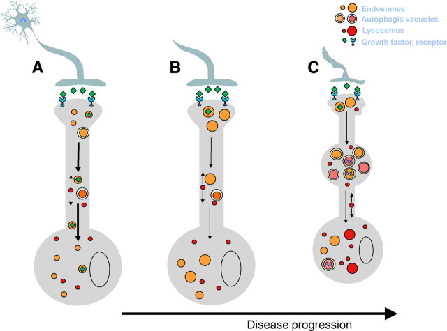
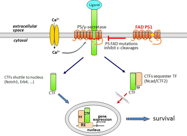
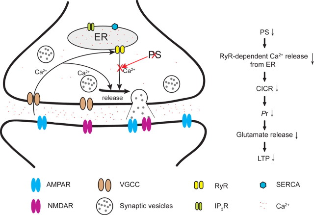
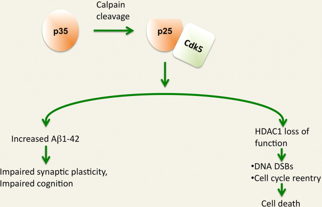
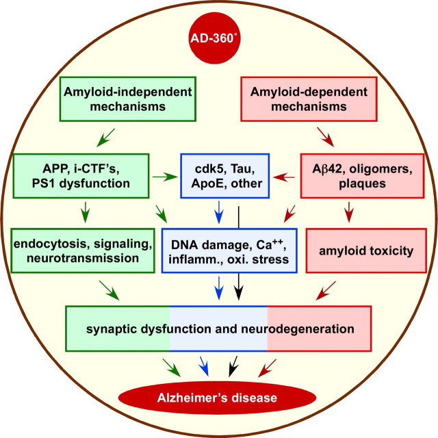
Despite the progress of the past two decades, the cause of Alzheimer's disease (AD) and effective treatments against it remain elusive. The hypothesis that amyloid-β (Aβ) peptides are the primary causative agents of AD retains significant support among researchers. Nonetheless, a growing body of evidence shows that Aβ peptides are unlikely to be the sole factor in AD etiology. Evidence that Aβ/amyloid-independent factors, including the actions of AD-related genes, also contribute significantly to AD pathogenesis was presented in a symposium at the 2010 Annual Meeting of the Society for Neuroscience. Here we summarize the studies showing how amyloid-independent mechanisms cause defective endo-lysosomal trafficking, altered intracellular signaling cascades, or impaired neurotransmitter release and contribute to synaptic dysfunction and/or neurodegeneration, leading to dementia in AD. A view of AD pathogenesis that encompasses both the amyloid-dependent and -independent mechanisms will help fill the gaps in our knowledge and reconcile the findings that cannot be explained solely by the amyloid hypothesis.


Figures

Figure 1.
Figure 1.
Dysfunction of autophagic and endocytic pathways to lysosomes driven by relevant genes and other risk factors in Alzheimer's disease. A–C, A normal neuron is depicted in A. At the earliest stages of AD (B), an abnormal acceleration of endocytosis, mediated partly by rab5, is known to be caused by App gene duplication (via β-CTF) in early onset FAD and Down syndrome and promoted by ApoE4 and elevated cholesterol in late-onset AD. Adverse consequences include endosome enlargement and defective endosome retrograde transport and neurotrophin signaling functions, which promote apoptotic pathway activation and neurodegeneration, particularly of cholinergic neuronal populations. Subsequently (C), failure of autophagy, prominently involving impaired lysosomal proteolysis, leads to massive selective accumulation of autophagic vacuoles (autophagosomes, autolysosomes etc,) containing partially digested autophagic and endocytic substrates within swollen “dystrophic” neurites. The diminished clearance by autophagy of toxic organelles and proteins, including Aβ, ubiquitinated proteins, activated caspases, and possibly tau, leads to neurodegeneration via multiple pathways. Failure of lysosomal proteolysis and autophagy in AD is driven directly by PS1 mutations in early-onset FAD, and is also promoted by normal aging, oxidative stress, Apo E4, intracellular Aβ, and other AD-related genetic and environmental risk factors.
Figure 2.
Figure 2.
The ε-cleavage of receptors is mediated by γ-secretase and inhibited by PS FAD mutations. This cleavage produces biologically active peptides containing the CTFs of substrates. ε-cleavages can be stimulated by ligand binding or calcium influx (Litterst et al., 2007). CTFs can travel to nucleus where they can regulate gene expression or sequester transcription factors (TF) in the cytoplasm. Many PS1 FAD mutants inhibit the ε-cleavage indicating FAD mutations cause a loss of γ-secretase function (Marambaud et al., 2003). PM, Plasma membrane; RE, response element.
Figure 3.
Figure 3.
A model depicting the role of presenilins in the regulation of neurotransmitter release. Upon stimulation, calcium concentration at the presynaptic terminal is drastically elevated due to calcium influx through VGCCs and calcium-induced calcium release (CICR) from intracellular stores, which is mediated through both ryanodine receptors and IP3 receptors. Loss of PS function in the presynaptic terminal specifically disrupts ryanodine receptor-mediated Ca2+ release from the ER store, thus reducing CICR and resulting in reduced increases of calcium-induced by action potentials in the presynaptic terminal. This reduction in calcium increases impairs the probability of neurotransmitter release, and the decreased glutamate release causes LTP impairment in PS-deficient presynaptic terminals. (Figure taken from supplementary information in Nature 460:632–636, 2009. Reprinted with permission.)
Figure 4.
Figure 4.
p25/Cdk5 in the pathogenesis of Alzheimer's disease. The p25/Cdk5 kinase exerts two parallel processes in the course of neurodegeneration. First, it increases β-amyloid production which contributes to synaptic impairment and memory loss. Second, p25/Cdk5 in the nucleus reduces HDAC1 activity, which leads to increased expression of cell cycle genes and DNA double-strand breaks. These pathologies eventually lead to neuronal loss and neurodegeneration. DSBs, double-strand breaks.
Figure 5.
Figure 5.
AD-360°. This global view of AD pathogenesis includes both the amyloid-independent (green pathway) and amyloid-dependent (red pathway) mechanisms. The Aβ/amyloid-independent mechanisms are mediated via APP, intracellular fragments (i-CTFs) and PS1 via the cellular processes discussed here (green arrows) while the amyloid mechanisms are mediated via Aβ42 or Aβ oligomers or plaques (red arrows). Cdk5 (blue box) may be influenced by or interacts with both pathways and its activation triggers DNA damage, cell cycle activation and neurodegeneration (blue arrows). Non-APP/PS factors such as Tau and ApoE also contribute to AD pathology (blue and black arrows) and there is strong evidence to suggest that cellular processes such as inflammation, oxidative stress and Ca2+ dysregulation implicated in AD pathogenesis can be triggered by both amyloid-dependent and amyloid-independent mechanisms. All of these pathways can lead to synaptic dysfunction and neurodegeneration and AD most likely results from the cumulative effects of multiple pathway.

References

    1. Arbel M, Yacoby I, Solomon B. Inhibition of amyloid precursor protein processing by beta-secretase through site-directed antibodies. Proc Natl Acad Sci U S A. 2005;102:7718–7723. - PMC - PubMed
    1. Baki L, Shioi J, Wen P, Shao Z, Schwarzman A, Gama-Sosa M, Neve R, Robakis NK. PS1 activates PI3K thus inhibiting GSK-3 activity and tau overphosphorylation: effects of FAD mutations. EMBO J. 2004;23:2586–2596. - PMC - PubMed
    1. Baki L, Neve RL, Shao Z, Shioi J, Georgakopoulos A, Robakis NK. Wild-type but not FAD mutant presenilin-1 prevents neuronal degeneration by promoting phosphatidylinositol 3-kinase neuroprotective signaling. J Neurosci. 2008;28:483–490. - PMC - PubMed
    1. Batelli S, Albani D, Prato F, Polito L, Franceschi M, Gavazzi A, Forloni G. Early-onset Alzheimer disease in an Italian family with presenilin-1 double mutation E318G and G394V. Alzheimer Dis Assoc Disord. 2008;22:184–187. - PubMed
    1. Beglopoulos V, Shen J. Regulation of CRE-dependent transcription by presenilins: prospects for therapy of Alzheimer's disease. Trends Pharmacol Sci. 2006;27:33–40. - PubMed

Publication types

Substances