Review
. 2010 Nov 10;30(45):14998-5004.
doi: 10.1523/JNEUROSCI.4190-10.2010.

Toward the second generation of optogenetic tools

Affiliations
Review

Toward the second generation of optogenetic tools

Thomas Knöpfel et al. J Neurosci. .

Abstract

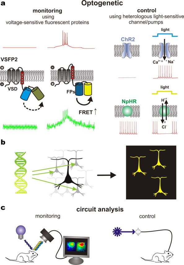
This mini-symposium aims to provide an integrated perspective on recent developments in optogenetics. Research in this emerging field combines optical methods with targeted expression of genetically encoded, protein-based probes to achieve experimental manipulation and measurement of neural systems with superior temporal and spatial resolution. The essential components of the optogenetic toolbox consist of two kinds of molecular devices: actuators and reporters, which respectively enable light-mediated control or monitoring of molecular processes. The first generation of genetically encoded calcium reporters, fluorescent proteins, and neural activators has already had a great impact on neuroscience. Now, a second generation of voltage reporters, neural silencers, and functionally extended fluorescent proteins hold great promise for continuing this revolution. In this review, we will evaluate and highlight the limitations of presently available optogenic tools and discuss where these technologies and their applications are headed in the future.


Figures

Figure 1.
Figure 1.
Genetically encoded, targetable actuator and reporter proteins that allow the use of light to either control or report molecular processes in specific cell populations within networks of heterologous cell types. a, Optical reporting and control are exemplified respectively by a voltage-sensitive fluorescent protein (VSFP2, left) and a light-sensitive ion channel (ChR2, top right) or ion pump [Np-halorhodopsin (NpHR), bottom right] that are directly gated by photoabsorption. Membrane potential traces are schematically indicated in red. VSFP2 reports action potential firing via an optical signal (green trace), while light is used to induce (ChR2) or inhibit (NpHR) action potential generation via the activity of actuator proteins. b, Optogenetic reporters and actuators can be selectively expressed in specific sets of neurons using genetic techniques. c, Light may be delivered (and collected) either by simple epi-illumination optics or more precisely targeted configurations, such as via fiber optics. Combinations of both methods can be used to investigate causality between the activity of specific neuronal populations and behavior.
Figure 2.
Figure 2.
Schematic representation of the three major GECI classes. a, Schematic of the single-FP GECI sensing mechanism. Upon calcium binding, conformational changes in the CaM/M13 complex induce fluorescence changes in the circularly permuted GFP (cpEGFP). b, The cameleon family of FRET-based GECIs. A calcium-dependent increase in FRET between a cyan fluorescent protein (CFP) and yellow fluorescent protein (YFP) FRET pair is coupled to the binding of calmodulin to the M13 peptide. c, Troponin C-based FRET GECIs. Binding of calcium to troponin C induces conformational changes and an increase in FRET between CFP and YFP. d, Imaging neural activity with GCaMP3 using a two-photon microscope in awake, behaving mice. GCaMP3 expression in layer 2/3 neurons of the primary motor cortex at 72 d postinjection (scale bar, 30 mm) and ΔF/F traces of individual cells (black lines). Relative treadmill movement indicated by red line. F, forward; B, backward; eCFP, enhanced CFP; ROI, region of interest. Adapted from Tian et al., 2009.
Figure 3.
Figure 3.
Quieting neural activity with light. a, Neural activity in a representative neuron expressing Arch (i) before, during, and after 5 s of illumination with yellow light, shown as a spike raster plot in which each spike is represented by a dot. This is displayed above a histogram that shows spike firing rate for each 20-ms-duration period throughout the light delivery process, averaged across trials. The bottom (ii) depicts the averaged instantaneous firing rate in a population of Arch-expressing neurons before, during and after yellow light illumination (black line, mean; gray lines, mean ± SE; data from 13 neurons). b, Multicolor silencing of two neural populations, enabled by ion pumps of different classes that are selectively induced by blue (Mac) or red (Halo) light. i, Action spectra of Mac versus Halo; rectangles indicate filter bandwidths used for multicolor silencing in vitro. Blue light illumination was achieved via a 470 + 20 nm filter at 5.3 mW/mm2, and red light illumination was achieved via a 630 + 15 nm filter at 2.1 mW/mm2. Action potentials evoked by current injection into patch-clamped cultured neurons transfected with Halo (ii) were selectively silenced by the red light but not the blue light, while the opposite was true for neurons expressing Mac (iii). Adapted from Chow et al., 2010.
Figure 4.
Figure 4.
Control of intracellular signal transduction by light. a, A plasma membrane-targeted phytochrome undergoes dimerization with a yellow fluorescent protein (YFP)-tagged PIF protein exposed to red light, which can be reversed by illumination with infrared light. b, This system can be used to translocate a targeted activator protein to the plasma membrane, enabling optogenetic control of a large number of biochemical signaling pathways.

References

    1. Ai HW, Shaner NC, Cheng Z, Tsien RY, Campbell RE. Exploration of new chromophore structures leads to the identification of improved blue fluorescent proteins. Biochemistry. 2007;46:5904–5910. - PubMed
    1. Akemann W, Mutoh H, Perron A, Rossier J, Knöpfel T. Imaging brain electric signals with genetically targeted voltage-sensitive fluorescent proteins. Nat Methods. 2010;7:643–649. - PubMed
    1. Ataka K, Pieribone VA. A genetically targetable fluorescent probe of channel gating with rapid kinetics. Biophys J. 2002;82:509–516. - PMC - PubMed
    1. Baird GS, Zacharias DA, Tsien RY. Circular permutation and receptor insertion within green fluorescent proteins. Proc Natl Acad Sci U S A. 1999;96:11241–11246. - PMC - PubMed
    1. Baker BJ, Mutoh H, Dimitrov D, Akemann W, Perron A, Iwamoto Y, Jin L, Cohen LB, Isacoff EY, Pieribone VA, Hughes T, Knöpfel T. Genetically encoded fluorescent sensors of membrane potential. Brain Cell Biol. 2008;36:53–67. - PMC - PubMed

Publication types

Substances

LinkOut - more resources