. 2010 Nov 10;30(45):15007-18.
doi: 10.1523/JNEUROSCI.1436-10.2010.

Early life stress enhances behavioral vulnerability to stress through the activation of REST4-mediated gene transcription in the medial prefrontal cortex of rodents

Affiliations

Early life stress enhances behavioral vulnerability to stress through the activation of REST4-mediated gene transcription in the medial prefrontal cortex of rodents

Shusaku Uchida et al. J Neurosci. .

Abstract

There is growing evidence suggesting that early life events have long-term effects on the neuroendocrine and behavioral developments of rodents. However, little is known about the involvement of early life events in the susceptibility to subsequent stress exposure during adulthood. The present study characterized the effect of maternal separation, an animal model of early life adversity, on the behavioral response to repeated restraint stress in adult rats and investigated the molecular mechanism underlying behavioral vulnerability to chronic stress induced by the maternal separation. Rat pups were separated from the dams for 180 min per day from postnatal day 2 through 14 (HMS180 rats). We found that, as young adults, HMS180 rats showed a greater hypothalamic-pituitary-adrenal axis response to acute restraint stress than nonseparated control rats. In addition, repeatedly restrained HMS180 rats showed increased depression-like behavior and an anhedonic response compared with nonrestrained HMS180 rats. Furthermore, HMS180 rats showed increased expression of REST4, a neuron-specific splicing variant of the transcriptional repressor REST (repressor element-1 silencing transcription factor), and a variety of REST target gene mRNAs and microRNAs in the medial prefrontal cortex (mPFC). Finally, REST4 overexpression in the mPFC of neonatal mice via polyethyleneimine-mediated gene transfer enhanced the expression of its target genes as well as behavioral vulnerability to repeated restraint stress. In contrast, REST4 overexpression in the mPFC of adult mice did not affect depression-like behaviors after repeated stress exposure. These results suggest that the activation of REST4-mediated gene regulation in the mPFC during postnatal development is involved in stress vulnerability.


Figures

Figure 1.
Figure 1.
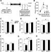
Maternal separation increases stress vulnerability in adults. A, Plasma corticosterone (CORT) levels in AFR, HMS15, and HMS180 rats before and 30 and 60 min after the initiation of a 30 min acute restraint stress (n = 6–8 for each group). HMS180 rats showed greater plasma CORT levels in response to restraint stress compared with the AFR and HMS15 rats. *p < 0.05. B, C, Bar graphs showing sucrose preference (B) and total fluid intake (C) in the sucrose preference test in AFR, HMS15, and HMS180 rats (n = 12–14 for each group) after the 14th repeated restraint stress sessions (RRS) or nonrepeated restraint stress (NRRS). HMS180 rats subjected to RRS showed significantly deceased sucrose preference compared with the nonrestrained HMS180 rats. *p < 0.05. D, E, Bar graphs showing the immobility time (D) and latency to immobility (E) in AFR, HMS15, and HMS180 rats subjected to a forced swim test (n = 12–14 for each group) after the RRS or NRRS. HMS180 rats subjected to RRS showed significantly increased immobility time (D) and decreased latency to immobility (E) compared with the nonrestrained HMS180 rats. *p < 0.05. F, G, Bar graphs showing latency to feed (F) and percentage body weight loss (G) in AFR, HMS15, and HMS180 rats subjected to a novelty-suppressed feeding test (n = 12–14 for each group) after the RRS or NRRS. *p < 0.05.
Figure 2.
Figure 2.
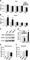
Maternal separation increases the REST4 expression in the mPFC of rats. A, B, Bar graphs showing the mRNA expressions of Rest (A) and Rest4 (B) in the mPFC of AFR, HMS15, and HMS180 rats at P7, P14, P21, P35, and P60 quantified by quantitative (Q)-PCR (n = 6 for all groups). The expression of Rest4 mRNA was significantly increased in the HMS180 rats at P7 and P14 compared with that in the age-matched HMS15 and control AFR rats (*p < 0.01). C, D, The protein expressions of REST and REST4 in the mPFC of AFR, HMS15, and HMS180 rats at P14 were quantified by Western blotting analysis (n = 4–5 for each group, 2–3 pooled tissues per n). The expression of REST4 protein was significantly increased in the HMS180 rats compared with that in the AFR and HMS15 rats (*p < 0.05). E, F, The mRNA expressions of Rest4 in the hippocampus (E) and the amygdala (F) of AFR, HMS15, and HMS180 rats at P14 were quantified by Q-PCR (n = 6 for all groups). There were no significant differences in Rest4 mRNA levels among the three groups in these brain regions.
Figure 3.
Figure 3.
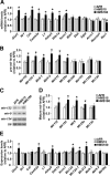
Expression analyses of mRNAs of RE-1-containing genes and brain-enriched pre-microRNAs in the mPFC of the maternally separated rats. A, B, The expression of mRNAs (A) and pre-microRNAs (B) of a variety of RE-1-containing genes in the mPFC of AFR, HMS15, and HMS180 rats at P14 were quantified by Q-PCR (n = 6 for all groups). C, D, The expression of mature microRNAs in the mPFC of AFR, HMS15, and HMS180 rats at P14 were quantified by Northern blotting analysis (n = 5–6 for each group). E, The mRNA and pre-microRNA expression of RE-1-containing genes in the mPFC of adult AFR, HMS15, and HMS180 rats were quantified by Q-PCR (n = 6 for all groups). *p < 0.05.
Figure 4.
Figure 4.
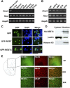
REST4 is specifically expressed in the brain and is localized in the nucleus of mPFC neurons. A, Ethidium bromide stained gels of products of reverse-transcription PCR with cDNA isolated from a variety of mouse tissues showing that Rest4 is expressed only in the brain, whereas Rest is ubiquitously expressed in the liver, lung, heart, spleen, kidney, testis and ovary. B, Within the brain, Rest4 mRNA was detected in the hippocampus (HP), mPFC, amygdala (AMY), striatum (STR), and hypothalamus (HYP). C, Fluorescence micrographs of Neuro2a cells transfected with either EGFP-REST4, EGFP-REST or EGFP expression vectors. EGFP fluorescence was detected in the nucleus of the EGFP-REST4- and EGFP-REST-transfected cells. Scale bar represents 50 μm. D, Western blots of nuclear and cytosolic fractions of Neuro2a cells transfected with pcDNA3-HA-REST4 then stained with antibodies against HA, β-actin (a marker for cytosolic protein), or histone H3 (a marker for nuclear protein). E, Anti-REST4 immunohistochemical analysis of rat coronal sections showing that REST4 immunoreactivity is colocalized with the neuronal nuclear marker NeuN in the mPFC and not with the astrocyte marker GFAP. Scale bars represent 500 and 50 μm for 2× and 20×, respectively.
Figure 5.
Figure 5.
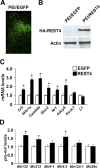
PEI-mediated REST4 overexpression in the mPFC of neonatal mice increases the expression of RE-1-containing genes. A, Fluorescence micrograph of neonatal mouse mPFC after being injected (P6–P7) with EGFP expression plasmids complexed with PEI. Three days after the injection, EGFP fluorescence was detected in the mPFC region. B, Western blotting with anti-HA antibody shows the transduction of HA-REST4 in the mPFC of mice 3 d after the injection of PEI/Rest4 complexes. C, D, Bar graphs showing Q-PCR of the levels of mRNAs (C) and microRNAs (D) in the mPFC of mice 3 d after injection of either the PEI/Egfp or PEI/Rest4 complexes (n = 6 for each group). *p < 0.05.
Figure 6.
Figure 6.
PEI-mediated REST4 overexpression in the mPFC of neonatal mice increases stress vulnerability. A, Schematic of experimental design. PEI/Rest4 or control PEI/Egfp complexes were injected to the mPFC of mice at P6–P7. Adult mice were subjected to repeated restraint stress (RRS) or nonrepeatedly restrained stress (NRRS) for 14 consecutive d, and then assessed for anxiety- and depression-like behaviors in the sucrose preference (SPT), forced swim (FST) and novelty-suppressed feeding (NSF) tests (n = 14–16 for each group; see Materials and Methods for details). B, Plasma corticosterone (CORT) levels before and 30 and 60 min after the initiation of restraint stress in mice injected with either PEI/Egfp or PEI/Rest4 complexes (n = 5–7 for each group). Plasma CORT levels in response to restraint stress were higher in the mice with REST4 overexpression compared with those with EGFP overexpression (*p < 0.05). C, D, Results of the sucrose preference test after RRS or NRRS, showing sucrose preference (C) and the total intake of fluids (D). REST4 overexpression mice subjected to RRS showed significantly deceased sucrose preference compared with nonrestrained REST4 overexpression mice (*p < 0.05). E, F, Results of the forced swim test after RRS or NRRS, showing immobility time (E) and the latency to first immobility (F). REST4 overexpression mice subjected to RRS showed significantly increased immobility time (E) but normal latency to first immobility (F) compared with the nonrestrained EGFP overexpression mice (*p < 0.01). G, H, Results of the novelty-suppressed feeding test after RRS or NRRS, showing the latency to feed (G) and the percentage body weight loss (H) in each group. REST4 overexpression mice with and without RRS showed significantly increased latencies to feed compared with corresponding EGFP overexpression mice (*p < 0.05).
Figure 7.
Figure 7.
Effects of REST4 overexpression in the mPFC of adult mice on stress vulnerability. A, Schematic of experimental design. rAAV-REST4 or control rAAV-EGFP were injected into the mPFC of adult mice at P56. Two weeks after the injection, mice were subjected to repeated restraint stress (RRS) or nonrepeatedly restrained stress (NRRS) for 14 consecutive days, and then assessed for anxiety- and depression-like behaviors in the sucrose preference (SPT), forced swim (FST), and novelty-suppressed feeding (NSF) tests (n = 11–14 for each group). B, Fluorescence micrograph of adult mouse mPFC after injection of AAV-REST4. Four weeks after the injection, HA-REST4 immunofluorescence was detected in the mPFC region. C, Western blotting with anti-HA antibody shows the overexpression of HA-REST4 in the mPFC of mice 4 weeks after the injection of AAV-REST4. D, E, Results of the sucrose preference test after RRS or NRRS, showing sucrose preference (D) and the total intake of fluids (E). F, Results of the forced swim test after RRS or NRRS, showing immobility time. G, H, Results of the novelty-suppressed feeding test after RRS or NRRS, showing the latency to feed (G) and the percentage body weight loss (H) in each group.

Similar articles

Cited by

References

    1. American Psychiatric Association. Diagnostic and Statistical Manual of Mental Disorders. Ed 4. Washington, DC: American Psychiatric Association; 1994.
    1. Andrés ME, Burger C, Peral-Rubio MJ, Battaglioli E, Anderson ME, Grimes J, Dallman J, Ballas N, Mandel G. CoREST: a functional corepressor required for regulation of neural-specific gene expression. Proc Natl Acad Sci U S A. 1999;96:9873–9878. - PMC - PubMed
    1. Armario A, Martí O, Vallès A, Dal-Zotto S, Ons S. Long-term effects of a single exposure to immobilization on the hypothalamic-pituitary-adrenal axis: neurobiologic mechanisms. Ann N Y Acad Sci. 2004;1018:162–172. - PubMed
    1. Arnsten AF. Stress signalling pathways that impair prefrontal cortex structure and function. Nat Rev Neurosci. 2009;10:410–422. - PMC - PubMed
    1. Ballas N, Battaglioli E, Atouf F, Andres ME, Chenoweth J, Anderson ME, Burger C, Moniwa M, Davie JR, Bowers WJ, Federoff HJ, Rose DW, Rosenfeld MG, Brehm P, Mandel G. Regulation of neuronal traits by a novel transcriptional complex. Neuron. 2001;31:353–365. - PubMed

Publication types

MeSH terms