. 2010 Nov 10;30(45):15019-29.
doi: 10.1523/JNEUROSCI.2071-10.2010.

Cleavage at the 586 amino acid caspase-6 site in mutant huntingtin influences caspase-6 activation in vivo

Affiliations

Cleavage at the 586 amino acid caspase-6 site in mutant huntingtin influences caspase-6 activation in vivo

Rona K Graham et al. J Neurosci. .

Abstract

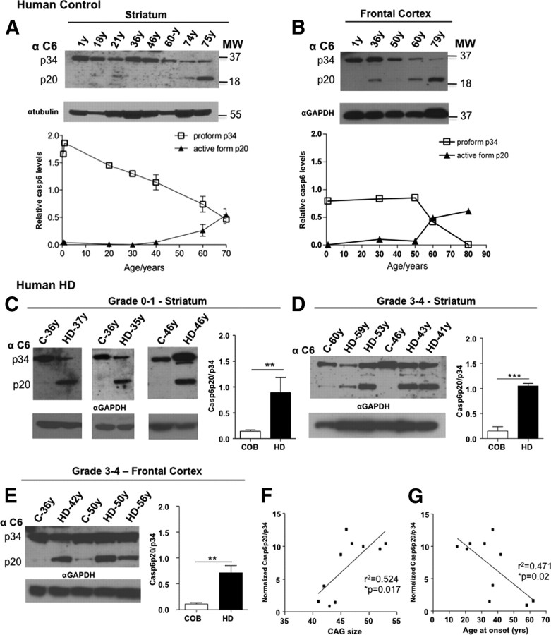
Caspase cleavage of huntingtin (htt) and nuclear htt accumulation represent early neuropathological changes in brains of patients with Huntington's disease (HD). However, the relationship between caspase cleavage of htt and caspase activation patterns in the pathogenesis of HD remains poorly understood. The lack of a phenotype in YAC mice expressing caspase-6-resistant (C6R) mutant htt (mhtt) highlights proteolysis of htt at the 586 aa caspase-6 (casp6) site as a key mechanism in the pathology of HD. The goal of this study was to investigate how proteolysis of htt at residue 586 plays a role in the pathogenesis of HD and determine whether inhibiting casp6 cleavage of mhtt alters cell-death pathways in vivo. Here we demonstrate that activation of casp6, and not caspase-3, is observed before onset of motor abnormalities in human and murine HD brain. Active casp6 levels correlate directly with CAG size and inversely with age of onset. In contrast, in vivo expression of C6R mhtt attenuates caspase activation. Increased casp6 activity and apoptotic cell death is evident in primary striatal neurons expressing caspase-cleavable, but not C6R, mhtt after NMDA application. Pretreatment with a casp6 inhibitor rescues the apoptotic cell death observed in this paradigm. These data demonstrate that activation of casp6 is an early marker of disease in HD. Furthermore, these data provide a clear link between excitotoxic pathways and proteolysis and suggest that C6R mhtt protects against neurodegeneration by influencing the activation of neuronal cell-death and excitotoxic pathways operative in HD.


Figures

Figure 1.
Figure 1.
Activation of caspase-6 is an early event in human HD striatum and correlates with CAG size. A, B, After the fifth decade, a decrease in the proform of casp6 is observed in the striatum and frontal cortex of human control tissue with a concomitant increase in the active p20 fragment of casp6. C, Increased levels of the active p20 form of casp6 are observed in early-grade human HD striatum compared with control brain (p < 0.01). D, E, Grade 3–4 human HD striatum and frontal cortex similarly demonstrate increased active p20 casp6 compared with control tissue (p < 0.001 and p < 0.01, respectively). Glyceraldehyde-3-phosphate dehydrogenase (GAPDH) demonstrates loading for all tissues. Representative immunoblots are shown for casp6. F, A positive correlation is observed between striatal expression of normalized active casp6 levels and CAG size (F (1,8) = 8.9, r 2 = 0.524, p = 0.017). G, Comparison of age of onset and normalized levels of active casp6 reveals an inverse correlation between these two variables (F (1,8) = 7.1, r 2 = 0.471, p = 0.02). Levels of casp6 have been corrected for equal loading with tubulin or GAPDH. Mean measurements and densitometric ratios of casp6p20/p34 and relative casp6 levels are ±SD. **p < 0.01; ***p < 0.001. MW, Molecular weight; COB, Control.
Figure 2.
Figure 2.
Accelerated activation of caspase-6 is observed in the striatum of YAC128 mice compared with wild-type and C6R mice. A, In WT murine brain, active casp6 is detected predominantly in medium-sized striatal neurons starting at 9 months (a–d). Active casp6 is detected in medium-sized striatal neurons of YAC128 mice by 3 months (e–h). In the striatum of two lines of mice expressing C6R mhtt (C6R7 and C6R13), low levels of activated casp6 are observed at 3 months with no change as the animals age (i–p). B–D, Increased casp6 activity is observed in YAC128 striatum compared with WT and C6R striatum at 3 months (B) (ANOVA, p = 0.004; WT vs YAC128, p < 0.05; C6R vs YAC128, p < 0.01), 12 months (C) (ANOVA, p = 0.003; WT vs YAC128, p < 0.01; C6R vs YAC128, p < 0.05), and 18 months (D) (ANOVA, p = 0.018; WT vs YAC128, p < 0.05; WT vs C6R, p > 0.05) of age. E, The neo-epitope antibody to casp6-cleaved tau strongly immunostains medium-sized striatal neurons in YAC128 striata and to a lesser extent in WT and C6R striatum (18 months). casp6 activity measurements are ±SEM. *p < 0.05; **p < 0.01. Magnification, 100×. Scale bar, 10 μm.
Figure 3.
Figure 3.
Caspase-6 activation is observed in YAC128 cortex subsequent to striatal activation. A, In murine WT cortex, expression of active casp6 is observed predominantly in layers 2–5 of the cortex commencing at 12 months of age (a–f). In contrast, at 9 months of age, YAC128 cortex demonstrates some immunopositive active casp6 stained neurons, and this increases with age (g–l). In mice expressing C6R mhtt (lines C6R7 and C6R13), a low level of immunopositive active casp6-stained neurons are observed at 12 and 18 months of age (m–x). B, C, A significant increase in casp6 activity is observed in YAC128 cortex compared with WT and C6R at 12 months (B) (ANOVA, p = 0.013; WT vs YAC128, p < 0.05; C6R vsYAC128, p < 0.05) and 18 months (C) (ANOVA, p = 0.002; WT vs YAC128, p < 0.01; C6R vsYAC128, p < 0.05) of age. Arrow points to active casp6 immunostaining on neurites. Casp6 activity measurements are ±SEM. *p < 0.05; **p < 0.01. Magnification, 40× and 100×. Scale bars, 10 μm.
Figure 4.
Figure 4.
Disease severity correlates with striatum and cortex caspase-6 activity in mouse models of HD. A, Onset and progression of HD is modulated by expression levels of mhtt in the YAC128 mouse models (Graham et al., 2006b). Western blot probed with N-terminal BKP1 htt antibody. B, At 3 months of age, no difference in casp6 activity is observed between WT or HD54 striatum (p = 0.34). One-way ANOVA linear trend post hoc test reveals a significant trend between WT, YAC128–HD54, and YAC128–HD53 for casp6 activity (ANOVA, p = 0.105, post hoc for linear trend: slope of 0.065, r 2 = 0.1493, p = 0.03). C, Assessment of casp6 activity in brain tissue from the BACHD model at 12 months of age reveals a significant increase in casp6 activity in both striatum and cortex of BACHD mice compared with WT (p = 0.003 and p = 0.02, respectively). casp6 activity measurements are ±SEM. *p < 0.05; **p < 0.01.
Figure 5.
Figure 5.
Activation of caspase-3 is not observed early in the pathogenesis of HD. A, No difference is observed in proform levels of casp3 in grade 0–1 human HD striatum compared with age-matched control tissue. B, In grade 3–4 HD striatum, there is a trend toward decreased p34 proform levels of casp3 compared with control tissue (p = 0.06). C, In grade 3–4 HD frontal cortex, an increase in p34 proform casp3 levels is observed compared with control tissue (p = 0.02). The active p20 casp3 fragments were not detected in control or HD striatal or frontal cortex tissue. D, No immunopositive active casp3 staining is observed in the striatum of WT or YAC128 brain. Immunopositive active casp3 staining is detected in the corpus callosum of both WT and YAC128 brain slices, but no difference in the extent of staining is observed between genotypes. E, No difference in casp3 activity is observed in WT versus YAC128 striatum at 3 or 12 months of age (3 months, p = 0.11; 12 months, p = 0.62). Mean measurements and densitometric ratios of casp3p34/GAPDH and casp3 activity measurements are ±SEM. *p < 0.05. Magnification, 100×. Scale bar, 10 μm.
Figure 6.
Figure 6.
NMDA-mediated excitotoxic stress triggers caspase-6 activation. A, B, Primary MSNs were double labeled with active casp6 neo-epitope antibody and Hoechst. In YAC128 MSNs, the percentage of MSNs with active casp6 immunoreactivity above background increased after NMDA treatment compared with WT at 6 h (p < 0.05) and a trend increased at 4 h (p = 0.07). C, A significant increase in casp6 activity is observed in neurons expressing mhtt compared with WT 3.5 h after NMDA application (t test, WT vs YAC128: p = 0.015 and p = 0.011, respectively). No increase in casp6 activity is observed in C6R MSNs after NMDA treatment (ANOVA, p = 0.04; WT vs C6R, p > 0.05; WT vs YAC128, p < 0.05). D, A significant decrease in NMDAR-mediated apoptotic cell death is observed in MSNs pretreated with z-VEID-fmk from WT (ANOVA, p = 0.014; percentage apoptotic neurons in NMDA vs NMDA plus casp6 inhibitor, p < 0.05) and YAC128 (ANOVA, p = 0.0001; percentage apoptotic neurons in BSS vs NMDA, p < 0.001; NMDA vs NMDA plus casp6 inhibitor, p < 0.001) striata. E, A significant decrease in NMDAR-mediated apoptotic cell death is observed in MSNs pretreated with z-DEVD-fmk from WT (ANOVA, p = 0.002; percentage apoptotic neurons in NMDA vs NMDA plus casp3 inhibitor, p < 0.01) and YAC128 (ANOVA, p = 0.0001; percentage apoptotic neurons in BSS vs NMDA, p < 0.001; NMDA vs NMDA plus casp3 inhibitor, p < 0.001) striata. Relative casp6 activity measurements are ±SD. Mean percentage apoptotic cell death is given ±SD. *p < 0.05; ***p < 0.001.

Similar articles

Cited by

References

    1. Albrecht S, Bourdeau M, Bennett D, Mufson EJ, Bhattacharjee M, LeBlanc AC. Activation of caspase-6 in aging and mild cognitive impairment. Am J Pathol. 2007;170:1200–1209. - PMC - PubMed
    1. Allsopp TE, McLuckie J, Kerr LE, Macleod M, Sharkey J, Kelly JS. Caspase 6 activity initiates caspase 3 activation in cerebellar granule cell apoptosis. Cell Death Differ. 2000;7:984–993. - PubMed
    1. Andrew SE, Goldberg YP, Theilmann J, Zeisler J, Hayden MR. A CCG repeat polymorphism adjacent to the CAG repeat in the Huntington disease gene: implications for diagnostic accuracy and predictive testing. Hum Mol Genet. 1994;3:65–67. - PubMed
    1. Aziz NA, van der Burg JM, Landwehrmeyer GB, Brundin P, Stijnen T, Roos RA EHDI Study Group. Weight loss in Huntington disease increases with higher CAG repeat number. Neurology. 2008;71:1506–1513. - PubMed
    1. Banwait S, Galvan V, Zhang J, Gorostiza OF, Ataie M, Huang W, Crippen D, Koo EH, Bredesen DE. C-terminal cleavage of the amyloid-beta protein precursor at Asp664: a switch associated with Alzheimer's disease. J Alzheimers Dis. 2008;13:1–16. - PMC - PubMed

Publication types

MeSH terms