. 2010 Nov 10;30(45):15113-23.
doi: 10.1523/JNEUROSCI.3947-10.2010.

IL-6- and NGF-induced rapid control of protein synthesis and nociceptive plasticity via convergent signaling to the eIF4F complex

Affiliations

IL-6- and NGF-induced rapid control of protein synthesis and nociceptive plasticity via convergent signaling to the eIF4F complex

Ohannes K Melemedjian et al. J Neurosci. .

Abstract

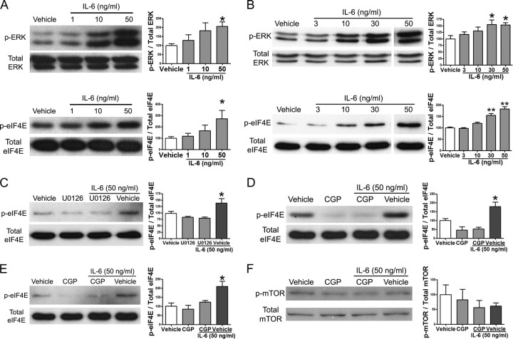
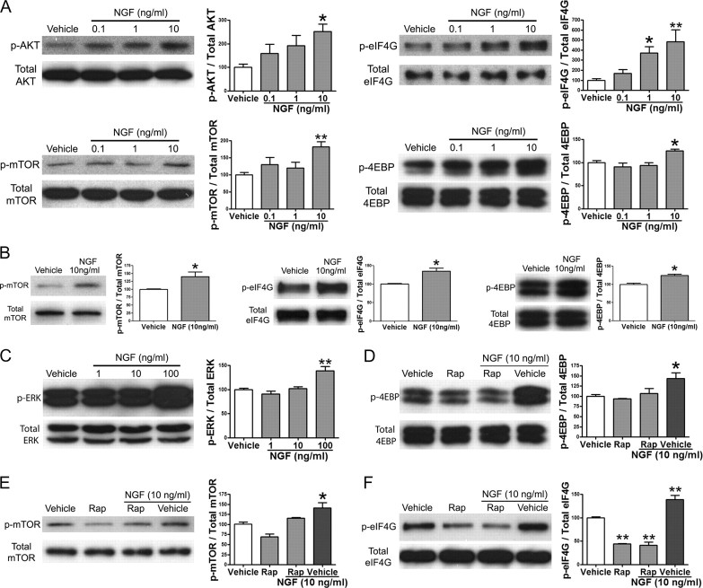
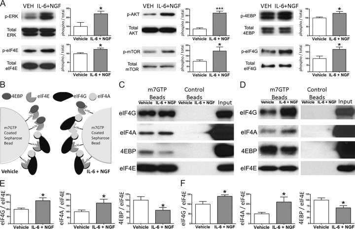
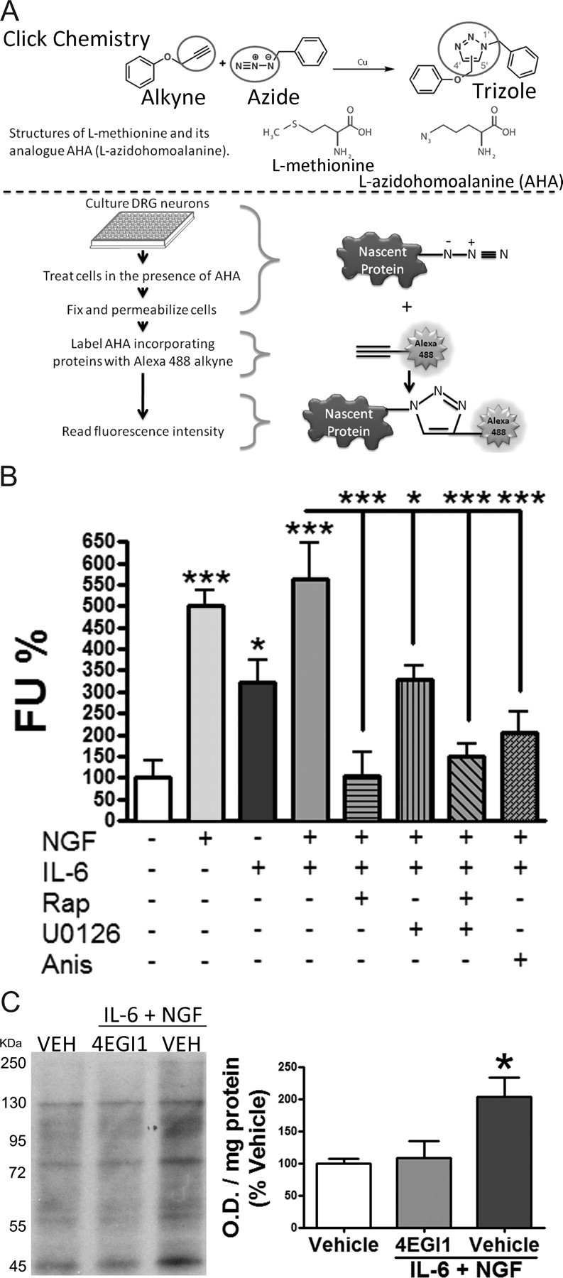
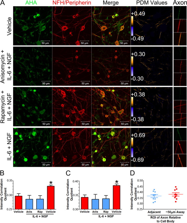
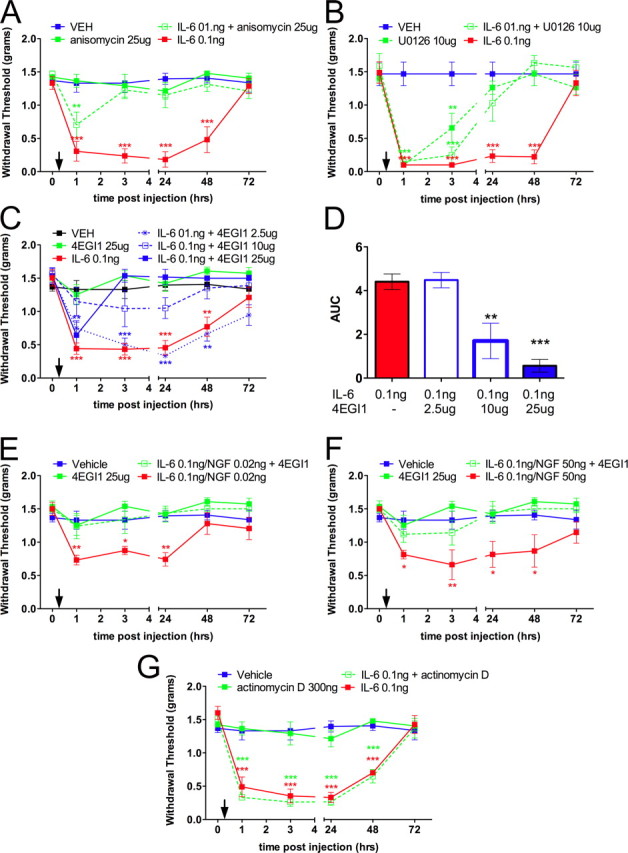
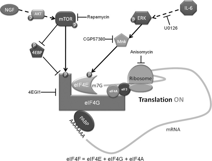
Despite the emergence of translational control pathways as mediators of nociceptive sensitization, effector molecules and mechanisms responsible for modulating activity in these pathways in pain conditions are largely unknown. We demonstrate that two major algogens, the cytokine interleukin 6 (IL-6) and the neurotrophin nerve growth factor (NGF), which are intimately linked to nociceptive plasticity across preclinical models and human pain conditions, signal primarily through two distinct pathways to enhance translation in sensory neurons by converging onto the eukaryotic initiation factor (eIF) eIF4F complex. We directly demonstrate that the net result of IL-6 and NGF signaling is an enhancement of eIF4F complex formation and an induction of nascent protein synthesis in primary afferent neurons and their axons. Moreover, IL-6- and NGF-induced mechanical nociceptive plasticity is blocked by inhibitors of general and cap-dependent protein synthesis. These results establish IL-6- and NGF-mediated cap-dependent translation of local proteins as a new model for nociceptive plasticity.


Figures

Figure 1.
Figure 1.
Interleukin-6 signals to the translational machinery through ERK and Mnk1. A, Western blot and quantification for p-ERK, total ERK, p-eIF4E, and total eIF4E following 15 min of treatment of DRG cultures with increasing doses of IL-6. B, Western blot and quantification for p-ERK, total ERK, p-eIF4E, and total eIF4E following 15 min of treatment of TG cultures with escalating doses of IL-6. C, Western blot and quantification for p-eIF4E and total eIF4E following pretreatment of DRG neurons in culture with the MEK inhibitor U0126 (10 μm) for 30 min and subsequent treatment with IL-6 (50 ng/ml) for 15 min. D, Western blot and quantification for p-eIF4E and total eIF4E following pretreatment of DRG cultures with the Mnk1 inhibitor CGP57380 (CGP, 50 μm) for 30 min and subsequent treatment with IL-6 (50 ng/ml) for 15 min. E, The Mnk1 inhibitor CGP57380 also inhibited IL-6-induced phosphorylation of eIF4E in TG neurons. F, IL-6 (50 ng/ml) did not stimulate mTOR phosphorylation in DRG neurons, and this was also not changed by CGP57380 (50 μm). Phosphorylated proteins were standardized relative to their respective totals and shown as percentage of vehicle. *p < 0.05, **p < 0.01.
Figure 2.
Figure 2.
NGF signals to the translational machinery by activating the mTOR pathway. A, Western blots and quantification for p-AKT, total AKT, p-mTOR, total mTOR, p-eIF4G, total eIF4G, p-4EBP, and total 4EBP following 15 min of treatment of DRG cultures with increasing doses of NGF. B, Western blot and quantification for p-mTOR, total mTOR, p-eIF4G, total eIF4G, p-4EBP, and total 4EBP following 15 min of treatment of TG cultures with NGF (10 ng/ml). C, Western blot and quantification for p-ERK and total ERK following 15 min of treatment of TG cultures with escalating doses of NGF. D–F, Western blots and quantification for p-4EBP and total 4EBP (D), p-mTOR and total mTOR (E), and p-eIF4G and total eIF4G (F) following pretreatment of DRG cultures with rapamycin (Rap, 500 nm) for 30 min and subsequent treatment with NGF (10 ng/ml) for 15 min. *p < 0.05, **p < 0.01.
Figure 3.
Figure 3.
Cotreatment with IL-6 and NGF signal through both ERK and mTOR pathways to stabilize the formation of eIF4F complex and dissociate 4EBP from eIF4E. A, Western blots and quantification for p-ERK, total ERK, p-eIF4E, total eIF4E, p-4EBP, total 4EBP, p-AKT, total AKT, p-mTOR, total mTOR, p-eIF4G, and total eIF4G following 15 min of treatment of DRG cultures with IL-6 (50 ng/ml) and NGF (10 ng/ml). B, A schematic diagram demonstrating that, in vehicle conditions (left panel), 7-methyl-GTP-conjugated Sepharose beads are associated with both the positive and negative regulators of cap-dependent translation, eIF4F (eIF4E, eIF4A, and eIF4G) and 4EBP, respectively. After cotreatment with IL-6 and NGF (right panel) the balance shifts toward the assembly of the eIF4F complex on the Sepharose beads. C, Western blots of eIF4G, eIF4A, 4EBP, and eIF4E following treatment of DRG neurons with IL-6 (50 ng/ml) and NGF (10 ng/ml) and coprecipitation with 7-methyl-GTP-conjugated Sepharose beads or control Sepharose beads. D, Western blots of eIF4G, eIF4A, 4EBP, and eIF4E following treatment of TG neurons with IL-6 (50 ng/ml) and NGF (10 ng/ml) and coprecipitation with 7-methyl-GTP-conjugated Sepharose beads or control Sepharose beads. E, eIF4G, eIF4A, and 4EBP coprecipitation from DRG were standardized relative to the levels of eIF4E and shown as percentage of vehicle. F, eIF4G, eIF4A, and 4EBP coprecipitation from TG were standardized relative to the levels of eIF4E and shown as percentage of vehicle. Cotreatment with IL-6 and NGF increased the association eIF4E to eIF4G and eIF4A while decreasing its association with 4EBP in both DRG and TG neurons. *p < 0.05.
Figure 4.
Figure 4.
IL-6 and NGF induce cap-dependent translation of nascent proteins in primary sensory neurons by activating ERK and mTOR pathways. A, A schematic diagram illustrating the experimental procedure. B, DRG neurons pretreated with rapamycin (500 nm, Rap), U0126 (10 μm), and anisomycin (50 μm, Anis) for 30 min followed by treatment with vehicle, IL-6 (50 ng/ml), and/or NGF (10 ng/ml) with and without inhibitors for 30 min. Fluorescence units were reported as percentage of vehicle. Treatment with IL-6 and/or NGF increases the incorporation of AHA, indicative of increased nascent protein synthesis, and this effect was inhibited by mTOR (rapamycin), MEK (U0126), and general protein synthesis (anisomycin) inhibitors. C, Western blot and quantification of primary sensory neurons in culture pretreated with vehicle or the inhibitor of eIF4F complex formation, 4EGI1 (20 μm), for 30 min followed by treatment with vehicle or IL-6 (50 ng/ml) and NGF (10 ng/ml) in AHA-containing media 30 min. *p < 0.05, ***p < 0.001.
Figure 5.
Figure 5.
IL-6 and NGF induce cap-dependent translation of nascent proteins in the axons of primary sensory neurons. A, Representative micrographs of intensity correlation analysis of fluorescently labeled AHA with neuronal markers (NF200 and peripherin). Neurons in cultures were treated with vehicle or with IL-6 (50 ng/ml) and NGF (10 ng/ml) with or without anisomycin (50 μm) or rapamycin (500 nm) pretreatment (30 min) and cotreatment. PDM values were used to generate an image that visualizes the extent of colocalization of AHA with NFH/peripherin in the axons (PDM Values panel). Intensity correlation analysis that excluded the cell bodies of the neurons resulted in higher PDM values with IL-6 and NGF treatment than in the vehicle group. PDM values are shown on the color bar on the far right. While the color bar scales are equivalent for all images, PDM values show the range of PDMs within that scale. Higher PDM values indicate higher intensity of colocalization between neuronal markers and AHA incorporation (more nascent protein synthesis). This increase was inhibited with anisomycin and rapamycin. High-magnification images of individual axons are shown on the far right (Axon). B, ICQ of fluorescently labeled AHA with neuronal markers (NF200 and peripherin) in axons quantified by ICA. Treatment with IL-6 (50 ng/ml) and NGF (10 ng/ml) results in a signification increase in the ICQ value in axons of primary sensory neurons in culture relative to the vehicle group. This increase was blocked with anisomycin (Anis) and rapamycin (Rap). C, ICQ analysis of the entire image (axons not excluded) showing that there is also an increase in nascent protein synthesis with IL-6 and NGF when axons and somas are considered. D, Analysis of proximal (adjacent to soma to 10 μm distal—Adjacent) versus distal (50–60 μm from the soma—∼50 μm Away) axon segments from IL-6- and NGF-treated cultures showing that the distribution of ICQ values are equivalent regardless of distance from the soma. *p < 0.05.
Figure 6.
Figure 6.
Intraplantar injection of IL-6 or coinjection of IL-6 and NGF into the hindpaw of mice induces long-lasting mechanical allodynia blocked by cotreatment with anisomycin, U0126, or 4EGI1. Mechanical thresholds were measured at the indicated time points before and after intraplantar injection (indicated by downward arrows). A, IL-6 (0.1 ng) induced a significant decrease in withdrawal thresholds that was blocked by coinjection of the general protein synthesis inhibitor anisomycin (25 μg). B, IL-6 (0.1 ng)-induced decreases in withdrawal thresholds were blocked by coinjection of the MEK inhibitor U0126 (10 μg). C, D, IL-6 (0.1 ng)-mediated allodynia was blocked with the inhibitor of eIF4F complex formation, 4EGI1 (2.5, 10, and 25 μg) in a dose-dependent manner. E, F, Coinjection of IL-6 (0.1 ng) and NGF [0.02 (E) and 50 (F) ng] resulted in a significant decrease in the mechanical thresholds at the indicated time points, and these effects were blocked by coinjection of 4EGI1 (25 μg). G, Coinjection of the transcription inhibitor actinomycin D (300 ng) had no effect on IL-6 (0.1 ng)-mediated allodynia. *p < 0.05, **p < 0.01, ***p < 0.001.
Figure 7.
Figure 7.
Schematic summarizing the present findings. IL-6 signals through the MEK/ERK/Mnk pathway to phosphorylate the cap-binding protein eIF4E. NGF signals primarily through the PI3 kinase/AKT/mTOR pathway to phosphorylate both 4EBP and eIF4G. Phosphorylation of 4EBP, which is a negative regulator of eIF4F complex formation, results in its dissociation from eIF4E, thus allowing the binding of eIF4E to eIF4G to occur. Phosphorylation of eIF4E and eIF4G enhances the formation of the eIF4F cap-binding complex. The outcome of IL-6- and NGF-mediated eIF4F cap complex formation is the enhancement of cap-dependent translation, leading to rapid changes in translational control of gene expression in sensory neurons, and this effect is linked to mechanical allodynia evoked by these algogens.

Similar articles

Cited by

References

    1. Akira S, Yoshida K, Tanaka T, Taga T, Kishimoto T. Targeted disruption of the IL-6 related genes: gp130 and NF-IL-6. Immunol Rev. 1995;148:221–253. - PubMed
    1. Amaya F, Wang H, Costigan M, Allchorne AJ, Hatcher JP, Egerton J, Stean T, Morisset V, Grose D, Gunthorpe MJ, Chessell IP, Tate S, Green PJ, Woolf CJ. The voltage-gated sodium channel Na(v)1.9 is an effector of peripheral inflammatory pain hypersensitivity. J Neurosci. 2006;26:12852–12860. - PMC - PubMed
    1. Anderson LC, Rao RD. Interleukin-6 and nerve growth factor levels in peripheral nerve and brainstem after trigeminal nerve injury in the rat. Arch Oral Biol. 2001;46:633–640. - PubMed
    1. Andratsch M, Mair N, Constantin CE, Scherbakov N, Benetti C, Quarta S, Vogl C, Sailer CA, Uceyler N, Brockhaus J, Martini R, Sommer C, Zeilhofer HU, Müller W, Kuner R, Davis JB, Rose-John S, Kress M. A key role for gp130 expressed on peripheral sensory nerves in pathological pain. J Neurosci. 2009;29:13473–13483. - PMC - PubMed
    1. Asante CO, Wallace VC, Dickenson AH. Formalin-induced behavioural hypersensitivity and neuronal hyperexcitability are mediated by rapid protein synthesis at the spinal level. Mol Pain. 2009;5:27. - PMC - PubMed

Publication types

MeSH terms

Substances