Context-specific grasp movement representation in macaque ventral premotor cortex
- PMID: 21068323
- PMCID: PMC6633833
- DOI: 10.1523/JNEUROSCI.3343-10.2010
Context-specific grasp movement representation in macaque ventral premotor cortex
Abstract
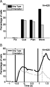
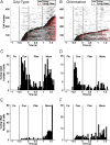
Hand grasping requires the transformation of sensory signals to hand movements. Neurons in area F5 (ventral premotor cortex) represent specific grasp movements (e.g., precision grip) as well as object features like orientation, and are involved in movement preparation and execution. Here, we examined how F5 neurons represent context-dependent grasping actions in macaques. We used a delayed grasping task in which animals grasped a handle either with a power or a precision grip depending on context information. Additionally, object orientation was varied to investigate how visual object features are integrated with context information. In 420 neurons from two animals, object orientation and grip type were equally encoded during the instruction epoch (27% and 26% of all cells, respectively). While orientation representation dropped during movement execution, grip type representation increased (20% vs 43%). According to tuning onset and offset, we classified neurons as sensory, sensorimotor, or motor. Grip type tuning was predominantly sensorimotor (28%) or motor (25%), whereas orientation-tuned cells were mainly sensory (11%) or sensorimotor (15%) and often also represented grip type (86%). Conversely, only 44% of grip-type tuned cells were also orientation-tuned. Furthermore, we found marked differences in the incidence of preferred conditions (power vs precision grips and middle vs extreme orientations) and in the anatomical distribution of the various cell classes. These results reveal important differences in how grip type and object orientation is processed in F5 and suggest that anatomically and functionally separable cell classes collaborate to generate hand grasping commands.
Figures


Similar articles
-
Context-specific grasp movement representation in the macaque anterior intraparietal area.J Neurosci. 2009 May 20;29(20):6436-48. doi: 10.1523/JNEUROSCI.5479-08.2009. J Neurosci. 2009. PMID: 19458215 Free PMC article.
-
Neural Dynamics of Variable Grasp-Movement Preparation in the Macaque Frontoparietal Network.J Neurosci. 2018 Jun 20;38(25):5759-5773. doi: 10.1523/JNEUROSCI.2557-17.2018. Epub 2018 May 24. J Neurosci. 2018. PMID: 29798892 Free PMC article.
-
Decoding the activity of grasping neurons recorded from the ventral premotor area F5 of the macaque monkey.Neuroscience. 2011 Aug 11;188:80-94. doi: 10.1016/j.neuroscience.2011.04.062. Epub 2011 May 14. Neuroscience. 2011. PMID: 21575688
-
The extended object-grasping network.Exp Brain Res. 2017 Oct;235(10):2903-2916. doi: 10.1007/s00221-017-5007-3. Epub 2017 Jul 26. Exp Brain Res. 2017. PMID: 28748312 Review.
-
Basal ganglia mechanisms underlying precision grip force control.Neurosci Biobehav Rev. 2009 Jun;33(6):900-8. doi: 10.1016/j.neubiorev.2009.03.004. Epub 2009 Mar 14. Neurosci Biobehav Rev. 2009. PMID: 19428499 Free PMC article. Review.
Cited by
-
Object vision to hand action in macaque parietal, premotor, and motor cortices.Elife. 2016 Jul 26;5:e15278. doi: 10.7554/eLife.15278. Elife. 2016. PMID: 27458796 Free PMC article.
-
Grasping actions and social interaction: neural bases and anatomical circuitry in the monkey.Front Psychol. 2015 Jul 14;6:973. doi: 10.3389/fpsyg.2015.00973. eCollection 2015. Front Psychol. 2015. PMID: 26236258 Free PMC article. Review.
-
Visually and Tactually Guided Grasps Lead to Different Neuronal Activity in Non-human Primates.Front Neurosci. 2021 Jul 19;15:679910. doi: 10.3389/fnins.2021.679910. eCollection 2021. Front Neurosci. 2021. PMID: 34349616 Free PMC article.
-
Neural model for learning-to-learn of novel task sets in the motor domain.Front Psychol. 2013 Oct 22;4:771. doi: 10.3389/fpsyg.2013.00771. eCollection 2013. Front Psychol. 2013. PMID: 24155736 Free PMC article.
-
The modulation of corticospinal excitability during motor imagery of actions with objects.PLoS One. 2011;6(10):e26006. doi: 10.1371/journal.pone.0026006. Epub 2011 Oct 13. PLoS One. 2011. PMID: 22022491 Free PMC article.
References
-
- Amemori K, Sawaguchi T. Rule-dependent shifting of sensorimotor representation in the primate prefrontal cortex. Eur J Neurosci. 2006;23:1895–1909. - PubMed
-
- Andersen RA, Buneo CA. Intentional maps in posterior parietal cortex. Annu Rev Neurosci. 2002;25:189–220. - PubMed
-
- Barbas H, Pandya DN. Architecture and frontal cortical connections of the premotor cortex (area 6) in the rhesus monkey. J Comp Neurol. 1987;256:211–228. - PubMed
-
- Battaglini PP, Muzur A, Galletti C, Skrap M, Brovelli A, Fattori P. Effects of lesions to area V6A in monkeys. Exp Brain Res. 2002;144:419–422. - PubMed
Publication types
MeSH terms
LinkOut - more resources
Full Text Sources
Miscellaneous
