Cortical GABAergic interneurons transiently assume a sea urchin-like nonpolarized shape before axon initiation
- PMID: 21068327
- PMCID: PMC6633849
- DOI: 10.1523/JNEUROSCI.1527-10.2010
Cortical GABAergic interneurons transiently assume a sea urchin-like nonpolarized shape before axon initiation
Abstract
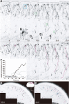
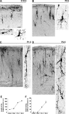
Mature neurons polarize by extending an axon and dendrites. In vitro studies of dissociated neurons have demonstrated that axons are initiated from a nonpolarized stage. Dissociated hippocampal neurons form four to five minor neurites shortly after plating but then one of them starts to elongate rapidly to become the future axon, whereas the rest constitutes the dendrites at later stages. However, neuroepithelial cells as well as migrating neurons in vivo are already polarized, raising the possibility that mature neurons inherit the polarities of immature neurons of neuroepithelial or migrating neurons. Here we show that the axon of interneurons in mouse cortical explant emerges from a morphologically nonpolarized shape. The morphological maturation of cortical interneurons labeled by electroporation at an embryonic stage was analyzed by time-lapse imaging during the perinatal stage. In contrast to earlier stages, most interneurons at this stage show sea urchin-like nonpolarized shapes with alternately extending and retracting short processes. Abruptly, one of these processes extends to give rise to an outstandingly long axon-like process. Given that the interneurons exhibit typical polarized shapes during embryonic development, the present results suggest that axon-dendrite polarity develops from a nonpolarized intermediate stage.
Figures


Similar articles
-
Excitatory cortical neurons with multipolar shape establish neuronal polarity by forming a tangentially oriented axon in the intermediate zone.Cereb Cortex. 2013 Jan;23(1):105-13. doi: 10.1093/cercor/bhr383. Epub 2012 Jan 19. Cereb Cortex. 2013. PMID: 22267309
-
Formation of axon-dendrite polarity in situ: initiation of axons from polarized and non-polarized cells.Dev Growth Differ. 2012 Apr;54(3):398-407. doi: 10.1111/j.1440-169X.2012.01344.x. Dev Growth Differ. 2012. PMID: 22524609 Review.
-
[Cholinergic neurons in chronically isolated associative cortical slab of the cat].Neirofiziologiia. 1989;21(1):60-6. Neirofiziologiia. 1989. PMID: 2725786 Russian.
-
Behaviour of small inhibitory interneurons in early postnatal mouse cerebellar microexplant cultures: a video time-lapse analysis.Eur J Neurosci. 1995 Jul 1;7(7):1449-59. doi: 10.1111/j.1460-9568.1995.tb01140.x. Eur J Neurosci. 1995. PMID: 7551171
-
Mechanisms of axon polarization in pyramidal neurons.Mol Cell Neurosci. 2020 Sep;107:103522. doi: 10.1016/j.mcn.2020.103522. Epub 2020 Jul 10. Mol Cell Neurosci. 2020. PMID: 32653476 Review.
Cited by
-
Neuronal polarization in the developing cerebral cortex.Front Neurosci. 2015 Apr 8;9:116. doi: 10.3389/fnins.2015.00116. eCollection 2015. Front Neurosci. 2015. PMID: 25904841 Free PMC article. Review.
-
From migration to settlement: the pathways, migration modes and dynamics of neurons in the developing brain.Proc Jpn Acad Ser B Phys Biol Sci. 2016;92(1):1-19. doi: 10.2183/pjab.92.1. Proc Jpn Acad Ser B Phys Biol Sci. 2016. PMID: 26755396 Free PMC article. Review.
-
Intrinsic and extrinsic mechanisms control the termination of cortical interneuron migration.J Neurosci. 2012 Apr 25;32(17):6032-42. doi: 10.1523/JNEUROSCI.3446-11.2012. J Neurosci. 2012. PMID: 22539863 Free PMC article.
-
Developmental trajectories of GABAergic cortical interneurons are sequentially modulated by dynamic FoxG1 expression levels.Proc Natl Acad Sci U S A. 2024 Apr 16;121(16):e2317783121. doi: 10.1073/pnas.2317783121. Epub 2024 Apr 8. Proc Natl Acad Sci U S A. 2024. PMID: 38588430 Free PMC article.
References
-
- Anderson SA, Eisenstat DD, Shi L, Rubenstein JL. Interneuron migration from basal forebrain to neocortex: dependence on Dlx genes. Science. 1997;278:474–476. - PubMed
-
- Arimura N, Kaibuchi K. Abstract Neuronal polarity: from extracellular signals to intracellular mechanisms. Nat Rev Neurosci. 2007;8:194–205. - PubMed
-
- Bradke F, Dotti CG. Establishment of neuronal polarity: lessons from cultured hippocampal neurons. Curr Opin Neurobiol. 2000;10:574–581. - PubMed
Publication types
MeSH terms
Substances
LinkOut - more resources
Full Text Sources
Miscellaneous
