. 2011 Jan;300(1):C164-75.
doi: 10.1152/ajpcell.00247.2010. Epub 2010 Nov 10.

The phosphorylation state of MRLC is polyamine dependent in intestinal epithelial cells

Affiliations

The phosphorylation state of MRLC is polyamine dependent in intestinal epithelial cells

Mitulkumar N Bavaria et al. Am J Physiol Cell Physiol. 2011 Jan.

Abstract

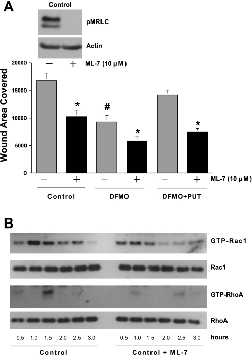
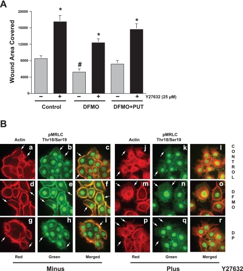
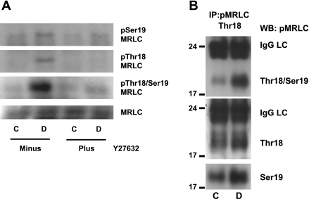
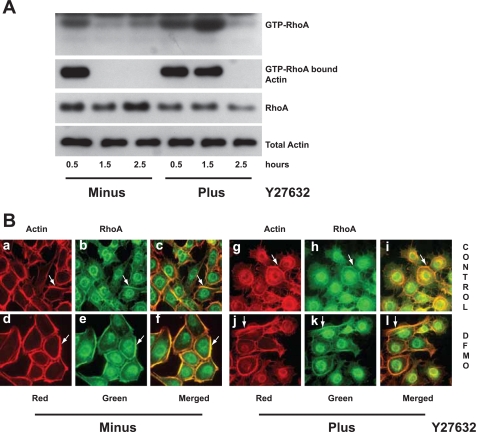
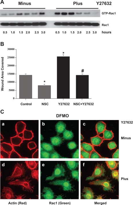
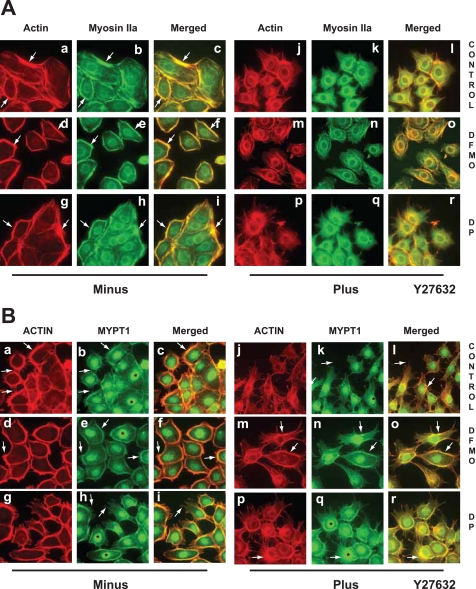
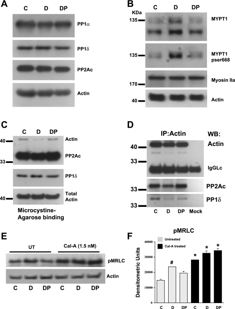
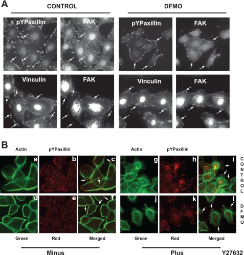
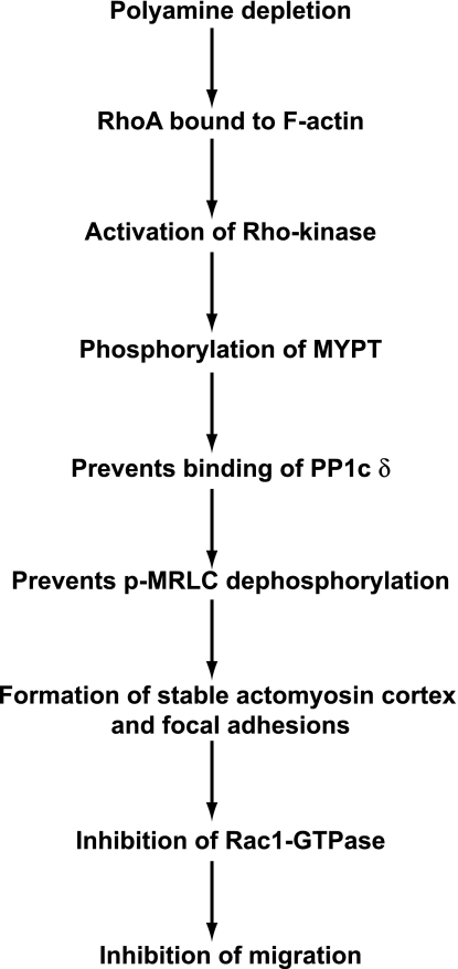
Cell migration is important to the integrity of the gastrointestinal tract for the normal movement of cells from crypt to villi and the healing of wounds. Polyamines are essential to cell migration, mucosal restitution, and, hence, healing. Polyamine depletion by α-difluoromethyl ornithine (DFMO) inhibited migration by decreasing lamellipodia and stress fiber formation and preventing the activation of Rho-GTPases. Polyamine depletion increased the association of the thick F-actin cortex with phosphorylated myosin regulatory light chain (pMRLC). In this study, we determined why MRLC is constitutively phosphorylated as part of the actin cortex. Inhibition of myosin light chain kinase (MLCK) decreased RhoA and Rac1 activities and significantly inhibited migration. Polyamine depletion increased phosphorylation of MRLC (Thr18/Ser19) and stabilized the actin cortex and focal adhesions. The Rho-kinase inhibitor Y27632 increased spreading and migration by decreasing the phosphorylation of MRLC, remodeling focal adhesions, and by activating Rho-GTPases. Thus phosphorylation of MRLC appears to be the rate-limiting step during the migration of IEC-6 cells. In addition, increased localization of RhoA with the actin cortex in polyamine-depleted cells appears to activate Rho-kinase. In the absence of polyamines, activated Rho-kinase phosphorylates myosin phosphatase targeting subunit 1 (MYPT1) at serine-668 leading to its inactivation and preventing the recruitment of phosphatase (protein phosphastase, PP1cδ) to the actomyosin cortex. In this condition, MRLC is constitutively phosphorylated and cycling does not occur. Thus activated myosin binds F-actin stress fibers and prevents focal adhesion turnover, Rho-GTPase activation, and the remodeling of the cytoskeleton required for migration.


Figures

Fig. 1.
Fig. 1.
Myosin light chain kinase (MLCK) activity is essential for migration. IEC-6 cells were grown to confluence in control, α-difluoromethyl ornithine (DFMO), and DFMO + putrescine (DFMO + PUT) containing media for 3 days followed by serum starvation for 24 h. Confluent monolayers were wounded with a gel-loading tip in the center of the plates, washed and left untreated (UT), or treated with 10 μM ML-7. A: migration was calculated as described in materials and methods. Values are means ± SE of triplicates. *Significantly different compared with respective UT groups. #Significantly different compared with UT control and DFMO + PUT. Inset, the effect of ML-7 on the levels of phosphorylated myosin regulatory light chain (pMRLC) in control cells. B: at indicated time intervals following wounding, cell extracts were assayed as described in materials and methods to determine the levels of total and active (GTP-RhoA and GTP-Rac1) protein by Western blot analysis. Representative blots from three observations are shown.
Fig. 2.
Fig. 2.
Inhibition of Rho-kinase stimulates migration by inhibiting pMRLC. IEC-6 cells grown as described in Fig. 1 were left untreated or treated with 25 μM Y27632. A: migration was calculated as described in materials and methods. Values are means ± SE of triplicates. *Significantly different compared with respective untreated samples. #Significantly different compared with untreated control and DFMO + PUT groups (DP). B: preconfluent IEC-6 cells grown on poly-l-lysine-coated coverslips in control, DFMO, and DFMO + PUT (DP)-containing media for 3 days followed by serum starvation for 24 h were left untreated or treated with 25 μM Y27632 in respective serum-free media for 3 h. Cells were fixed and stained for the localization of pMRLC (Thr18/Ser19) and F-actin. Coverslips were mounted on glass slides and images were captured using CCD camera attachment with a Nikon microscope at ×40 magnification. Representative images from three experiments carried out in triplicate are shown. See text for more details of individual panels.
Fig. 3.
Fig. 3.
Y27632 inhibits phosphorylation of MRLC in polyamine-depleted cells. IEC-6 cells grown in control and DFMO medium as described in Fig. 2A were left untreated or treated with 25 μM Y27632 in respective serum-free media and incubated for 3 h. C, control; D, DFMO cells were washed with dPBS and lysed using mammalian protein extraction reagent (MPER) containing protease and phosphatase inhibitors. A: equal amounts of protein were subjected to SDS-PAGE electrophoresis followed by Western blot analysis using pSer19MRLC, pThr18MRLC, pThr18/Ser19MRLC, and MRLC antibodies. B: equal amounts of protein were immunoprecipitated using anti-goat pThr18MRLC antibody and separated by SDS-PAGE electrophoresis followed by Western blot analysis using anti-rabbit pSer19-MRLC, anti-goat pThr18-MRLC, anti-goat pThr18/Ser19-MRLC antibodies. Anti-goat antibodies showed light chain band (IgG LC). Representative blots from three observations are shown.
Fig. 4.
Fig. 4.
Effect of Rho-kinase inhibition on the activation and localization of RhoA. A: IEC-6 cells grown in control medium were wounded and left untreated (minus) or treated (plus) with 25 μM Y27632 in respective serum-free media. At indicated time intervals, cell extracts were assayed as described in materials and methods to determine the levels of active (GTP-RhoA) protein by Western blot analysis. Whole cell extracts were used to determine the total amounts of RhoA and actin. Membranes from the pull-down assay were stripped and probed for the detection of actin using a specific antibody. Representative blots from three observations are shown. B: IEC-6 cells grown, treated, and stained for RhoA and F-actin as described in Fig. 2B. Images were captured using CCD camera attachment with a Nikon microscope at ×40 magnification. Representative images from three experiments carried out in triplicate are shown. See text for more details about individual panels.
Fig. 5.
Fig. 5.
Rac1 activation is essential for migration induced in response to Rho-kinase inhibition. A: IEC-6 cells were grown and treated as described in Fig. 4A. At indicated time intervals, cell extracts were assayed as described in materials and methods to determine the levels of active (GTP-Rac1) protein by Western blot analysis. Whole cell extracts were used to determine the total amounts of Rac1. Representative blots from three observations are shown. B: confluent monolayers were wounded with a gel-loading tip in the center of the plates, washed, and left untreated or treated with Y27632 (25 μM) in the presence or absence of 120 μM NSC23766. Migration was measured as described in materials and methods. Values are means ± SE of triplicates. *Significantly different compared with control. #Significantly different compared with Y27632. C: IEC-6 cells grown in DFMO-containing medium were treated and stained for Rac1 and F-actin as described in Fig. 2B. Images were captured using CCD camera attachment with a Nikon microscope at ×40 magnification. Representative images from three experiments carried out in triplicate are shown. See text for more details about individual panels.
Fig. 6.
Fig. 6.
Effect of Rho-kinase inhibition on myosin IIa and myosin phosphatase targeting protein 1 (MYPT1) localization. IEC-6 cells grown and treated as described in Fig. 2B were stained for myosin IIa and F-actin (A) and MYPT1 and F-actin (B). Images were captured using CCD camera attachment with a Nikon microscope at ×40 magnification. Representative images from three experiments carried out in triplicate are shown. See text for more details about individual panels.
Fig. 7.
Fig. 7.
Serine threonine phosphatases and MRLC phosphorylation. IEC-6 cells were grown to confluence in control (C), DFMO- (D), and DFMO + PUT (DP)-containing media for 3 days followed by serum starvation for 24 h. Confluent monolayers were washed with dPBS and lysed using MPER containing protease and phosphatase inhibitors. Equal amounts of proteins were subjected to SDS-PAGE electrophoresis followed by Western blot analysis using specific antibodies to determine the levels of protein phosphatase 1c-α (PP1cα), protein phosphatase 1cδ (PP1cδ), protein phosphatase 2Ac (PP2Ac) (A), and β-actin, MYPT1, pSer668-MYPT1, myosin IIa, and β-actin (B). Equal amounts of cell extracts (200 μg) from control (C), DFMO (D), and DFMO + PUT (DP) groups were incubated with 50 μl microcystine-agarose (MC-agarose) and immunoprecipitated using anti-actin antibody (2 μg) for 2 h at 4°C. Proteins bound to microcystine-agarose (C) and actin (D) were eluted, dissolved in sample buffer, resolved by SDS-PAGE, and transferred to PVDF membranes. Western blot analysis was carried out to detect PP2Ac, PP1cδ, and β-actin. Whole cell extracts were used to determine the total β-actin content. E: confluent monolayers left untreated or treated with 1.5 nM calyculin A (Cal-A) for 3 h were washed with dPBS and lysed using MPER-containing protease and phosphatase inhibitors (E). Equal amounts of proteins were subjected to SDS-PAGE electrophoresis followed by Western blot analysis to determine the levels of pThr18/Ser19-MRLC and β-actin. F: densitometric analysis of pMRLC levels from blots from E. *Significantly different compared with respective untreated samples. #Significantly different compared with untreated C and DP groups.
Fig. 8.
Fig. 8.
Organization of focal adhesions in polyamine-depleted cells. A: IEC-6 cells grown in control and DFMO-containing media as described in Fig. 2B were stained for focal adhesion kinase (FAK), vinculin, and pYPaxillin. B: IEC-6 cells grown in control and DFMO-containing media were treated and stained for pYPaxillin and F-actin as described in Fig. 2B. Images were captured using CCD camera attachment with a Nikon microscope at ×40 magnification. Representative images from three experiments carried out in triplicate are shown. See text for more details about individual panels.
Fig. 9.
Fig. 9.
Role of myosin activation during migration of intestinal epithelial cells. Schematic representation of the signaling events leading to the constitutive phosphorylation of MRLC, formation of actomyosin cortex, and inhibition of migration in polyamine-depleted IEC-6 cells.

Similar articles

Cited by

References

    1. Alessi D, MacDougall LK, Sola MM, Ikebe M, Cohen P. The control of protein phosphatase-1 by targeting subunits. The major myosin phosphatase in avian smooth muscle is a novel form of protein phosphatase-1. Eur J Biochem 1210: 1023–1035, 1992 - PubMed
    1. Amano M, Ito M, Kimura K, Fukata Y, Chihara K, Nakano T, Matsuura Y, Kaibuchi K. Phosphorylation and activation of myosin by Rho-associated kinase (Rho-kinase). J Biol Chem 271: 20246–20249, 1996 - PubMed
    1. Babbin BA, Koch S, Bachar M, Conti MA, Parkos CA, Adelstein RA, Nusrat A, Ivanov A. Non-muscle myosin IIA differentially regulates intestinal epithelial cell restitution and matrix invasion. Am J Pathol 174: 436–448, 2009 - PMC - PubMed
    1. Berkes J, Viswanathan VK, Savkovic SD, Hecht G. Intestinal epithelial responses to enteric pathogens: Effects on the tight junction barrier, ion transport and inflammation. Gut 52: 439–51, 2003 - PMC - PubMed
    1. Bhadriraju K, Yang M, Ruiz SA, Pirone D, Tan J, Chen CS. Activation of ROCK by RhoA is regulated by cell adhesion, shape, and cytoskeletal tension. Exp Cell Res 313: 3616–3626, 2007 - PMC - PubMed

Publication types

MeSH terms