. 2010 Nov 30;107(48):20768-73.
doi: 10.1073/pnas.1009201107. Epub 2010 Nov 10.

An endogenous aryl hydrocarbon receptor ligand acts on dendritic cells and T cells to suppress experimental autoimmune encephalomyelitis

Affiliations

An endogenous aryl hydrocarbon receptor ligand acts on dendritic cells and T cells to suppress experimental autoimmune encephalomyelitis

Francisco J Quintana et al. Proc Natl Acad Sci U S A. .

Abstract

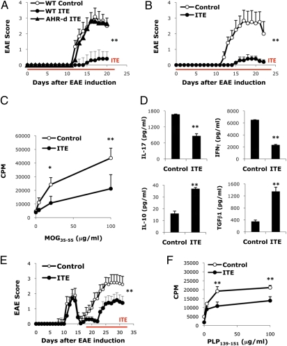
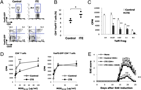
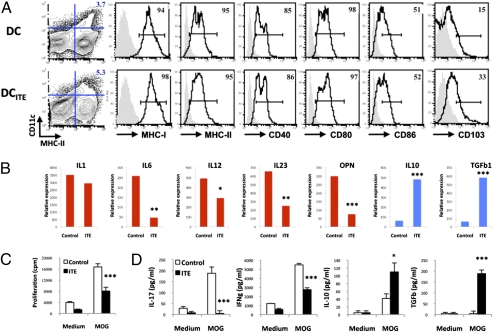
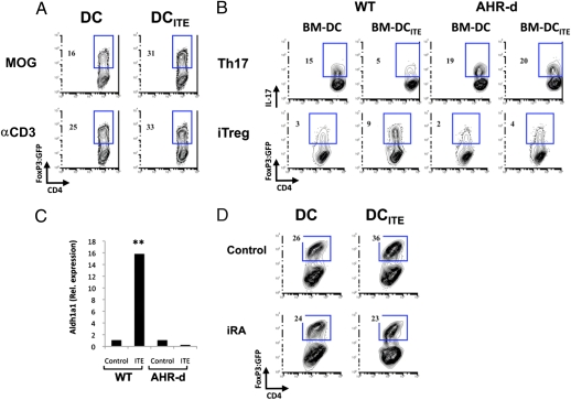
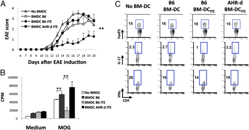
The ligand-activated transcription factor aryl hydrocarbon receptor (AHR) participates in the differentiation of FoxP3(+) T(reg), Tr1 cells, and IL-17-producing T cells (Th17). Most of our understanding on the role of AHR on the FoxP3(+) T(reg) compartment results from studies using the toxic synthetic chemical 2,3,7,8-tetrachlorodibenzo-p-dioxin. Thus, the physiological relevance of AHR signaling on FoxP3(+) T(reg) in vivo is unclear. We studied mice that carry a GFP reporter in the endogenous foxp3 locus and a mutated AHR protein with reduced affinity for its ligands, and found that AHR signaling participates in the differentiation of FoxP3(+) T(reg) in vivo. Moreover, we found that treatment with the endogenous AHR ligand 2-(1'H-indole-3'-carbonyl)-thiazole-4-carboxylic acid methyl ester (ITE) given parenterally or orally induces FoxP3(+) T(reg) that suppress experimental autoimmune encephalomyelitis. ITE acts not only on T cells, but also directly on dendritic cells to induce tolerogenic dendritic cells that support FoxP3(+) T(reg) differentiation in a retinoic acid-dependent manner. Thus, our work demonstrates that the endogenous AHR ligand ITE promotes the induction of active immunologic tolerance by direct effects on dendritic and T cells, and identifies nontoxic endogenous AHR ligands as potential unique compounds for the treatment of autoimmune disorders.


Conflict of interest statement

The authors declare no conflict of interest.

Figures

Fig. 1.
Fig. 1.
AHR activation by the nontoxic endogenous ligand ITE suppresses EAE. (A) EAE was induced in naive wild-type B6 or AHR-d mice, and ITE or vehicle as control was administered i.p. daily from the day of immunization until the termination of the experiment. The course of EAE is shown as the mean EAE score + SEM. Shown are representative data of one of three experiments that produced similar results. **P < 0.001 compared to control-treated WT mice or ITE treated AHR-d mice. (B) EAE was induced in naive wild-type mice and ITE or vehicle as control was administered orally daily from the day of immunization until the termination of the experiment. The course of EAE is shown as the mean EAE score + SEM. **P < 0.001 compared to control-treated WT mice. (C) Proliferative response to MOG35-55 of lymph node cells from ITE or control treated animals 10 d after immunization with MOG35-55 in CFA. Cell proliferation is indicated as cpm + SD in triplicate wells. *P < 0.05 and **P < 0.001 compared to cells from control-treated mice. (D) Cytokine secretion triggered by MOG35-55 in lymph node cells from ITE or control treated animals 10 d after immunization with MOG35-55 in CFA. **P < 0.001 compared to cells taken from control-treated mice. (E) EAE was induced in naive SJL mice, and ITE or vehicle as control was administered i.p. daily from day 17 after the immunization till the termination of the experiment. The course of EAE in these mice is shown as the mean EAE score + SEM. **P < 0.001 when compared to control-treated WT mice. (F) Proliferative response to PLP139-151 of splenocytes taken from ITE or control treated SJL mice 30 days after immunization with PLP139-151/CFA. Cell proliferation is indicated as cpm + SD in triplicate wells. **P < 0.001 when compared to cells taken from control-treated mice. Representative data of 1 of at least 2 experiments that produced similar results.
Fig. 2.
Fig. 2.
AHR activation by the nontoxic endogenous ligand ITE expands the FoxP3+ Treg compartment. (A) Frequency of CD4+ Foxp3:GFP+ Treg in splenocytes from ITE- or control-treated mice, 21 d after EAE induction. (B) CD4+ Foxp3:GFP T cells from naive Foxp3gfp were transferred into RAG-1 deficient hosts, the recipients were immunized with MOG35–55 in IFA, treated daily with ITE for 1 wk, and the frequency of CD4+ FoxP3:GFP+ Treg was analyzed in the spleen 3 wk after immunization. *P < 0.05 compared with mice transferred with WT cells. (C) Suppressive activity of CD4+ Foxp3:GFP Treg from ITE- or control-treated Foxp3gfp mice coincubated with naive 2D2 Foxp3:GFP T cells activated with MOG35–55 and irradiated APC. **P < 0.001 compared with cells taken from control-treated mice. (D) Recall response to MOG35–55 of CD4+ T cells and CD4+ Foxp3:GFP T cells lymph node cells from ITE- or control-treated Foxp3gfp mice 10 d after immunization with MOG35–55 in CFA. Cell proliferation is indicated as cpm ± SD in triplicate wells. *P < 0.05 and **P < 0.001 compared with cells taken from control-treated mice. (E) CD4+ or CD4+CD25 T cells (5 × 10⁶) were purified from ITE- or control-treated mice 10 d after immunization with MOG35–55 in CFA and transferred into naive mice. After 1 d, EAE was induced in the recipients with MOG35–55 in CFA. The course of EAE in these mice is shown as the mean EAE score ± SEM. **P < 0.001 compared with mice transferred with CD4+ T cells from control-treated mice or CD4+ CD25 T cells from ITE-treated cells. Representative data of one of at least three experiments that produced similar results.
Fig. 3.
Fig. 3.
AHR activation by the nontoxic endogenous ligand ITE induces tolerogenic DC. (A) FACS analysis of splenic DC from ITE- (DCITE) or control- (DC) treated mice. Numbers indicate the percent of positive cells; the staining obtained with isotype control antibodies is shown in gray. (B) Quantitative PCR analysis of cytokine expression by DC or DCITE. *P < 0.05; **P < 0.01; and ***P < 0.001 compared with DC. (C and D) Naive 2D2+ CD4+ FoxP3:GFP T cells were stimulated with MOG35–55 and DC or DCITE and, and proliferation (C) and cytokine secretion (D) was analyzed. *P < 0.05; **P < 0.01; and ***P < 0.001 compared with T cells incubated with control DC. Representative data of one of at least three experiments that produced similar results.
Fig. 4.
Fig. 4.
DCITE promote FoxP3+ Treg differentiation by an RA-dependent mechanism. (A) Naive 2D2+ CD4+ FoxP3:GFP T cells were stimulated with DC or DCITE, MOG35–55, and TGF-β1 + IL-2, and the frequency of FoxP3:GFP+ T cells was analyzed. (B) Naive 2D2+ CD4+ FoxP3:GFP T cells were stimulated with MOG35–55 and control (BM-DC) or ITE-treated BM-DC (BM-DCITE) derived from WT or AHR-d mice, in the presence of TGF-β1 + IL-6 or TGF-β1 + IL-2 and the frequency of IL-17+ T cells and FoxP3:GFP+ T cells was analyzed, respectively. (C) Quantitative PCR analysis of aldh1a2 expression by DC or DCITE from WT or AHR-d mice; results are presented relative to GAPDH mRNA. **P < 0.001 compared with DC from WT mice or DCITE from AHR-d mice. (D) Naive 2D2+ CD4+ FoxP3:GFP T cells were stimulated with DC or DCITE, MOG35–55 and TGF-β1 + IL-2, with or without the specific inhibitor of RA signaling LE135 (iRA). Representative data of one of at least three experiments that produced similar results.
Fig. 5.
Fig. 5.
Passive transfer of BM-DCITE suppresses EAE. (A) Naive mice received BM-DC and BM-DCITE (2 × 10⁶ per mouse, three times every 4 d), derived from WT or AHR-d mice and EAE was induced. The course of EAE is shown as the mean EAE score ± SEM. **P < 0.001 compared with mice transferred with BM-DC from WT mice or BM-DCITE from AHR-d mice. (B) Recall response to MOG35–55 in splenocytes 21 d after EAE induction. Cell proliferation is indicated as cpm ± SD in triplicate wells. **P < 0.001 compared with cells from mice transferred with BM-DC from WT mice or BM-DCITE from AHR-d mice. (C) Frequency of CD4+Foxp3+ Treg in splenocytes 21 d after EAE induction, and frequency of CD4+ IL-17+ T cells and CD4+ IFN-γ+ T cells in splenocytes 21 d after EAE, following activation with MOG35–55 for 5 d. Representative data of one of at least two experiments that produced similar results.

Comment in

Similar articles

Cited by

References

    1. Sakaguchi S. Naturally arising CD4+ regulatory t cells for immunologic self-tolerance and negative control of immune responses. Annu Rev Immunol. 2004;22:531–562. - PubMed
    1. Bluestone JA, Thomson AW, Shevach EM, Weiner HL. What does the future hold for cell-based tolerogenic therapy? Nat Rev Immunol. 2007;7:650–654. - PubMed
    1. Quintana FJ, et al. Control of T(reg) and T(H)17 cell differentiation by the aryl hydrocarbon receptor. Nature. 2008;453:65–71. - PubMed
    1. Hauben E, et al. Activation of the aryl hydrocarbon receptor promotes allograft-specific tolerance through direct and dendritic cell-mediated effects on regulatory T cells. Blood. 2008;112:1214–1222. - PubMed
    1. Kimura A, Naka T, Nohara K, Fujii-Kuriyama Y, Kishimoto T. Aryl hydrocarbon receptor regulates Stat1 activation and participates in the development of Th17 cells. Proc Natl Acad Sci USA. 2008;105:9721–9726. - PMC - PubMed

Publication types

MeSH terms

LinkOut - more resources