. 2011 Jan 7;286(1):555-66.
doi: 10.1074/jbc.M110.188805. Epub 2010 Nov 10.

Cox25 teams up with Mss51, Ssc1, and Cox14 to regulate mitochondrial cytochrome c oxidase subunit 1 expression and assembly in Saccharomyces cerevisiae

Affiliations

Cox25 teams up with Mss51, Ssc1, and Cox14 to regulate mitochondrial cytochrome c oxidase subunit 1 expression and assembly in Saccharomyces cerevisiae

Flavia Fontanesi et al. J Biol Chem. .

Abstract

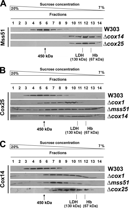
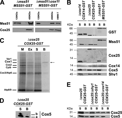
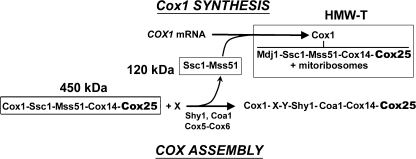
In the yeast Saccharomyces cerevisiae, mitochondrial cytochrome c oxidase (COX) biogenesis is translationally regulated. Mss51, a specific COX1 mRNA translational activator and Cox1 chaperone, drives the regulatory mechanism. During translation and post-translationally, newly synthesized Cox1 physically interacts with a complex of proteins involving Ssc1, Mss51, and Cox14, which eventually hand over Cox1 to the assembly pathway. This step is probably catalyzed by assembly chaperones such as Shy1 in a process coupled to the release of Ssc1-Mss51 from the complex. Impaired COX assembly results in the trapping of Mss51 in the complex, thus limiting its availability for COX1 mRNA translation. An exception is a null mutation in COX14 that does not affect Cox1 synthesis because the Mss51 trapping complexes become unstable, and Mss51 is readily available for translation. Here we present evidence showing that Cox25 is a new essential COX assembly factor that plays some roles similar to Cox14. A null mutation in COX25 by itself or in combination with other COX mutations does not affect Cox1 synthesis. Cox25 is an inner mitochondrial membrane intrinsic protein with a hydrophilic C terminus protruding into the matrix. Cox25 is an essential component of the complexes containing newly synthesized Cox1, Ssc1, Mss51, and Cox14. In addition, Cox25 is also found to interact with Shy1 and Cox5 in a complex that does not contain Mss51. These results suggest that once Ssc1-Mss51 are released from the Cox1 stabilization complex, Cox25 continues to interact with Cox14 and Cox1 to facilitate the formation of multisubunit COX assembly intermediates.


Figures

FIGURE 1.
FIGURE 1.
S. cerevisiae COX25 codes for a protein required for respiratory growth. A, sequence alignment of COX25 from S. cerevisiae (Sc) and other fungi: Neurospora crassa (Nc), Aspergillus nidulans (An), Podospora anserina (Pa), Gibberella zeae (Gz), Kluiveromyces lactis (Kl), Pichia pastoris (Pp), and Yarrowia lipolytica (Yl). TM indicates a predicted transmembrane domain. B, the respiratory competent wild-type strains BY4741 and W303 and strains carrying a null allele of cox25 in both genetic backgrounds were grown overnight in liquid YPD media. 10-fold serial dilutions of the four strains were plated on solid YPD or YPEG media and incubated at 30 °C. The pictures were taken after 3 days of incubation. C, wild-type W303 and mutant Δcox25 cells overexpressing or not COX25 were grown overnight in liquid YPD media. Serial dilution growth test was performed as for B.
FIGURE 2.
FIGURE 2.
Biochemical properties of Δcox25 cells. Respiratory assays A, KCN-sensitive endogenous cell respiration measured polarographically in the presence of galactose. B, rate of NADH and succinate oxidation in isolated mitochondria. C, total mitochondrial cytochrome spectra. Mitochondria from the wild-type strain W303 and the null mutant Δcox25 cells were extracted at a protein concentration of 5 mg/ml with potassium deoxycholate under conditions that quantitatively solubilize all of the cytochromes (36). Difference spectra of the reduced (sodium dithionite) versus oxidized (potassium ferricyanide) extracts were recorded at room temperature. The absorption bands corresponding to cytochromes a and a3 have maxima at 603 nm (a and a3); the maxima for cytochrome b (b) and for cytochrome c and c1 (c and c1) are 560 and 550 nm, respectively. D, mitochondrial respiratory chain enzyme spectrophotometric measurements in isolated mitochondria. COX, NADH cytochrome c reductase (NCCR), succinate cytochrome c reductase (SCCR), and ATP synthase activities were measured as described under “Experimental Procedures.” E, steady state concentrations of mitochondrial respiratory chain complexes IV (COX), II (Sdh), and III (bc1 complex) and ATP synthase subunits estimated by Western blot analyses of proteins separated in a 12% Tris-glycine SDS-PAGE. F, steady state concentrations of COX subunit 5 isoforms estimated by Western blot analyses of proteins separated in a 16.5% Tris-Tricine SDS-PAGE. Two expositions of the film are shown. In E and F, an antibody against porin was used to normalize the signals for protein loading.
FIGURE 3.
FIGURE 3.
In vivo synthesis and turnover of mitochondrial gene products in Δcox25 cells. A, wild type (either BY4741 or W303), null mutants of cox25 in both backgrounds and a double mutant Δcox25Δcox14 were labeled with [35S]methionine at 30 °C for the indicated times in the presence of cycloheximide. Equivalent amounts of protein were separated by SDS-PAGE on a 17.5% polyacrylamide gel, transferred to a nitrocellulose membrane, and analyzed by autoradiography. #1 and #2 indicate two independent Δcox25 clones. B, in vivo labeling of mitochondrial products from the indicated strains with [35S]methionine at 30 °C for 5 and 10 min in the presence of cycloheximide. C, steady state levels of Cox14 and Mss51 in mitochondria isolated from wild-type, Δcox25, and Δcox14 cells analyzed by Western blotting. D, the indicated strains were labeled for 15 min at 30 °C with [35S]methionine in the presence of cycloheximide. Labeling was terminated by the addition of 80 μmol of cold methionine and 12 μg/ml puromycin (0 time). The samples were collected after the indicated times of incubation at 30 °C and processed as in A.
FIGURE 4.
FIGURE 4.
Mitochondrial localization of Cox25. A, Cox25 is a mitochondrial protein. Mitochondria (M) and the post-mitochondrial supernatant (PMS) fraction were isolated from the wild-type W303 strain. Samples of the two fractions corresponding to 40 μg of protein were analyzed by Western blotting using antibodies against Cox25, the cytosolic marker 3-phosphoglycerate kinase subunit 1 (Pgk1), and the mitochondrial marker porin. The specificity of the signal detected with the anti-Cox25 antibody was tested by including a sample of Δcox25 mitochondria. B, Cox25 is a membrane protein. As described under “Experimental Procedures,” soluble (S) and membrane-bound (P) mitochondrial proteins were separated from 40 μg of total wild-type mitochondria. The pellet was submitted to alkaline extraction to allow the separation of the extrinsic proteins present in the supernatant (CS) from the intrinsic proteins in the pellet (CP). The samples were analyzed by Western blotting using antibodies against Cox25, Cox14, the inner membrane extrinsic protein Mss51, the soluble intermembrane space protein Cox17, and the inner membrane intrinsic protein Cox3. C, Cox25 is an inner mitochondrial membrane protein. Isolated mitochondria were fractionated into inner and outer membranes by sonication plus sucrose gradient sedimentation as described (37). Inner membranes (IM) and outer membranes (OM) were analyzed by Western blotting using antibodies against porin (outer membrane marker), Cox1 (inner membrane marker), Cox14, and Cox25. D, Cox25 is a membrane protein facing the matrix. Four aliquots of 40 μg of mitochondrial protein were pelleted and resuspended in buffer containing either 20 mm HEPES or 0.6 m sorbitol with 20 mm HEPES. One aliquot in each buffer was supplemented with final 100 μg/ml proteinase K (PK) and incubated on ice for 60 min. The reaction was stopped with 2 mm PMSF. Mitochondria (Mt) and mitoplasts (Mp) were recovered by centrifugation at 50,000 × gav for 15 min at 4 °C. The samples were analyzed by Western blotting using antibodies against Cox25, Cox14, Cmc2 (protein facing the inner membrane space), and Mss51 (protein facing the matrix). E, sequence alignment of S. cerevisiae Cox25 and Cox14. F, Kyte-Doolittle hydrophobicity plots for these proteins. TM indicates a predicted transmembrane domain. G, topology of Cox25 and Cox14 in the inner membrane. Mitochondria were prepared from Δcox25 or Δcox14 strains, respectively, expressing Cox25 or Cox14 fused with GST at their C terminus and used for proteinase protection assays as in D. H, cartoon depicting the topology of Cox25 and Cox14 in the mitochondrial inner membrane.
FIGURE 5.
FIGURE 5.
Native molecular mass and steady state levels of Cox25. A, sedimentation properties of Mss51 in a linear 7–20% sucrose gradient using mitochondrial extracts from the indicated strains. The gradient was calibrated with hemoglobin (Hb, 67 kDa) and lactate dehydrogenase (LDH, 130 kDa). The distribution of Mss51 was assayed by Western blot analysis. B and C, sedimentation properties of Cox25 (B) and of Cox14 (C) in a linear 7–20% sucrose gradient using mitochondrial extracts from the indicated strains, analyzed as in A.
FIGURE 6.
FIGURE 6.
Cox25 physically interacts with Cox1 and Cox1 biogenesis and assembly factors. A, Cox25 interacts with Mss51 in the 450-kDa complex and in the high molecular mass Cox1 translational complex (HMW-T) but not in the 120-kDa complex previously described (8). Complexes containing Mss51 were purified by sucrose gradient followed by GST pulldown of mitochondrial extracts from either a Δmss51 or a Δmss51Δcox11 both expressing a GST-tagged version of Mss51 (8). Samples from the different complexes were analyzed by Western blotting using antibodies against Mss51 and Cox25. B, mitochondria were prepared from Δmss51 cells expressing Mss51-GST, Δcox14 cells expressing Cox14-GST and Δcox25 cells expressing Cox25-GST, extracted with 1% digitonin, 150 mm KCl, and 1.2 mm MgCl2 in buffer containing 20 mm HEPES and 0.5 mm PMSF and used for GST pulldown assays. Samples of material bound to GST-Sepharose beads (lanes B) or remaining in the supernatant (lanes S) were separated in a 12% Tris-glycine SDS-PAGE and analyzed by Western blotting using specific antibodies against the indicated proteins. C, interaction of Cox25 with newly synthesized Cox1. Mitochondria isolated from a Δcox25 strain with a chromosomally integrated plasmid expressing Cox25-GST fusion protein were labeled with [35S]methionine for 30 min, extracted, and submitted to GST pulldown as described under “Experimental Procedures.” Mitochondria (M) corresponding to 20 μg of protein, equivalent volumes of the extract (Ex), the supernatant from the glutathione-Sepharose beads (S), and the washed beads (B) were separated on a 17.5% polyacrylamide gel by SDS-PAGE. D, Cox25-GST pulldown samples were also separated in a 16.5% Tris-Tricine SDS-PAGE and analyzed by Western blotting using and anti-Cox5 antibody. E, GST pulldown analyses performed as in C, using mitochondrial extracts from Δcox25, Δcox25Δcox14, and Δcox25Δshy1 cells all expressing COX25-GST. Material unbound (S) or bound to the GST-Sepharose beads was analyzed by Western blotting using antibodies against Cox25 and Cox5.
FIGURE 7.
FIGURE 7.
Scheme of a model depicting the role of Ssc1 and Cox14 on translational regulation of cytochrome c oxidase biogenesis by interacting with Mss51 in several high molecular mass complexes. Cox25 is a partner in some of these complexes. See the explanation in the text.

Similar articles

Cited by

References

    1. Fontanesi F., Soto I. C., Horn D., Barrientos A. (2006) Am. J. Physiol. Cell Physiol. 291, C1129–C1147 - PubMed
    1. Fontanesi F., Soto I. C., Barrientos A. (2008) IUBMB Life 60, 557–568 - PMC - PubMed
    1. Langer T., Käser M., Klanner C., Leonhard K. (2001) Biochem. Soc. Trans. 29, 431–436 - PubMed
    1. Perez-Martinez X., Broadley S. A., Fox T. D. (2003) EMBO J. 22, 5951–5961 - PMC - PubMed
    1. Barrientos A., Korr D., Tzagoloff A. (2002) EMBO J. 21, 43–52 - PMC - PubMed

Publication types

MeSH terms