. 2011 Mar;4(2):228-39.
doi: 10.1242/dmm.006130. Epub 2010 Nov 10.

Development of severe skeletal defects in induced SHP-2-deficient adult mice: a model of skeletal malformation in humans with SHP-2 mutations

Affiliations

Development of severe skeletal defects in induced SHP-2-deficient adult mice: a model of skeletal malformation in humans with SHP-2 mutations

Timothy J Bauler et al. Dis Model Mech. 2011 Mar.

Abstract

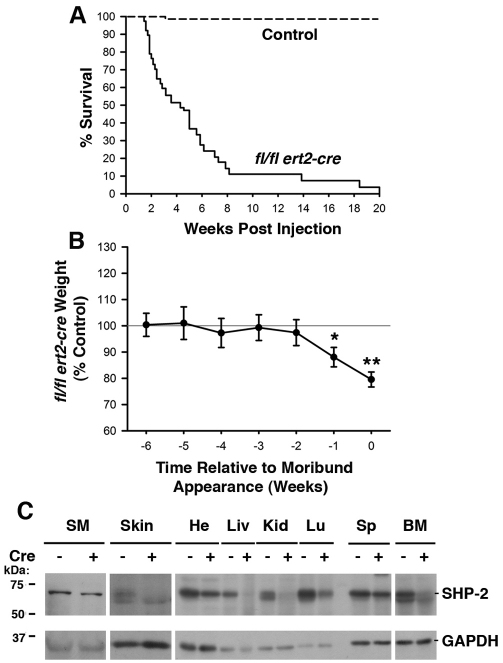
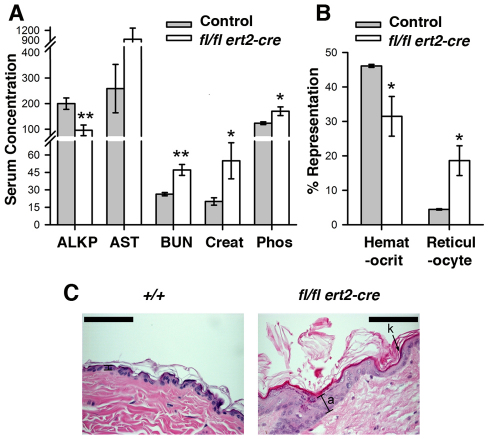
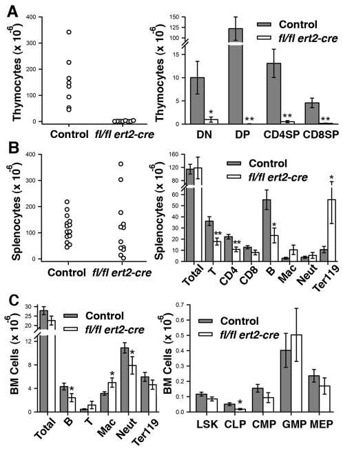
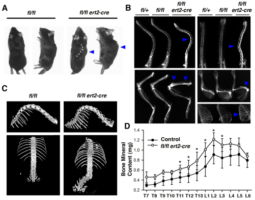
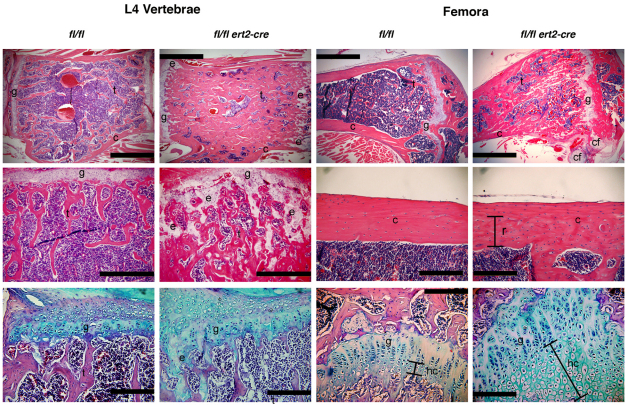
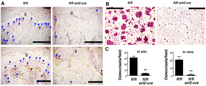
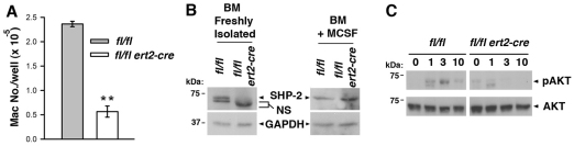
SHP-2 (encoded by PTPN11) is a ubiquitously expressed protein tyrosine phosphatase required for signal transduction by multiple different cell surface receptors. Humans with germline SHP-2 mutations develop Noonan syndrome or LEOPARD syndrome, which are characterized by cardiovascular, neurological and skeletal abnormalities. To study how SHP-2 regulates tissue homeostasis in normal adults, we used a conditional SHP-2 mouse mutant in which loss of expression of SHP-2 was induced in multiple tissues in response to drug administration. Induced deletion of SHP-2 resulted in impaired hematopoiesis, weight loss and lethality. Most strikingly, induced SHP-2-deficient mice developed severe skeletal abnormalities, including kyphoses and scolioses of the spine. Skeletal malformations were associated with alterations in cartilage and a marked increase in trabecular bone mass. Osteoclasts were essentially absent from the bones of SHP-2-deficient mice, thus accounting for the osteopetrotic phenotype. Studies in vitro revealed that osteoclastogenesis that was stimulated by macrophage colony-stimulating factor (M-CSF) and receptor activator of nuclear factor kappa B ligand (RANKL) was defective in SHP-2-deficient mice. At least in part, this was explained by a requirement for SHP-2 in M-CSF-induced activation of the pro-survival protein kinase AKT in hematopoietic precursor cells. These findings illustrate an essential role for SHP-2 in skeletal growth and remodeling in adults, and reveal some of the cellular and molecular mechanisms involved. The model is predicted to be of further use in understanding how SHP-2 regulates skeletal morphogenesis, which could lead to the development of novel therapies for the treatment of skeletal malformations in human patients with SHP-2 mutations.


Figures

Fig. 1.
Fig. 1.
Induced SHP-2 deficiency in adult mice results in weight loss and rapid mortality. (A) Kaplan-Meier plot depicting the survival of ptpn11fl/fl ert2-cre mice (n=38) and pooled ptpn11+/+, ptpn11fl/+, ptpn11fl/+ ert2-cre and ptpn11fl/fl littermate control mice (n=75) following tamoxifen injection at 6–8 weeks of age. (B) Weight of tamoxifen-injected ptpn11fl/fl ert2-cre mice (n=19) expressed as a mean percentage ± 1 s.e.m. of the weight of tamoxifen-injected littermate controls (n=33) at time points at and prior to the time that ptpn11fl/fl ert2-cre mice appear moribund. Mice were injected with tamoxifen at 6–8 weeks of age. Statistical significance was determined using a one sample Student’s t-test. In A and B, all control mice were similar with regards to survival and weight following tamoxifen injection and no effect of haploinsufficiency of SHP-2 in ptpn11fl/+ ert2-cre mice was observed. For ptpn11fl/fl ert2-cre mice, no influence of mouse gender upon survival or weight loss was apparent. *P<0.05; **P<0.005. (C) Western blots showing expression of SHP-2 in the indicated organs from ptpn11fl/fl ert2-cre (+) and littermate control ptpn11fl/fl mice, both injected with tamoxifen 15 days previously (at 6–8 weeks of age). SM, skeletal muscle; He, heart; Liv, liver; Kid, kidney; Lu, lung; Sp, spleen; BM, bone marrow. Blots were stripped and reprobed with an anti-GAPDH antibody to demonstrate equivalent protein loading. Shown are representative experiments of three repeats.
Fig. 2.
Fig. 2.
Altered levels of serum biomarkers, anemia and skin abnormalities in induced SHP-2-deficient mice. (A) Shown are the mean levels of the indicated biomarkers in serum ± 1 s.e.m. of tamoxifen-injected (at 6–8 weeks) moribund ptpn11fl/fl ert2-cre mice and littermate control mice (n=6 each genotype). Age range of mice at the time of analysis was 9–12 weeks. ALKP and AST concentrations are shown in U/l; BUN, mg/dl; phosphorous, μg/ml; creatinine, μg/cl. Statistical significance was determined using a two sample Student’s t-test. *P<0.05; **P<0.005. (B) Depicted are mean hematocrit (percent blood volume) and reticulocyte counts (percent blood cells) ± 1 s.e.m. performed on heparinized whole blood from mice in A. Statistical significance was determined using a two sample Student’s t-test. (C) Representative H&E-stained skin section (n=6) of a tamoxifen-injected (at 6 weeks of age) moribund ptpn11fl/fl ert2-cre mouse and a littermate ptpn11+/+ control showing hyperkeratosis (k) and acanthosis (a) in the former. Analysis was performed 4 weeks after tamoxifen injection. Scale bars: 200 μm.
Fig. 3.
Fig. 3.
Altered hematopoiesis in induced SHP-2-deficient mice. (A) Numbers of total thymocytes (left) and indicated thymocyte subpopulations (right) in thymi isolated from tamoxifen-injected moribund ptpn11fl/fl ert2-cre mice (n=8) and littermate controls (n=8). DN, CD4–CD8–double negative; DP, CD4+CD8+ double positive; SP, CD4+CD8– and CD4–CD8+ single positive T cells. (B) Numbers of total splenocytes (left) and indicated splenocyte subpopulations (right) in spleens isolated from tamoxifen-injected moribund ptpn11fl/fl ert2-cre mice (n=12) and littermate controls (n=14). T cells (T), B cells (B), macrophages (Mac) and neutrophils (Neut) were identified as TCRβ+, B220+, CD11bint/GR1int and CD11bhi/GR1hi, respectively. Ter119+ cells represent erythrocyte precursors. (C) Total numbers and numbers of indicated lineage-positive cells (left) and numbers of indicated lineage-negative progenitor cells (right) in bone marrow of tamoxifen-injected moribund ptpn11fl/fl ert2-cre mice and littermate controls (for each genotype, n=11 for lineage-positive and n=5 for lineage-negative). LSK, LinSca-1+c-Kit+ cells, which are enriched for HSCs; CLP, LinSca-1loc-KitloCD127+; CMP, LinSca-1c-Kit+CD34+CD16/32lo; GMP, LinSca-1c-Kit+CD34+CD16/32hi; MEP, LinSca-1c-Kit+CD34CD16/32–/lo. In scatter plots, each symbol represents an individual mouse. In bar graphs, the mean ± 1 s.e.m. is depicted. All mice were injected with tamoxifen at 6–8 weeks of age. Age range of mice at time of analysis was 10–16 weeks. Statistical significance was determined by two sample Student’s t-test. *P<0.05; **P<0.005.
Fig. 4.
Fig. 4.
Spinal curvature and increased bone mineral content in induced SHP-2-deficient mice. (A) Gross morphology of moribund ptpn11fl/fl ert2-cre mice and ptpn11fl/fl littermate control mice. Note lateral spinal curvature (dotted line) and hump (arrowhead) in the ptpn11fl/fl ert2-cre mouse. (B) X-rays of tamoxifen-injected moribund ptpn11fl/fl ert2-cre mice showing kyphosis (top left) and scoliosis (top right), compared with the indicated littermate controls. Note also increased radiodensity of vertebral bodies of spines, metaphyses of femur (bottom left) and humerus (middle right), and entire rib bones (bottom right) of the ptpn11fl/fl ert2-cre mice (all indicated with arrowheads). In A and B, mice were treated with tamoxifen 5 weeks previously, at 6–8 weeks of age. (C) μCT images of the isosurfaces of spines of ptpn11fl/fl ert2-cre mice and ptpn11fl/fl littermate control mice injected with tamoxifen 5 weeks previously, at 7 weeks of age. Images show scoliosis with rotated vertebral bodies. (D) Mean bone mineral content ± 1 s.e.m. of individual thoracic (T) and lumbar (L) vertebrae from ptpn11fl/fl ert2-cre mice and littermate controls determined by μCT scanning (n=4 for each mouse strain). All mice were treated with tamoxifen 5 weeks previously, at 7 weeks of age. Statistical significance was determined by paired Student’s t-test. *P<0.05.
Fig. 5.
Fig. 5.
Increased and disorganized bone and cartilage in induced SHP-2-deficient mice. Shown are representative images of L4 vertebrae and femora of moribund tamoxifen-injected ptpn11fl/fl ert2-cre mice and ptpn11fl/fl littermate controls. Mice were injected with tamoxifen at 7 weeks of age and analysis was performed at 12 weeks of age. Top and middle panels are stained with H&E, and bottom panels are stained with Alcian blue to highlight cartilage. Scale bars: 1 mm in top panels, 500 μm in L4 vertebrae middle panels, and 200 μm in femora middle panels and all bottom panels. The amount of trabecular bone (t) is dramatically increased in ptpn11fl/fl ert2-cre vertebrae and femora. A region of remodeled (r) cortical bone (c) in femora of ptpn11fl/fl ert2-cre mice shows disorganized bone in this region compared with the same region in the control. Remains of ectopic cartilaginous elements (e) were identified in the trabecular bone region and ectopic cartilage formation (cf) was identified next to growth plates (g) in ptpn11fl/fl ert2-cre mice. The columnar formation of growth plates was disorganized in ptpn11fl/fl ert2-cre mice and the area of hypertrophic chondrocytes (hc) was elongated.
Fig. 6.
Fig. 6.
Impaired osteoclastogenesis in induced SHP-2-deficient mice. (A) Femur sections of tamoxifen-injected moribund ptpn11fl/fl ert2-cre mice and ptpn11fl/fl littermate controls were stained for TRAP to visualize osteoclasts (red color). Images are from 11-week-old mice injected with tamoxifen 5 weeks previously. The location of osteoclasts beneath growth plates (g) and within the secondary spongiosa are indicated with blue arrowheads. Note the paucity of osteoclasts in ptpn11fl/fl ert2-cre mice. Scale bars: top panels, 500 μm; bottom panels, 200 μm. (B) Bone marrow cells from ptpn11fl/fl ert2-cre mice and ptpn11fl/fl littermate control mice, both treated with tamoxifen 3 weeks previously (at 7 weeks of age), were cultured with M-CSF and RANKL for 7 days on glass coverslips. Osteoclasts were identified by TRAP staining. Note the abundance of multinucleated osteoclasts in control cultures and absence from ptpn11fl/fl ert2-cre cultures. Scale bars: 100 μm. (C) Shown are the mean numbers of osteoclasts + 1 s.e.m. per field identified in bone sections (in situ) and in vitro osteoclast differentiation experiments described in A and B. For in situ analysis, the field size was as shown in the top panels of A and encompassed the growth plate and secondary spongiosa regions. Data are derived from randomly selected fields of femur heads from moribund ptpn11fl/fl ert2-cre mice and ptpn11fl/fl littermate controls injected with tamoxifen at 6 weeks of age (n=3 in each genotype). Mice were 10–11 weeks of age at the time of analysis. Size of fields in in vitro experiments are as indicated in B and were selected randomly on coverslips. Bone marrow was derived from ptpn11fl/fl ert2-cre mice and ptpn11fl/fl littermate mice that were injected with tamoxifen at 6–8 weeks of age (n=6 in each genotype). Osteoclast differentiation experiments were initiated 1–3 weeks thereafter. Statistical significance was determined by paired Student’s t-test. **P<0.005.
Fig. 7.
Fig. 7.
Blocked M-CSF signal transduction in induced SHP-2-deficient mice. (A) Bone marrow cells from a ptpn11fl/fl ert2-cre mouse and a littermate ptpn11fl/fl mouse (both treated with tamoxifen 10 days beforehand at 7 weeks of age) were cultured in wells of a 24-well plate (1×10⁶ cells/well) in the presence of M-CSF. After 5 days, macrophages in wells were harvested and counted. Depicted is the mean number of macrophages ± 1 s.e.m. (n=4). Results are representative of four repeat experiments. Statistical significance was determined by two sample Student’s t-test. **P<0.005. (B) Western blots showing expression of SHP-2 in bone marrow cells (freshly isolated or after culture in M-CSF for 5 days) from a ptpn11fl/fl ert2-cre mouse and a littermate ptpn11fl/fl mouse (both treated with tamoxifen 3 weeks beforehand at 6 weeks of age). NS, non-specific band. Blots were stripped and reprobed with an anti-GAPDH antibody to verify equivalent protein loading. Similar results were obtained in five independent experiments. (C) Lineage-negative bone marrow cells from a ptpn11fl/fl ert2-cre mouse and a littermate ptpn11fl/fl mouse (both treated with tamoxifen 10 days beforehand at 6 weeks of age) were stimulated with M-CSF for the indicated times (in minutes). Activation of AKT was determined by western blotting of whole-cell lysates using a phospho-specific anti-AKT antibody. Blots were reprobed with an anti-AKT antibody to verify equal loading. Similar results were obtained in three repeat experiments.

References

    1. Asagiri M., Takayanagi H. (2007). The molecular understanding of osteoclast differentiation. Bone 40, 251–264 - PubMed
    1. Bard-Chapeau E. A., Yuan J., Droin N., Long S., Zhang E. E., Nguyen T. V., Feng G. S. (2006). Concerted functions of Gab1 and Shp2 in liver regeneration and hepatoprotection. Mol. Cell. Biol. 26, 4664–4674 - PMC - PubMed
    1. Cai T., Nishida K., Hirano T., Khavari P. A. (2002). Gab1 and SHP-2 promote Ras/MAPK regulation of epidermal growth and differentiation. J. Cell Biol. 159, 103–112 - PMC - PubMed
    1. Chan R. J., Johnson S. A., Li Y., Yoder M. C., Feng G. S. (2003). A definitive role of Shp-2 tyrosine phosphatase in mediating embryonic stem cell differentiation and hematopoiesis. Blood 102, 2074–2080 - PubMed
    1. Chan R. J., Li Y., Hass M. N., Walter A., Voorhorst C. S., Shelley W. C., Yang Z., Orschell C. M., Yoder M. C. (2006). Shp-2 heterozygous hematopoietic stem cells have deficient repopulating ability due to diminished self-renewal. Exp. Hematol. 34, 1230–1239 - PubMed

Publication types

MeSH terms

Substances