. 2010 Nov 10;2(57):57ra82.
doi: 10.1126/scitranslmed.3001510.

TLR9 differentiates rapidly from slowly progressing forms of idiopathic pulmonary fibrosis

Affiliations

TLR9 differentiates rapidly from slowly progressing forms of idiopathic pulmonary fibrosis

Glenda Trujillo et al. Sci Transl Med. .

Abstract

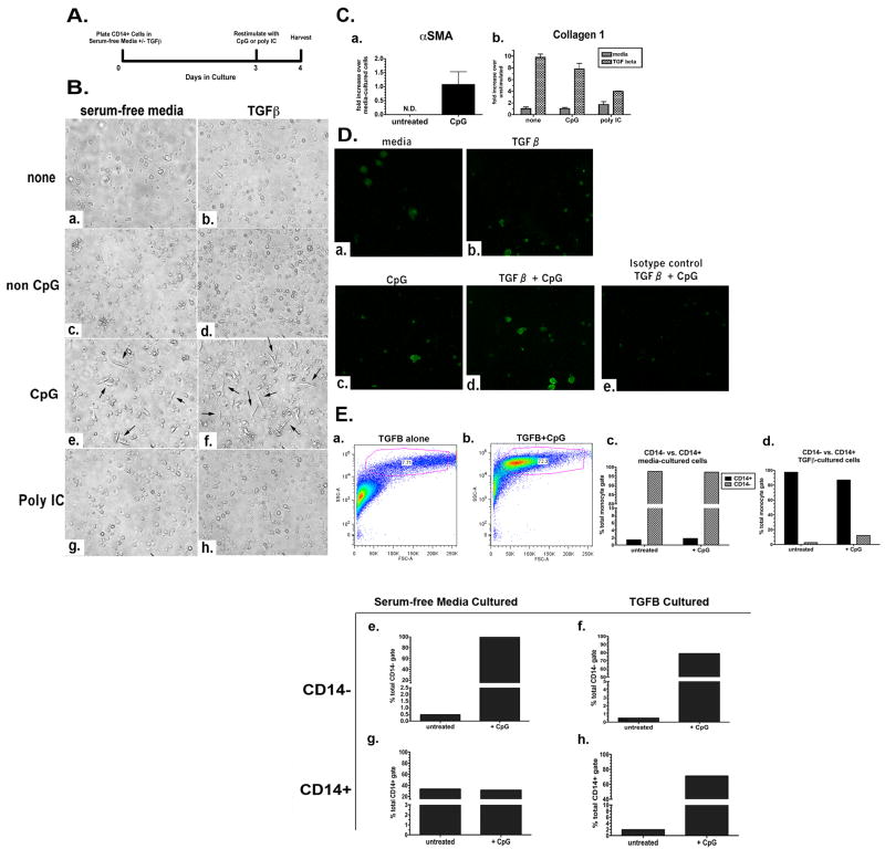
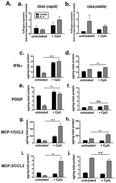
Idiopathic pulmonary fibrosis is characterized by diffuse alveolar damage and severe fibrosis, resulting in a steady worsening of lung function and gas exchange. Because idiopathic pulmonary fibrosis is a generally progressive disorder with highly heterogeneous disease progression, we classified affected patients as either rapid or slow progressors over the first year of follow-up and then identified differences between the two groups to investigate the mechanism governing rapid progression. Previous work from our laboratory has demonstrated that Toll-like receptor 9 (TLR9), a pathogen recognition receptor that recognizes unmethylated CpG motifs in bacterial and viral DNA, promotes myofibroblast differentiation in lung fibroblasts cultured from biopsies of patients with idiopathic pulmonary fibrosis. Therefore, we hypothesized that TLR9 functions as both a sensor of pathogenic molecules and a profibrotic signal in rapidly progressive idiopathic pulmonary fibrosis. Indeed, TLR9 was present at higher concentrations in surgical lung biopsies from rapidly progressive patients than in tissue from slowly progressing patients. Moreover, fibroblasts from rapid progressors were more responsive to the TLR9 agonist, CpG DNA, than were fibroblasts from slowly progressing patients. Using a humanized severe combined immunodeficient mouse, we then demonstrated increased fibrosis in murine lungs receiving human lung fibroblasts from rapid progressors compared with mice receiving fibroblasts from slowly progressing patients. This fibrosis was exacerbated by intranasal CpG challenges. Furthermore, CpG induced the differentiation of blood monocytes into fibrocytes and the epithelial-to-mesenchymal transition of A549 lung epithelial cells. These data suggest that TLR9 may drive the pathogenesis of rapidly progressive idiopathic pulmonary fibrosis and may serve as a potential indicator for this subset of the disease.


Figures

Figure 1
Figure 1. Clinical Features of Patients with Rapid and Slowly Progressive Forms of IPF and TLR9 Expression
A. The survival of IPF patients classified as rapid (red line) or slow progressors (black line). B. Representative histology of IPF in a patient with slow (a,b) and rapid (c,d) progression shown at 20× and 400× magnification. C. Quantitative TaqMan PCR analysis of TLR9 gene expression in upper lobe SLBs from rapid and slow progressors. The data shown are the mean of all the combined upper lobe mRNA values compared to the mean of normal SLBs mRNA values (standardized to GAPDH housekeeping gene). The error bar shows the SEM of all the data in the rapid (n=10) and stable (n=13) progressor patient groups. The two-tailed P value was determined by the unpaired t test with Welch correction. D. Representative immunohistochemical staining of TLR9 in SLBs from a total of 7 slow (a) and 5 rapid (c) progressors shown at 20× magnification. Corresponding fields stained with isotype control (IgG) shown in b and d.
Figure 2
Figure 2. Induction by CpG of CD14+ Human Monocytes to Differentiate into Fibrocyte-like Cells. A
Experimental scheme for the in vitro differentiation of CD14+ monocytes. B. Photomicrographs of monocytes cultured in serum-free media or serum-free media containing 10 ng/ml TGFβ and stimulated with nothing (a,b), 50 μg/mL non CpG (c,d), 50 μg/mL CpG (e,f), or 50 μg/mL poly IC (g, h) on Day 3. C. qRT-PCR analysis of fibrocyte markers. αSMA gene expression in monocytes cultured for 3 d in serum-free media +/- CpG for 24 h (a). Collagen 1 gene expression in monocytes cultured for 3 d in serum-free media or TGFβ, +/- CpG or poly I:C (b). D. Fluorescent ICC for collagen 1 in monocytes (40× magnification) cultured in serum-free media (a) or TGFβ (b); serum-free media + CpG (c), or TGFβ + CpG (d). Isotype control for monocytes cultured in TGFβ + CpG (e). Representative (n=3) FC for collagen 1 protein as percent of CD14+ cells in CD45+ gate from monocytes cultured in serum-free media or TGFβ, and serum-free media + CpG or TGFβ + CpG (f). E. Forward and side scatter FC of monocytes cultured in serum-free media containing TGFβ (a) or TGFβ + CpG (b). Representative (n=3) FC for CD14 as percent of total cells from monocytes cultured in serum-free media (c) or monocytes cultured in serum-free media containing TGFβ (d) stained with anti-CD14. F. Representative (n=3) FC for CD45 as percent of CD14- cells from monocytes cultured in serum-free media (e) or monocytes cultured in serum-free media containing TGFβ (f) stained with anti-CD45 and gated with respect to CD14 expression. Representative data (n=3) is graphed as percent of CD14+ cells from monocytes cultured in serum-free media (g) and monocytes cultured in serum-free media containing TGFβ (h) stained with anti-CD45 and gated with respect to CD14 expression.
Figure 3
Figure 3. CpG-induced EMT in Human A549 Cells
A. Representative photomicrographs (n=5) of A549 cells cultured in media (DMEM + 10% FCS) (a), TGFβ (b), and increasing CpG concentrations of 5 μg/mL (c), 10 μg/mL (d), 50 μg/mL (e), 100 μg/mL (f), and 200 μg/mL (g) for 96 h. B. qRT-PCR analysis of αSMA (a), vimentin (b), and e-cadherin (c) in A549 cells cultured with increasing concentrations of CpG for 96 hours. C. qRT-PCR analysis of IFNα in A549 cells cultured with increasing concentrations of CpG for 96 h. D. Fluorescent ICC for collagen 1 in A549 cells (40× magnification) that were cultured for 96 h in media (a), 10 μg/mL CpG (b), 50 μg/mL CpG (c), and 100 μg/mL CpG (d). Isotype control for collagen 1 antibody using cells cultured with 100 μg/mL CpG (e). E. siRNA knockdown of TLR9 in A549 cells in a CpG EMT assay: Western Blot analysis of TLR9 protein and β-actin loading control in A549 cell lysates after siRNA treatment with a non targeting control siRNAs, cyclophilin B control siRNAs (a), and TLR9 siRNAs; photomicrographs of A549 cells before CpG-DNA treatment cultured in media and transfection agent alone (b), with non target siRNA (c), and with TLR9 siRNA (d); representative photomicrographs (n=4) of A549 cells after siRNA treatment and stimulated with media and transfection agent alone (e), non target siRNA + 75 μg/ml CpG (f), and TLR9 siRNA + 75 μg/ml CpG-DNA for 72 hrs (g); qRT-PCR analysis of vimentin (h) and e-cadherin (i) in siRNA-treated A549 cells and cultured with 75 μg/ml CpG for 72 hours. Data are mean ± SD. *** p < 0.0001.
Figure 4
Figure 4. TLR9 Expression in Rapid and Slowly Progressive IPF Lung Fibroblasts and Response to CpG
A. qRT-PCR analysis of TLR9 gene expression in representative rapid UIP/IPF (n=5-8) (a) and slow IPF (n=5-8) (b) fibroblast cell lines treated for 24 h without (untreated) or with CpG-ODN (10 μg/ml) in the presence or absence of IL-4 (10 ng/ml). Fold increase is calculated within each group of disease compared with the respective untreated fibroblasts. Bioplex analyses of rapid or slow IPF fibroblast conditioned media for IFNα (c and d), PDGF (e and f), MCP-1/CCL2 (g and h), and MCP-3/CCL3 (i and j). Fibroblast cell lines were treated for 24 h without (untreated) or with CpG-ODN (10 μg/ml) in presence or absence of IL-4 (10 ng/ml). Data is representative of at least 5 slow IPF and 5 rapid IPF fibroblast cell lines. Data are mean ± SEM. ** p<0.001 and *** p < 0.0001.
Figure 5
Figure 5. Exacerbation of Fibrosis by CpG in a Human-SCID Mouse Model of IPF Induced by Rapidly Progressive Human Lung Fibroblasts
A. Experimental scheme for establishing a human-SCID model of AE-IPF. B. Representative mouse lung sections stained with Masson's trichrome to depict degree of fibrosis from mice that received normal human lung fibroblasts and intranasally challenged on Day 35 with saline (a) or CpG (b), rapid UIP/IPF human lung fibroblasts intranasally challenged on Day 35 with saline (c) or CpG (d), and slow UIP/IPF human lung fibroblasts intranasally challenged on Day 35 with saline (e) or CpG (f). C. Hydroxyproline levels in half lung homogenates from saline-challenged or CpG-challenged mice that received rapid UIP/IPF human lung fibroblasts (a) and stable UIP/IPF human lung fibroblasts (b). Data are mean ± SEM from five mice at each time point. Data are mean ± SEM. ** p<0.001.

References

    1. Wynn TA. Cellular and molecular mechanisms of fibrosis. J Pathol. 2008;214:199. published online EpubJan (10.1002/path.2277 [doi]) - PMC - PubMed
    1. Laurent GJ, McAnulty RJ, Hill M, Chambers R. Escape from the matrix: multiple mechanisms for fibroblast activation in pulmonary fibrosis. Proc Am Thorac Soc. 2008;5:311. published online EpubApr 15 (5/3/311 [pii] 10.1513/pats.200710-159DR [doi]) - PMC - PubMed
    1. Mehrad B, Burdick MD, Zisman DA, Keane MP, Belperio JA, Strieter RM. Circulating peripheral blood fibrocytes in human fibrotic interstitial lung disease. Biochem Biophys Res Commun. 2007;353:104. published online EpubFeb 2 (S0006-291X(06)02616-7 [pii] 10.1016/j.bbrc.2006.11.149 [doi]) - PubMed
    1. Ishida Y, Kimura A, Kondo T, Hayashi T, Ueno M, Takakura N, Matsushima K, Mukaida N. Essential roles of the CC chemokine ligand 3-CC chemokine receptor 5 axis in bleomycin-induced pulmonary fibrosis through regulation of macrophage and fibrocyte infiltration. Am J Pathol. 2007;170:843. published online EpubMar (170/3/843 [pii] 10.2353/ajpath.2007.051213 [doi]) - PMC - PubMed
    1. Moore BB, Murray L, Das A, Wilke CA, Herrygers AB, Toews GB. The role of CCL12 in the recruitment of fibrocytes and lung fibrosis. Am J Respir Cell Mol Biol. 2006;35:175. published online EpubAug (2005-0239OC [pii] 10.1165/rcmb.2005-0239OC [doi]) - PMC - PubMed

Publication types