. 2010 Dec 2;468(7324):696-700.
doi: 10.1038/nature09536. Epub 2010 Nov 10.

Oxidant stress evoked by pacemaking in dopaminergic neurons is attenuated by DJ-1

Affiliations

Oxidant stress evoked by pacemaking in dopaminergic neurons is attenuated by DJ-1

Jaime N Guzman et al. Nature. .

Erratum in

Abstract

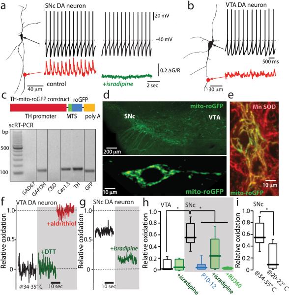
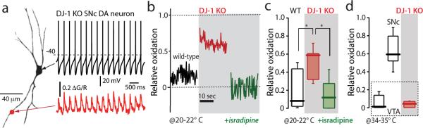
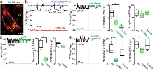
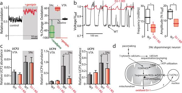
Parkinson's disease is a pervasive, ageing-related neurodegenerative disease the cardinal motor symptoms of which reflect the loss of a small group of neurons, the dopaminergic neurons in the substantia nigra pars compacta (SNc). Mitochondrial oxidant stress is widely viewed as being responsible for this loss, but why these particular neurons should be stressed is a mystery. Here we show, using transgenic mice that expressed a redox-sensitive variant of green fluorescent protein targeted to the mitochondrial matrix, that the engagement of plasma membrane L-type calcium channels during normal autonomous pacemaking created an oxidant stress that was specific to vulnerable SNc dopaminergic neurons. The oxidant stress engaged defences that induced transient, mild mitochondrial depolarization or uncoupling. The mild uncoupling was not affected by deletion of cyclophilin D, which is a component of the permeability transition pore, but was attenuated by genipin and purine nucleotides, which are antagonists of cloned uncoupling proteins. Knocking out DJ-1 (also known as PARK7 in humans and Park7 in mice), which is a gene associated with an early-onset form of Parkinson's disease, downregulated the expression of two uncoupling proteins (UCP4 (SLC25A27) and UCP5 (SLC25A14)), compromised calcium-induced uncoupling and increased oxidation of matrix proteins specifically in SNc dopaminergic neurons. Because drugs approved for human use can antagonize calcium entry through L-type channels, these results point to a novel neuroprotective strategy for both idiopathic and familial forms of Parkinson's disease.


Figures

Figure 1
Figure 1. Calcium influx through L-type calcium channels during pacemaking increases mitochondrial oxidant stress in SNc dopaminergic neurons
(a) Somatic whole cell recording from a SNc dopaminergic neuron (shown to the left as a projection image) in pacemaking mode. At the bottom of the panel, a 2PLSM measurement of dendritic Fluo-4 fluorescence (red trace) is shown. To the right are shown a similar set of measurements after application of isradipine; note the absence of a change in pacemaking rate but the loss of dendritic calcium transients (green trace)(n=10 neurons, P<0.05). (b) A similar set of measurements in a VTA dopaminergic neuron; these neurons consistently lacked dendritic calcium oscillations (n=6 neurons). (c) Schematic of the TH-mito-roGFP construct. Below the construct, single-cell RT-PCR analysis of mito-roGFP expressing SNc dopaminergic neuron showing expression of tyrosine hydroxylase (TH) and Cav1.3 calcium channel mRNA, but not calbindin (CBD) or GAD67 mRNA; similar results were obtained in all 5 neurons examined. (d) Top, a low magnification image of the mesencephalon of a transgenic TH-mito-roGFP mouse showing expression in SNc and VTA neurons. Bottom, a higher magnification image of a SNc neuron showing cytoplasmic but not nuclear labeling. (e) Overlay of Mn-SOD immunostaining (red) showing colocalization with mito-roGFP in cultured roGFP SNc neurons. (f) Mito-roGFP measurements from a VTA neuron; before (control, black trace), and after application of dithiothreitol (DTT) (green trace), and aldrithiol (red trace). Relative oxidation is plotted as described in Methods. (g) Mito-roGFP measurements from a SNc dopaminergic neuron (black trace) revealed a higher basal oxidation; treatment with isradipine diminished mitochondrial oxidation (green trace). (h) Left, box-plots summarizing mean redox measurements in control (n=9) and isradipine treated (n=5) VTA dopaminergic neurons; in control (n=14), isradipine (n=9) and Ru360 (n=8) treated SNc dopaminergic neurons; SNc neurons were significantly more oxidized than VTA neurons (P<0.05); both isradipine and Ru360 significantly reduced oxidation (P<0.05). SNc dopaminergic neurons from young mito-roGFP mice (P10-P12) were consistently less oxidized than adult neurons (P<0.05, n=8). (i) Box plots summarizing mean mito-roGFP measurements in SNc dopaminergic neurons in brain slices held at near physiological temperature (34-35°C) (n=14) and at room temperature (20-22°C) (n=5); oxidation was significantly less at room temperature (P<0.05). Statistical significance in all plots is shown by an asterisk and determined using a non-parametric test for comparing multiple groups (Kruskal-Wallis ANOVA with Dunnet's post hoc test).
Figure 2
Figure 2. Oxidant stress is elevated in SNc dopaminergic neurons from DJ-1 knockout mice
(a) Somatic whole cell recording from a SNc dopaminergic neuron in a brain slice from a DJ-1 knockout mouse, showing normal pacemaking (top) and intracellular calcium oscillations (bottom); similar results were obtained in all five neurons examined. (b) Mitochondrial mito-roGFP oxidation was higher (red trace) than in control SNc dopaminergic neurons (black trace); isradipine pretreatment normalized oxidation of mito-roGFP (green trace); experiments were done at 20-22 °C. (c) Box plot summarizing mean mito-roGFP measurements in wild-type SNc neurons (n=9), DJ-1 knockout SNc neurons (n=6) and DJ-1 knockout neurons after isradipine pretreatment (n=7); differences between wild-type and DJ-1 knockout were significant (P<0.05), as were differences between knockouts with and without isradipine treatment (P<0.05). (d) Box plot summarizing mean mito-roGFP measurements from wild-type VTA dopaminergic neurons (n=9), wild-type SNc dopaminergic neurons (n=14) and DJ-1 knockout VTA dopaminergic neurons (red box) (n=4) at 34-35 °C. VTA dopaminergic neurons were unaffected by DJ-1 deletion (P>0.05).
Figure 3
Figure 3. Mitochondrial flickering is dependent on superoxide production and recruitment of mitochondrial uncoupling proteins
(a) SNc dopaminergic neuron in a brain slice incubated with tetramethyl rhodamine methyl ester (TMRM) to label mitochondria; a region of interest (ROI) from which fluorescence measurements were taken is shown (yellow circle). (b) Representative fluorescence time-series from a SNc dopaminergic neuron (black trace) before and after rotenone (red trace) application; similar results were seen in all cells examined (n>20). For comparison, a time series from a typical VTA dopaminergic neuron is shown (blue trace) (n=10). (c) Left, TMRM fluorescence measurements before (black trace) and after bath application of isradipine (green trace); Right, box plots summarizing the mean frequency of flickering in control cells before and after application of isradipine (n=5) or Ru360 (n=6), both significantly slowed flickering frequency (P<0.05); box plots summarizing the mean amplitude of the relative voltage change inferred from the fluorescence measurement; the amplitudes were similar in all conditions (P>0.05). (d) Left, fluorescence time series before and after bath application of MPG (green trace); Right, box plots summarizing mean frequency and amplitude measurements (n=5); MPG significantly reduced the frequency (P<0.05) but not the amplitude (P>0.05) of flickering. (e) Left, fluorescence time series before and after application of genipin (green trace); genipin decreased the frequency and amplitude of the flickering; Right, box plots summarizing mean amplitude and frequency measurements before and after genipin (n=5); genipin significantly decreased both parameters (P<0.05). Both measurements were normal in cyclophilin D knockouts (n=10; P>0.05).
Figure 4
Figure 4. Loss of DJ-1 attenuated UCP-dependent flickering in mitochondrial membrane potential
(a) Left, mito-roGFP measurements in a SNc dopaminergic neuron (as in Figure 1) before and after application of genipin (red trace). Right, box plots summarizing mean mito-roGFP measurements following isradipine application (green box) (n=9) or genipin (red box) (n=6) to SNc dopaminergic neurons; isradipine significantly decreased oxidation, whereas genipin increased oxidation (P<0.05). Also show is a box plot of mito-roGFP measurements from VTA dopaminergic neurons following genipin; genipin had no effect on these measurements (n=5, P>0.05). (b) Left, TMRM fluorescence measurement from a wild-type SNc dopaminergic neuron (black trace) and a DJ-1 knockout (red trace). Right, box plots of mean frequency and amplitude data from wild-type (n=21) and DJ-1 knockout neurons (n=7); both amplitude and frequency of flickering were decreased in DJ-1 knockouts (P<0.05). (c) Quantitative PCR analysis of UCP expression in DJ-1 knockout mice: UCP2 mRNA abundance (relative to GAPDH) was not significantly altered in VTA and SNc (P>0.05, n=9). To the right, bar graphs plot the abundance of UCP4 and UCP5 mRNAs, normalized by that of UCP2 in each sample. The relative abundance of UCP4 and UCP5 mRNA was decreased in the SNc of DJ-1 knockout mice (P<0.05, n=9). UCP4 mRNA abundance was higher in VTA from DJ-1 knockout mice (P<0.05, n=9), while UCP5 mRNA was unchanged. (d) Schematic summary of the results presented linking calcium entry through L-type channels during pacemaking with elevated mitochondrial oxidant stress and opening of UCPs. The model also proposes that oxidized DJ-1 translocates to the nucleus and increases the transcription of UCP4 and UCP5, leading to increased UCP protein in the IMM.

Comment in

References

    1. Albin RL, Young AB, Penney JB. The functional anatomy of disorders of the basal ganglia. Trends in neurosciences. 1995;18:63–64. - PubMed
    1. Schapira AH. Mitochondria in the aetiology and pathogenesis of Parkinson's disease. Lancet Neurol. 2008;7:97–109. - PubMed
    1. Puopolo M, Raviola E, Bean BP. Roles of subthreshold calcium current and sodium current in spontaneous firing of mouse midbrain dopamine neurons. J Neurosci. 2007;27:645–656. - PMC - PubMed
    1. Chan CS, et al. ‘Rejuvenation’ protects neurons in mouse models of Parkinson's disease. Nature. 2007;447:1081–1086. doi:nature05865 [pii]10.1038/nature05865. - PubMed
    1. Khaliq ZM, Bean BP. Pacemaking in dopaminergic ventral tegmental area neurons: depolarizing drive from background and voltage-dependent sodium conductances. J Neurosci. 2010;30:7401–7413. doi:30/21/7401 [pii]10.1523/JNEUROSCI.0143-10.2010. - PMC - PubMed

Publication types

MeSH terms

Substances