Review
. 2010 Nov 11;468(7321):232-43.
doi: 10.1038/nature09613.

Glial and neuronal control of brain blood flow

Affiliations
Review

Glial and neuronal control of brain blood flow

David Attwell et al. Nature. .

Abstract

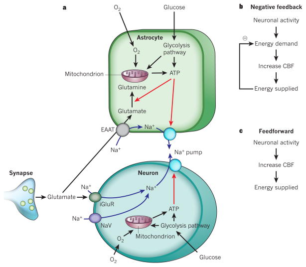
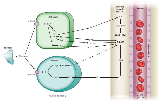
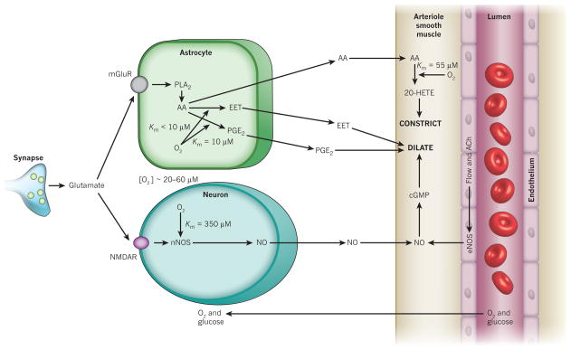
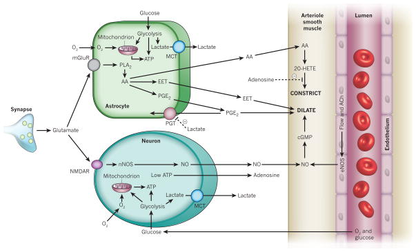
Blood flow in the brain is regulated by neurons and astrocytes. Knowledge of how these cells control blood flow is crucial for understanding how neural computation is powered, for interpreting functional imaging scans of brains, and for developing treatments for neurological disorders. It is now recognized that neurotransmitter-mediated signalling has a key role in regulating cerebral blood flow, that much of this control is mediated by astrocytes, that oxygen modulates blood flow regulation, and that blood flow may be controlled by capillaries as well as by arterioles. These conceptual shifts in our understanding of cerebral blood flow control have important implications for the development of new therapeutic approaches.


Conflict of interest statement

The authors declare no competing financial interests.

Figures

Figure 1
Figure 1. Energy supply, usage and blood flow regulation in the brain
a, ATP is generated from glycolysis and mitochondrial oxidative phosphorylation in neurons and glia. ATP is mainly consumed (red arrows) by ion pumping in neurons, to maintain the ion gradients underlying synaptic and action potentials, following Na+ entry (blue arrows) through ionotropic glutamate receptors (iGluR) and voltage-gated Na+ channels (NaV). It is also used in glia for Na+-coupled neurotransmitter uptake by excitatory amino acid transporters (EAAT) and for metabolic processing (shown for conversion of glutamate to glutamine), and on maintaining the cells’ resting potentials. b, The negative-feedback control hypothesis for vascular energy supply, in which a fall in energy level induces an increased cerebral blood flow (CBF). c, The feedforward regulation hypothesis for vascular energy supply.
Figure 2
Figure 2. Major pathways by which glutamate regulates cerebral blood flow
Pathways from astrocytes and neurons (left) that regulate blood flow by sending messengers (arrows) to influence the smooth muscle around the arterioles that supply oxygen and glucose to the cells (right, shown as the vessel lumen surrounded by endothelial cells and smooth muscle). In neurons, synaptically released glutamate acts on N-methyl-D-aspartate receptors (NMDAR) to raise [Ca2+]i, causing neuronal nitric oxide synthase (nNOS) to release NO, which activates smooth muscle guanylate cyclase. This generates cGMP to dilate vessels. Raised [Ca2+]i may also (dashed line) generate arachidonic acid (AA) from phospholipase A2 (PLA2), which is converted by COX2 to prostaglandins (PG) that dilate vessels. Glutamate raises [Ca2+]i in astrocytes by activating metabotropic glutamate receptors (mGluR), generating arachidonic acid and thus three types of metabolite: prostaglandins (by COX1/3, and COX2 in pathological situations) and EETs (by P450 epoxygenase) in astrocytes, which dilate vessels, and 20-HETE (by ω-hydroxylase) in smooth muscle, which constricts vessels. A rise of [Ca2+]i in astrocyte endfeet may activate Ca2+-gated K+ channels (gK(Ca)), releasing K+, which also dilates vessels.
Figure 3
Figure 3. Arachidonic acid metabolites that may contribute to control of cerebral blood flow
Arachidonic acid is formed from membrane phospholipids by Ca2+-dependent and Ca2+-independent lipases. Metabolites shown in green are vasodilators, red metabolites are vasoconstrictors, and blue denotes the location of some of the relevant enzymes. COX, cyclooxygenase; CYP, cytochrome P450 superfamily of enzymes; EET, epoxyeicosatrienoic acid; 20-HETE, 20-hydroxyeicosatetraenoic acid; HPETE, hydroperoxy-eicosatetraenoic acid.
Figure 4
Figure 4. Nitric oxide inhibits the production of key arachidonic acid-derived messengers
NO inhibits (dashed lines) the production of both the vasoconstricting 20-HETE and the vasodilating EETs,. NO also weakly stimulates COX1 and inhibits COX2 (not shown). Endothelial nitric oxide synthase (eNOS) can be activated by flow-induced shear stress or by acetylcholine (ACh). Other abbreviations as in earlier figures.
Figure 5
Figure 5. Oxygen differentially affects the synthesis of neurovascular messengers
The concentration of O2 in the extracellular space is 20–60 μM. This is significantly higher than the effective Km for O2 activating the enzymes synthesizing EETs and prostaglandins, but is in a range in which changes in O2 concentration will modulate the production of NO and 20-HETE. Abbreviations as in earlier figures.
Figure 6
Figure 6. Lactate and adenosine affect neurovascular signalling at low [O2]
Low O2 concentrations lead to mitochondrial oxidative phosphorylation failing to consume all the pyruvate produced by glycolysis, resulting in an export of lactate by monocarboxylate transporters (MCTs). Extracellular lactate inhibits the reuptake of PGE2 by the prostaglandin transporter (PGT), promoting vasodilation. Low energy levels also lead to the formation of adenosine, which inhibits 20-HETE-mediated arteriolar constriction by acting on adenosine A2A receptors. Other abbreviations as in earlier figures.

Similar articles

Cited by

References

    1. Attwell D, Laughlin SB. An energy budget for signaling in the grey matter of the brain. J Cereb Blood Flow Metab. 2001;21:1133–1145. - PubMed
    1. Leffler CW, Busija DW, Mirro R, Armstead WM, Beasley DG. Effects of ischemia on brain blood flow and oxygen consumption of newborn pigs. Am J Physiol. 1989;257:H1917–H1926. - PubMed
    1. Girouard H, Iadecola C. Neurovascular coupling in the normal brain and in hypertension, stroke, and Alzheimer disease. J Appl Physiol. 2006;100:328–335. - PubMed
    1. Baptiste DC, Fehlings M. Pharmacological approaches to repair the injured spinal cord. J Neurotrauma. 2006;23:318–334. - PubMed
    1. Tian R, et al. Role of extracellular and intracellular acidosis for hypercapnia-induced inhibition of tension of isolated rat cerebral arteries. Circ Res. 1995;76:269–275. - PubMed

Publication types

MeSH terms