

Biological implications of preformed mast cell mediators
- PMID: 21069421
- PMCID: PMC11114649
- DOI: 10.1007/s00018-010-0587-0
Biological implications of preformed mast cell mediators
Abstract
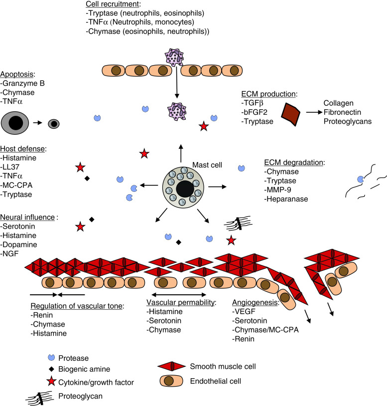
Mast cells store an impressive array of preformed compounds (mediators) in their secretory granules. When mast cells degranulate, these are released and have a profound impact on any condition in which mast cell degranulation occurs. The preformed mast cell mediators include well-known substances such as histamine, proteoglycans, proteases, and preformed cytokines, as well as several recently identified compounds. Mast cells have recently been implicated in a large number of novel pathological settings in addition to their well-established contribution to allergic reactions, and there is consequently a large current interest in the molecular mechanisms by which mast cells act in the context of a given condition. In many cases, preformed mast cell mediators have been shown to account for functions ascribed to mast cells, and these compounds are hence emerging as major players in numerous pathologies. In this review we summarize the current knowledge of preformed mast cell mediators.
Figures
Similar articles
-
Mast cell secretory granules: armed for battle.Nat Rev Immunol. 2014 Jul;14(7):478-94. doi: 10.1038/nri3690. Epub 2014 Jun 6. Nat Rev Immunol. 2014. PMID: 24903914 Review.
-
Mast cell proteoglycans.J Histochem Cytochem. 2012 Dec;60(12):950-62. doi: 10.1369/0022155412458927. Epub 2012 Aug 16. J Histochem Cytochem. 2012. PMID: 22899859 Free PMC article. Review.
-
Different mast cell mediators produced by different mast cell phenotypes.Ciba Found Symp. 1989;147:36-45; discussion 45-52. doi: 10.1002/9780470513866.ch4. Ciba Found Symp. 1989. PMID: 2695309 Review.
-
Distorted secretory granule composition in mast cells with multiple protease deficiency.J Immunol. 2013 Oct 1;191(7):3931-8. doi: 10.4049/jimmunol.1301441. Epub 2013 Aug 23. J Immunol. 2013. PMID: 23975861
-
Diverse exocytic pathways for mast cell mediators.Biochem Soc Trans. 2018 Apr 17;46(2):235-247. doi: 10.1042/BST20170450. Epub 2018 Feb 22. Biochem Soc Trans. 2018. PMID: 29472369 Free PMC article. Review.
Cited by
-
Bile and urine peptide marker profiles: access keys to molecular pathways and biological processes in cholangiocarcinoma.J Biomed Sci. 2020 Jan 3;27(1):13. doi: 10.1186/s12929-019-0599-5. J Biomed Sci. 2020. PMID: 31900160 Free PMC article.
-
Tryptase as a polyfunctional component of mast cells.Histochem Cell Biol. 2018 May;149(5):461-477. doi: 10.1007/s00418-018-1659-8. Epub 2018 Mar 12. Histochem Cell Biol. 2018. PMID: 29532158 Review.
-
Granzyme D is a novel murine mast cell protease that is highly induced by multiple pathways of mast cell activation.Infect Immun. 2013 Jun;81(6):2085-94. doi: 10.1128/IAI.00290-13. Epub 2013 Mar 25. Infect Immun. 2013. PMID: 23529614 Free PMC article.
-
Cyclic Hypoxia Induces Transcriptomic Changes in Mast Cells Leading to a Hyperresponsive Phenotype after FcεRI Cross-Linking.Cells. 2022 Jul 19;11(14):2239. doi: 10.3390/cells11142239. Cells. 2022. PMID: 35883682 Free PMC article.
-
Nuclear receptor 4a3 (nr4a3) regulates murine mast cell responses and granule content.PLoS One. 2014 Feb 20;9(2):e89311. doi: 10.1371/journal.pone.0089311. eCollection 2014. PLoS One. 2014. PMID: 24586680 Free PMC article.
References
-
- Marshall JS. Mast-cell responses to pathogens. Nat Rev Immunol. 2004;4:787–799. - PubMed
-
- Lu LF, Lind EF, Gondek DC, Bennett KA, Gleeson MW, Pino-Lagos K, Scott ZA, Coyle AJ, Reed JL, Van Snick J, Strom TB, Zheng XX, Noelle RJ. Mast cells are essential intermediaries in regulatory T-cell tolerance. Nature. 2006;442:997–1002. - PubMed
-
- Grimbaldeston MA, Nakae S, Kalesnikoff J, Tsai M, Galli SJ. Mast cell-derived interleukin 10 limits skin pathology in contact dermatitis and chronic irradiation with ultraviolet B. Nat Immunol. 2007;8:1095–1104. - PubMed
-
- Blank U, Rivera J. The ins and outs of IgE-dependent mast-cell exocytosis. Trends Immunol. 2004;25:266–273. - PubMed
Publication types
MeSH terms
Substances
LinkOut - more resources
Full Text Sources
