Review
. 2011 Feb;68(4):599-612.
doi: 10.1007/s00018-010-0550-0. Epub 2010 Oct 31.

The physiological roles of phosducin: from retinal function to stress-dependent hypertension

Affiliations
Review

The physiological roles of phosducin: from retinal function to stress-dependent hypertension

Nadine Beetz et al. Cell Mol Life Sci. 2011 Feb.

Abstract

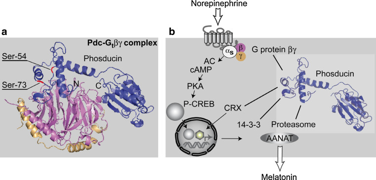
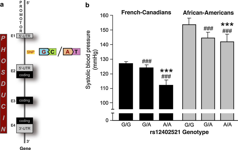
In the time since its discovery, phosducin's functions have been intensively studied both in vivo and in vitro. Phosducin's most important biochemical feature in in vitro studies is its binding to heterotrimeric G protein βγ-subunits. Data on phosducin's in vivo relevance, however, have only recently been published but expand the range of biological actions, as shown both in animal models as well as in human studies. This review gives an overview of different aspects of phosducin biology ranging from structure, phylogeny of phosducin family members, posttranscriptional modification, biochemical features, localization and levels of expression to its physiological functions. Special emphasis will be placed on phosducin's function in the regulation of blood pressure. In the second part of this article, findings concerning cardiovascular regulation and their clinical relevance will be discussed on the basis of recently published data from gene-targeted mouse models and human genetic studies.


Figures

Fig. 1
Fig. 1
Sequence alignment of phosducin or phosducin-like orphan protein from different species. Sequences were aligned using ClustalW alignment tool available from EMBL–EBI (http://www.ebi.ac.uk/Tools/clustaw/index.html) [143]. Colour code marks degree of homology between species
Fig. 2
Fig. 2
a Structure of phosducin-Gtβγ complex in the retina as assessed after crystallography by X-ray analysis, phosducin (blue), G protein β-subunit (magenta), γ-subunit (beige) [43]. Phosducin’s N- and C-terminal domains are marked with N and C, respectively. Note the extensive interaction area between N-terminal phosducin and the top surface of the β-subunit. In the N-terminal domain, Ser-54 and Ser-73, as the two most important phosphorylation sites, are indicated in red. b Schematic illustration of phosducin’s proposed biochemical features and its putative binding partners in pinealocytes or other cells (βγ, CRX, 14-3-3ζ, proteasomal subunits) [56, 83, 84]
Fig. 3
Fig. 3
Phosducin’s function in mice. a, b Post-operative systolic and diastolic blood pressure after surgery for implantation of telemetric blood pressure devices; *p < 0.05, n = 5–9 per genotype. c Schematic presentation of the fate of norepinephrine after its exocytosis into the synaptic cleft (DHPG dihydroxyphenylglycol, NMN normetanephrine, MAO monoaminoxidase, COMT catechol-O-methyltransferase). d Circulating plasma norepinephrine levels; ***p < 0.001, n = 6–9 per genotype. e, f Neuronal firing in isolated sympathetic neurons upon acetylcholine administration, representative traces of a wild-type and a knockout neuron are shown (ACh acetylcholine). g Potassium conductivity in sympathetic neurons depolarized by current injection; *p < 0.05, n = 5–8 per genotype. h Sympathetic neurons isolated from the superior cervical ganglion, cultured and stained with an antibody against tyrosine hydroxylase. (Figure reproduced with permission of the American Society for Clinical Investigation [40])
Fig. 4
Fig. 4
Overview of the sympathetic nervous system neuronal chain
Fig. 5
Fig. 5
Phosducin’s function in essential hypertension. a Schematic illustration of the phosducin gene, the SNP rs12402521 is indicated between exon 1 and 2. b Average wake systolic blood pressure in French-Canadian and African-American populations according to the SNP rs12402521 genotype. ### p < 0.0001 versus G/G, ***p < 0.0001 versus G/A genotype. (Figure reproduced with permission of the American Society for Clinical Investigation [40])

Similar articles

Cited by

References

    1. Lolley RN, Brown BM, Farber DB. Protein phosphorylation in rod outer segments from bovine retina: cyclic nucleotide-activated protein kinase and its endogenous substrate. Biochem Biophys Res Commun. 1977;78:572–578. doi: 10.1016/0006-291X(77)90217-0. - DOI - PubMed
    1. Lee RH, Brown BM, Lolley RN. Light-induced dephosphorylation of a 33K protein in rod outer segments of rat retina. Biochemistry. 1984;23:1972–1977. doi: 10.1021/bi00304a014. - DOI - PubMed
    1. Kuo CH, Akiyama M, Miki N. Isolation of a novel retina-specific clone (MEKA cDNA) encoding a photoreceptor soluble protein. Brain Res Mol Brain Res. 1989;6:1–10. doi: 10.1016/0169-328X(89)90022-3. - DOI - PubMed
    1. Bauer PH, Muller S, Puzicha M, Pippig S, Obermaier B, Helmreich EJ, Lohse MJ. Phosducin is a protein kinase A-regulated G-protein regulator. Nature. 1992;358:73–76. doi: 10.1038/358073a0. - DOI - PubMed
    1. Lee RH, Fowler A, McGinnis JF, Lolley RN, Craft CM. Amino acid and cDNA sequence of bovine phosducin, a soluble phosphoprotein from photoreceptor cells. J Biol Chem. 1990;265:15867–15873. - PubMed

Publication types

MeSH terms