. 2011 Oct;129(3):691-701.
doi: 10.1007/s10549-010-1239-7. Epub 2010 Nov 11.

Local anesthetics inhibit kinesin motility and microtentacle protrusions in human epithelial and breast tumor cells

Affiliations

Local anesthetics inhibit kinesin motility and microtentacle protrusions in human epithelial and breast tumor cells

Jennifer R Yoon et al. Breast Cancer Res Treat. 2011 Oct.

Abstract

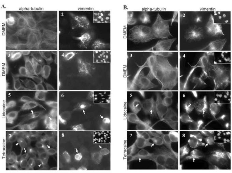
Detached breast tumor cells produce dynamic microtubule protrusions that promote reattachment of cells and are termed tubulin microtentacles (McTNs) due to their mechanistic distinctions from actin-based filopodia/invadopodia and tubulin-based cilia. McTNs are enriched with vimentin and detyrosinated α-tubulin, (Glu-tubulin). Evidence suggests that vimentin and Glu-tubulin are cross-linked by kinesin motor proteins. Using known kinesin inhibitors, Lidocaine and Tetracaine, the roles of kinesins in McTN formation and function were tested. Live-cell McTN counts, adhesion assays, immunofluorescence, and video microscopy were performed to visualize inhibitor effects on McTNs. Viability and apoptosis assays were used to confirm the non-toxicity of the inhibitors. Treatments of human non-tumorigenic mammary epithelial and breast tumor cells with Lidocaine or Tetracaine caused rapid collapse of vimentin filaments. Live-cell video microscopy demonstrated that Tetracaine reduces motility of intracellular GFP-kinesin and causes centripetal collapse of McTNs. Treatment with Tetracaine inhibited the extension of McTNs and their ability to promote tumor cell aggregation and reattachment. Lidocaine showed similar effects but to a lesser degree. Our current data support a model in which the inhibition of kinesin motor proteins by Tetracaine leads to the reductions in McTNs, and provides a novel mechanism for the ability of this anesthetic to decrease metastatic progression.


Figures

FIGURE 1
FIGURE 1. Tetracaine is an effective inhibitor of McTN protrusions in MCF10A and MDA-MB-436 cells
MCF10A human mammary epithelial cells and MDA-MB-436 human breast cancer cells were transfected with GFP-Membrane and scored for microtentacle (McTN) generation. A-C) Representation of McTN types counted and not counted A) DMEM (positive) B) 5μM LA (positive) C) 0.25mM Tetracaine + LA (negative) D) MCF10A n=4 E) MDA-MB-436 n=4 McTN counts D,E) MCF10A and MDA-MB-436 cells either under No Treatment, 5μM LA, 1mM Lidocaine +/− 5μMLA, or 0.25mM Tetracaine +/− 5μMLA. Cells were blindly scored positive when exhibiting two or more McTNs that extended greater than the radius of the cell body. Three independent experiments with at least 100 GFP-positive cells were counted for each bar. In MCF10A cells, 1mM Lidocaine and 0.25mM Tetracaine were effective in inhibiting McTN +/− LA. In MDA-MB-436 cells, only 0.25mM Tetracaine was effective in inhibiting McTNs +/− LA but not effective in 1mM Lidocaine. * indicates significant difference compared to that of No Treatment. ** indicates significant difference compared to that of 5μM LA treatment. Statistical analyses were done with an ANOVA test, p<0.05.
FIGURE 2
FIGURE 2. Lidocaine and Tetracaine do not affect cellular viability or induce PARP at the concentrations that inhibit microtentacle protrusions
A,B) XTT Viability Assay on MCF10A, MCF10A-Bcl2, and MDA-MB-436 cells in a 96-well plate for 4hrs. Cells are viable after 4 hours of treatment at the concentrations 1mM Lidocaine and 0.25mM Tetracaine. (n=3, each trial done in triplicate.) C) Western Blot: MCF10A, MCF10A-Bcl2, and MDA-MB-436 samples with/without Lidocaine and Tetracaine treatments were run on 12% polyacrylamide gels and blotted for PARP cleavage, Vimentin, Bcl2, and actin. MDA-MB-436 was additionally treated with 1μg/ml TRAIL for two hours as a positive control for cleaved PARP. Lidocaine and Tetracaine do not cause significant PARP cleavage in the three cell lines tested at the working concentrations of 1mM Lidocaine and .25mM Tetracaine after 4 hours of treatment.
FIGURE 3
FIGURE 3. Anesthetic treatment causes collapse of vimentin intermediate filaments
MCF10A cells (A) and MDA-MB-436 cells (B) were fixed and immunostained for α-tubulin and vimentin. 1,2) T=0 in DMEM 3,4) T=30 minutes in DMEM 5,6) DMEM+1mM Lidocaine 7,8) DMEM+0.25mM Tetracaine. Arrows point to areas of vimentin collapse at T=30 minutes with 1mM Lidocaine and 0.25mM Tetracaine treatment with corresponding arrows in α-tubulin. Vimentin collapse occurs in both MCF10A and MDA-MB-436 cells at T=30 minutes in both 1mM Lidocaine and 0.25mM Tetracaine treatment. Arrowheads (A,7) point to focal points of α-tubulin collapse seen in MCF10A cells. Hoescht DNA stain in top, right insets.
FIGURE 4
FIGURE 4. Anesthetic treatment of suspended MDA-MB-436 cells cause the collapse of vimentin and α-tubulin
Suspended MDA-MD-436 cells were left untreated (A,B) or treated with 0.25mM Tetracaine (C,D) fixed and immunostained for vimentin (A,C) or α–tubulin (B,D) after 15 minutes of suspension. Arrows point to microtentacles with vimentin and α–tubulin staining in the control cells (A, B). Suspended cells treated with Tetracaine show decreased vimentin extension and microtentacle protrusions (C, D).
FIGURE 5
FIGURE 5. Lidocaine and Tetracaine impede reattachment of human MECs
MCF10A (A), MCF10A-Bcl2 (B), and MDA-MB-436 breast tumor cells (C). Attachment was measured through impedance of electrical current using the xCelligence RTCA SP real-time cell sensing instrument in the presence of Lidocaine (1mM) or Tetracaine (0.25mM) as indicated.
FIGURE 6
FIGURE 6. Tetracaine causes the reabsorption of microtentacle protrusions
Still, time-lapse images of suspended MDA-MD-436 cells in Phenol red-free DMEM. Cells have been allowed to partially attach to the bottom of a glass culture dish and filmed using DIC microscopy. In control cells, microtentacles persist up to 12 minutes but when treated with 0.125mM Tetracaine, microtentacles collapse and are reabsorbed completely by 12 minutes. Time-lapse movies in supplemental data.
FIGURE 7
FIGURE 7. GFP-KIF5C forward motion is inhibited by Tetracaine
MCF10A human MECs were transfected with a GFP-tagged full-length kinesin-1 motor protein, GFP-KIF5C. GFP-KIF5C movements were observed using live-cell microscopy within MCF10A cells before treatment (A) and after treatment with Tetracaine (B) where velocities of selected GFP speckles were observed and tracked. Significant differences in velocities are observed. (n=3, P<0.05, t-test) (C).

References

    1. Nicolson GL, Fidler IJ, Poste G. Effects of tertiary amine local anesthetics on the blood-borne implantation and cell surface properties of metastatic mouse melanoma cells. J Natl Cancer Inst. 1986;76:511–9. - PubMed
    1. Whipple RA, Cheung AM, Martin SS. Detyrosinated microtubule protrusions in suspended mammary epithelial cells promote reattachment. Exp Cell Res. 2007;313:1326–36. - PMC - PubMed
    1. Whipple RA, Balzer EM, Cho EH, Matrone MA, Yoon JR, Martin SS. Vimentin filaments support extension of tubulin-based microtentacles in detached breast tumor cells. Cancer Res. 2008;68:5678–88. - PMC - PubMed
    1. Korb T, Schluter K, Enns A, Spiegel HU, Senninger N, Nicolson GL, Haier J. Integrity of actin fibers and microtubules influences metastatic tumor cell adhesion. Exp Cell Res. 2004;299:236–47. - PubMed
    1. Machesky LM. Lamellipodia and filopodia in metastasis and invasion. FEBS Lett. 2008 - PubMed

Publication types

MeSH terms