

A new generation fatty acid amide hydrolase inhibitor protects against kainate-induced excitotoxicity
- PMID: 21069475
- PMCID: PMC4124033
- DOI: 10.1007/s12031-010-9472-4
A new generation fatty acid amide hydrolase inhibitor protects against kainate-induced excitotoxicity
Abstract
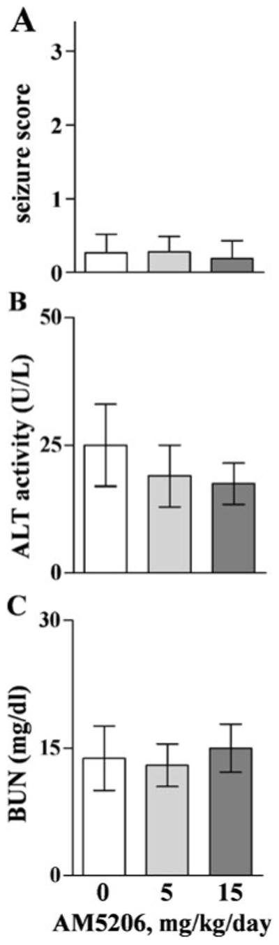
Endocannabinoids, including anandamide (AEA), have been implicated in neuroprotective on-demand responses. Related to such a response to injury, an excitotoxic kainic acid (KA) injection (i.p.) was found to increase AEA levels in the brain. To modulate the endocannabinoid response during events of excitotoxicity in vitro and in vivo, we utilized a new generation compound (AM5206) that selectively inhibits the AEA deactivating enzyme fatty acid amide hydrolase (FAAH). KA caused calpain-mediated spectrin breakdown, declines in synaptic markers, and disruption of neuronal integrity in cultured hippocampal slices. FAAH inhibition with AM5206 protected against the neurodegenerative cascade assessed in the slice model 24 h postinsult. In vivo, KA administration induced seizures and the same neurodegenerative events exhibited in vitro. When AM5206 was injected immediately after KA in rats, the seizure scores were markedly reduced as were levels of cytoskeletal damage and synaptic protein decline. The pre- and postsynaptic proteins were protected by the FAAH inhibitor to levels comparable to those found in healthy control brains. These data support the idea that endocannabinoids are released and converge on pro-survival pathways that prevent excitotoxic progression.
Figures


Similar articles
-
Endocannabinoid enhancement protects against kainic acid-induced seizures and associated brain damage.J Pharmacol Exp Ther. 2007 Sep;322(3):1059-66. doi: 10.1124/jpet.107.120147. Epub 2007 Jun 1. J Pharmacol Exp Ther. 2007. PMID: 17545313
-
Inhibitor of Endocannabinoid Deactivation Protects Against In Vitro and In Vivo Neurotoxic Effects of Paraoxon.J Mol Neurosci. 2017 Sep;63(1):115-122. doi: 10.1007/s12031-017-0963-4. Epub 2017 Aug 12. J Mol Neurosci. 2017. PMID: 28803438
-
Equipotent inhibition of fatty acid amide hydrolase and monoacylglycerol lipase - dual targets of the endocannabinoid system to protect against seizure pathology.Neurotherapeutics. 2012 Oct;9(4):801-13. doi: 10.1007/s13311-011-0100-y. Neurotherapeutics. 2012. PMID: 22270809 Free PMC article.
-
Fatty acid amide hydrolase inhibitors--progress and potential.CNS Neurol Disord Drug Targets. 2011 Aug;10(5):545-58. doi: 10.2174/187152711796234989. CNS Neurol Disord Drug Targets. 2011. PMID: 21631410 Review.
-
Enhancement of endocannabinoid signaling by fatty acid amide hydrolase inhibition: a neuroprotective therapeutic modality.Life Sci. 2010 Apr 10;86(15-16):615-23. doi: 10.1016/j.lfs.2009.06.003. Epub 2009 Jun 13. Life Sci. 2010. PMID: 19527737 Free PMC article. Review.
Cited by
-
Commonalities for comorbidity: Overlapping features of the endocannabinoid system in depression and epilepsy.Front Psychiatry. 2022 Oct 19;13:1041460. doi: 10.3389/fpsyt.2022.1041460. eCollection 2022. Front Psychiatry. 2022. PMID: 36339877 Free PMC article. Review.
-
Impaired 2-AG Signaling in Hippocampal Glutamatergic Neurons: Aggravation of Anxiety-Like Behavior and Unaltered Seizure Susceptibility.Int J Neuropsychopharmacol. 2015 Aug 1;19(2):pyv091. doi: 10.1093/ijnp/pyv091. Int J Neuropsychopharmacol. 2015. PMID: 26232789 Free PMC article.
-
Binge Alcohol Exposure Transiently Changes the Endocannabinoid System: A Potential Target to Prevent Alcohol-Induced Neurodegeneration.Brain Sci. 2017 Nov 29;7(12):158. doi: 10.3390/brainsci7120158. Brain Sci. 2017. PMID: 29186065 Free PMC article.
-
Disease-modifying effects of RHC80267 and JZL184 in a pilocarpine mouse model of temporal lobe epilepsy.CNS Neurosci Ther. 2014 Oct;20(10):905-15. doi: 10.1111/cns.12302. Epub 2014 Jul 3. CNS Neurosci Ther. 2014. PMID: 24989980 Free PMC article.
-
Early Synaptic Alterations and Selective Adhesion Signaling in Hippocampal Dendritic Zones Following Organophosphate Exposure.Sci Rep. 2019 Apr 25;9(1):6532. doi: 10.1038/s41598-019-42934-z. Sci Rep. 2019. PMID: 31024077 Free PMC article.
References
-
- Araujo BH, Torres LB, Cossa AC, Naffah-Mazzacoratti Mda G, Cavalheiro EA. Hippocampal expression and distribution of CB1 receptors in the Amazonian rodent Proechimys: an animal model of resistance to epilepsy. Brain Res. 2010;1335:35–40. - PubMed
-
- Arida RM, Scorza FA, de Amorim CR, Cavalheiro EA. Proechimys guyannensis: an animal model of resistance to epilepsy. Epilepsia. 2005;46(Suppl 5):189–197. - PubMed
-
- Bahr BA. Long-term hippocampal slices: a model system for investigating synaptic mechanisms and pathologic processes. J Neurosci Res. 1995;42:294–305. - PubMed
-
- Bahr BA, Abai B, Gall CM, Vanderklish PW, Hoffman KB, Lynch G. Induction of β-amyloid-containing polypeptides in hippocampus: evidence for a concomitant loss of synaptic proteins and interactions with an excitotoxin. Exp Neurol. 1994;129:81–94. - PubMed
-
- Bahr BA, Bendiske J, Brown QB, Munirathinam S, Caba E, Rudin M, Urwyler S, Sauter A, Rogers G. Survival signaling and selective neuroprotection through glutamatergic transmission. Exp Neurol. 2002;174:37–47. - PubMed
Publication types
MeSH terms
Substances
Grants and funding
LinkOut - more resources
Full Text Sources
